WB
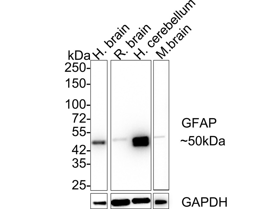
Western blot analysis of GFAP on different lysates with Rabbit anti-GFAP antibody at 1/5,000 dilution. Lane 1: Human brain tissue lysate, Lane 2: Rat brain tissue lysate, Lane 3: Human cerebellum tissue lysate, Lane 4: Mouse brain tissue lysate, Lysates/proteins at 20 µg/Lane. Exposure time: 11 seconds; 4-20% SDS-PAGE gel. Proteins were transferred to a PVDF membrane and blocked with 5% NFDM/TBST for 1 hour at room temperature. The primary antibody at 1/5,000 dilution was used in 5% NFDM/TBST at 4℃ overnight. Goat Anti-Rabbit IgG - HRP Secondary Antibody at 1/50,000 dilution was used for 1 hour at room temperature.IHC
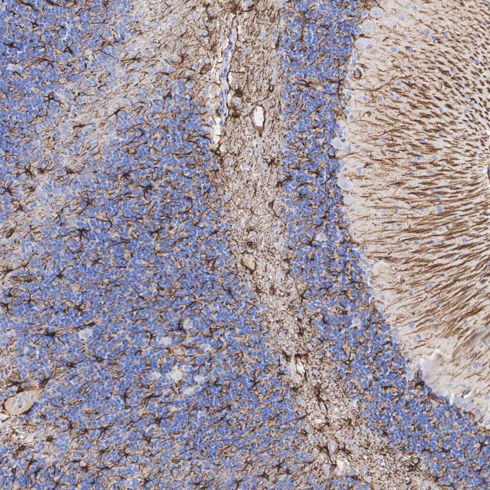
Immunohistochemical analysis of paraffin-embedded rat cerebellum tissue with Rabbit anti-GFAP antibody at 1/1,000 dilution. The section was pre-treated using heat mediated antigen retrieval with Tris-EDTA buffer (pH 9.0) for 20 minutes. The tissues were blocked in 1% BSA for 20 minutes at room temperature, washed with ddH2O and PBS, and then probed with the primary antibody at 1/1,000 dilution for 1 hour at room temperature. The detection was performed using an HRP conjugated compact polymer system. DAB was used as the chromogen. Tissues were counterstained with hematoxylin and mounted with DPX.IHC
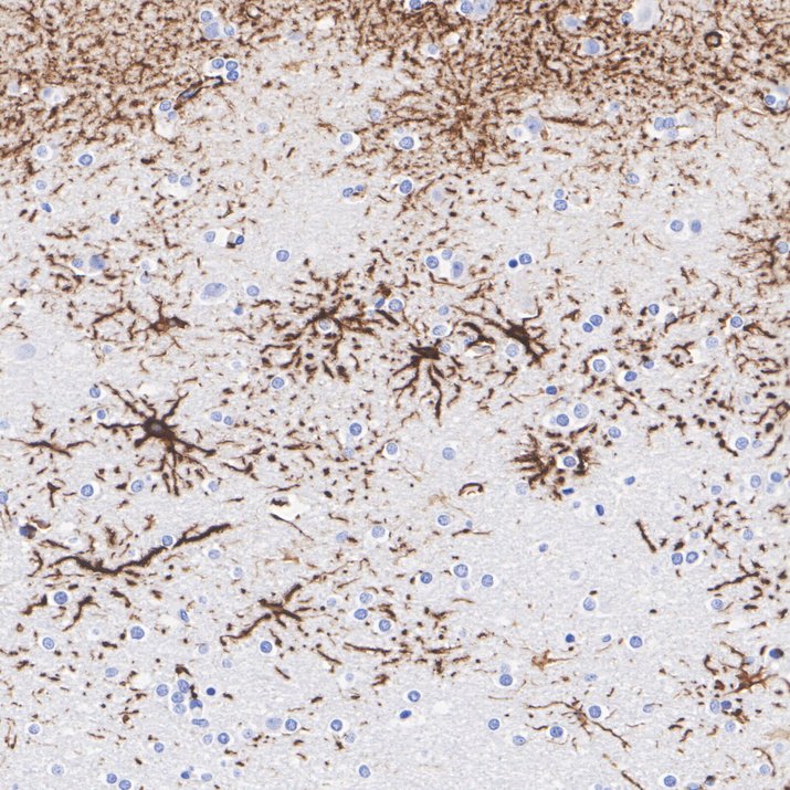
Immunohistochemical analysis of paraffin-embedded human brain tissue with Rabbit anti-GFAP antibody at 1/1,000 dilution. The section was pre-treated using heat mediated antigen retrieval with Tris-EDTA buffer (pH 9.0) for 20 minutes. The tissues were blocked in 1% BSA for 20 minutes at room temperature, washed with ddH2O and PBS, and then probed with the primary antibody at 1/1,000 dilution for 1 hour at room temperature. The detection was performed using an HRP conjugated compact polymer system. DAB was used as the chromogen. Tissues were counterstained with hematoxylin and mounted with DPX.IHC
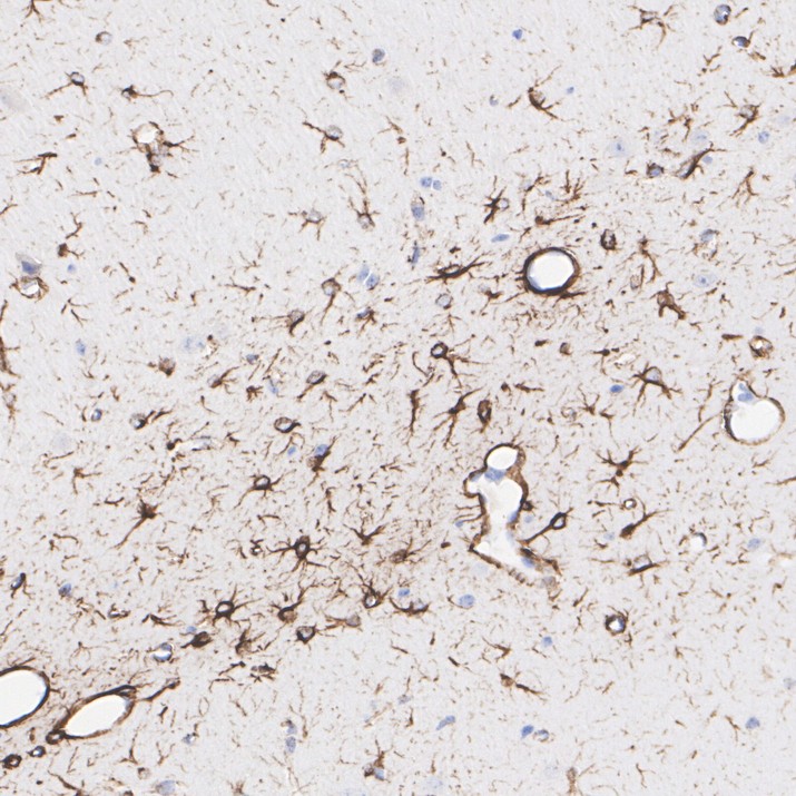
Immunohistochemical analysis of paraffin-embedded mouse cerebral cortex tissue with Rabbit anti-GFAP antibody at 1/1,000 dilution. The section was pre-treated using heat mediated antigen retrieval with Tris-EDTA buffer (pH 9.0) for 20 minutes. The tissues were blocked in 1% BSA for 20 minutes at room temperature, washed with ddH2O and PBS, and then probed with the primary antibody at 1/1,000 dilution for 1 hour at room temperature. The detection was performed using an HRP conjugated compact polymer system. DAB was used as the chromogen. Tissues were counterstained with hematoxylin and mounted with DPX.IF-P
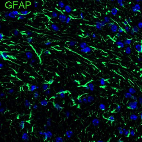
Species: Mouse, Site: Cerebellum, Sample: Paraffin-embedded section, Antibody concentration: 1/500.IF-P
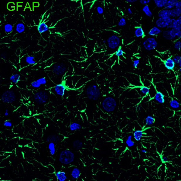
Species: Rat, Site: Cerebral cortex, Sample: Paraffin-embedded section, Antibody concentration: 1/500.IF-F
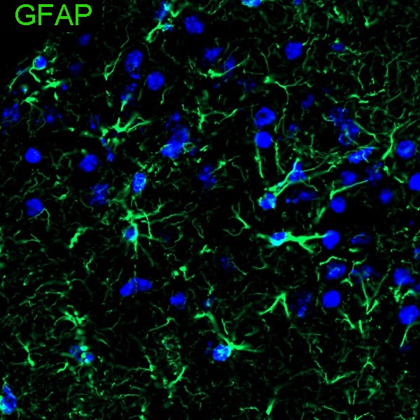
Species: Mouse, Site: Cerebral cortex, Sample: Frozen section (Fresh), Antibody concentration: 1/200, Antigen retrieval: Not required.IF-F
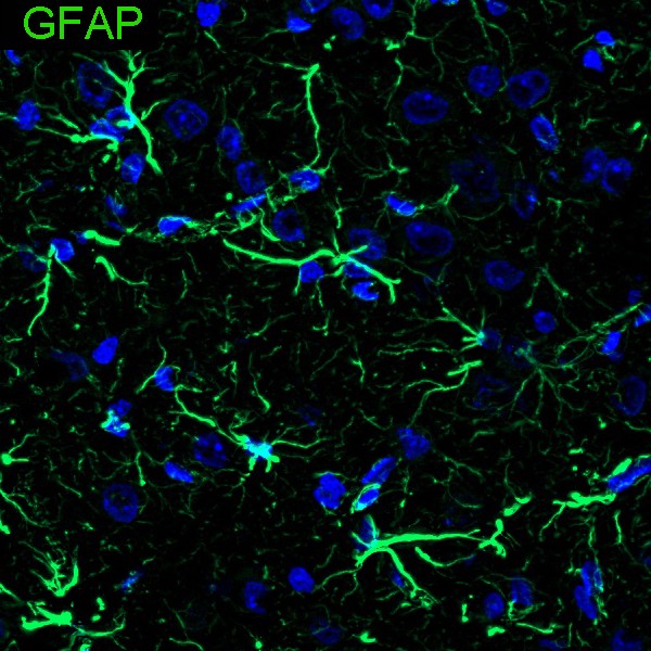
Species: Rat, Site: Cerebral cortex, Sample: Frozen section, Antibody concentration: 1/500, Antigen retrieval: Not required.| Product Name | GFAP Recombinant Rabbit Monoclonal Antibody |
|---|---|
| Antibody Type | Primary Antibodies |
| Product description | GFAP, a class-III intermediate filament, is a cell-specific marker that, during the development of the central nervous system, distinguishes astrocytes from other glial cells. In particular, vimentin filaments are present at early developmental stages, while GFAP filaments are characteristic of differentiated and mature brain astrocytes. In addition, GFAP intermediate filaments are also present in nonmyelin-forming Schwann cells in the peripheral nervous system. |
| Immunogen | Synthetic peptide within Human GFAP aa 1-50 / 432. |
| Clonality | monoclonal |
|---|---|
| Isotype | IgG |
| Host Species | Recombinant rabbit |
| Tested Applications | IF-FIF-PIHCWB |
| WB:1:5000 IHC:1:500-1:1000 IF-P:1:200-1:500 IF-F:1:200-1:500 |
|
| Species Reactivity | HumanMouseRat |
| Concentration | 1mg/ml |
| Purification | Protein A |
| Alternative Names | wu:fb34h11 antibody ALXDRD antibody cb345 antibody etID36982.3 antibody FLJ42474 antibody FLJ45472 antibody GFAP antibody GFAP_HUMAN antibody gfapl antibody Glial fibrillary acidic protein antibody Intermediate filament protein antibody wu:fk42c12 antibody xx:af506734 antibody zgc:110485 antibody |
|---|---|
| Molecular Weight(MW) | 50 kDa |
| Cellular Localization | Cytoplasm |
WB
Western blot analysis of GFAP on different lysates with Rabbit anti-GFAP antibody at 1/5,000 dilution. Lane 1: Human brain tissue lysate, Lane 2: Rat brain tissue lysate, Lane 3: Human cerebellum tissue lysate, Lane 4: Mouse brain tissue lysate, Lysates/proteins at 20 µg/Lane. Exposure time: 11 seconds; 4-20% SDS-PAGE gel. Proteins were transferred to a PVDF membrane and blocked with 5% NFDM/TBST for 1 hour at room temperature. The primary antibody at 1/5,000 dilution was used in 5% NFDM/TBST at 4℃ overnight. Goat Anti-Rabbit IgG - HRP Secondary Antibody at 1/50,000 dilution was used for 1 hour at room temperature.
IHC
Immunohistochemical analysis of paraffin-embedded rat cerebellum tissue with Rabbit anti-GFAP antibody at 1/1,000 dilution. The section was pre-treated using heat mediated antigen retrieval with Tris-EDTA buffer (pH 9.0) for 20 minutes. The tissues were blocked in 1% BSA for 20 minutes at room temperature, washed with ddH2O and PBS, and then probed with the primary antibody at 1/1,000 dilution for 1 hour at room temperature. The detection was performed using an HRP conjugated compact polymer system. DAB was used as the chromogen. Tissues were counterstained with hematoxylin and mounted with DPX.
IHC
Immunohistochemical analysis of paraffin-embedded human brain tissue with Rabbit anti-GFAP antibody at 1/1,000 dilution. The section was pre-treated using heat mediated antigen retrieval with Tris-EDTA buffer (pH 9.0) for 20 minutes. The tissues were blocked in 1% BSA for 20 minutes at room temperature, washed with ddH2O and PBS, and then probed with the primary antibody at 1/1,000 dilution for 1 hour at room temperature. The detection was performed using an HRP conjugated compact polymer system. DAB was used as the chromogen. Tissues were counterstained with hematoxylin and mounted with DPX.
IHC
Immunohistochemical analysis of paraffin-embedded mouse cerebral cortex tissue with Rabbit anti-GFAP antibody at 1/1,000 dilution. The section was pre-treated using heat mediated antigen retrieval with Tris-EDTA buffer (pH 9.0) for 20 minutes. The tissues were blocked in 1% BSA for 20 minutes at room temperature, washed with ddH2O and PBS, and then probed with the primary antibody at 1/1,000 dilution for 1 hour at room temperature. The detection was performed using an HRP conjugated compact polymer system. DAB was used as the chromogen. Tissues were counterstained with hematoxylin and mounted with DPX.
IF-P
Species: Mouse, Site: Cerebellum, Sample: Paraffin-embedded section, Antibody concentration: 1/500.
IF-P
Species: Rat, Site: Cerebral cortex, Sample: Paraffin-embedded section, Antibody concentration: 1/500.
IF-F
Species: Mouse, Site: Cerebral cortex, Sample: Frozen section (Fresh), Antibody concentration: 1/200, Antigen retrieval: Not required.
IF-F
Species: Rat, Site: Cerebral cortex, Sample: Frozen section, Antibody concentration: 1/500, Antigen retrieval: Not required.| Positive Control | Rat spinal cord tissue, rat brain tissue, mouse spinal cord tissue, mouse brain tissue. |
|---|---|
| Application Notes | WB:1:5000 IHC:1:500-1:1000 IF-P:1:200-1:500 IF-F:1:200-1:500 |
| Form | Liquid |
|---|---|
| Storage Instructions | Store at +4℃ after thawing. Aliquot store at -20℃ or -80℃. Avoid repeated freeze / thaw cycles. |
| Storage Buffer | 1*TBS (pH7.4), 1%BSA, 40%Glycerol. Preservative: 0.05% Sodium Azide. |


