WB
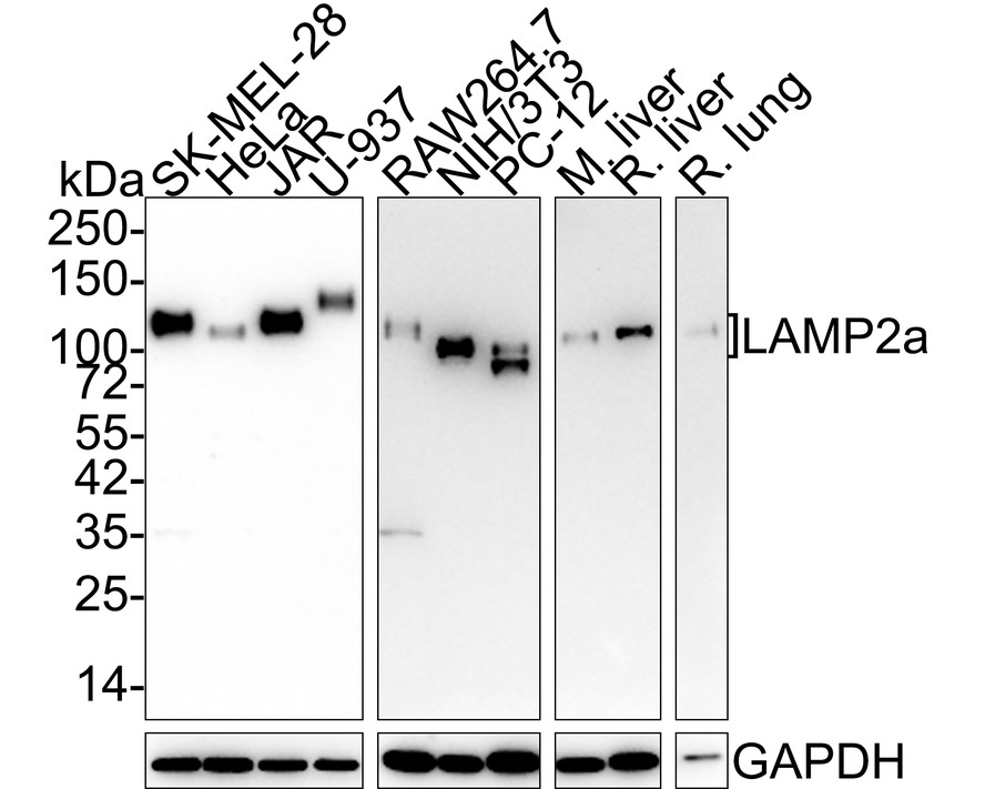
Western blot analysis of LAMP2a on different lysates with Rabbit anti-LAMP2a antibody at 1/5,000 dilution. Lane 1: SK-MEL-28 cell lysate (20 µg/Lane), Lane 2: HeLa cell lysate (20 µg/Lane), Lane 3: JAR cell lysate (20 µg/Lane), Lane 4: U-937 cell lysate (20 µg/Lane), Lane 5: RAW264.7 cell lysate (15 µg/Lane), Lane 6: NIH/3T3 cell lysate (15 µg/Lane), Lane 7: PC-12 cell lysate (15 µg/Lane), Lane 8: Mouse liver tissue lysate (30 µg/Lane), Lane 9: Rat liver tissue lysate (30 µg/Lane), Lane 10: Rat lung tissue lysate (30 µg/Lane), Exposure time: Lane 1-4: 2 minutes 37 seconds; Lane 5-10: 5 minutes; 4-20% SDS-PAGE gel. Proteins were transferred to a PVDF membrane and blocked with 5% NFDM/TBST for 1 hour at room temperature. The primary antibody at 1/5,000 dilution was used in 5% NFDM/TBST at 4℃ overnight. Goat Anti-Rabbit IgG - HRP Secondary Antibody at 1:50,000 dilution was used for 1 hour at room temperature.IHC
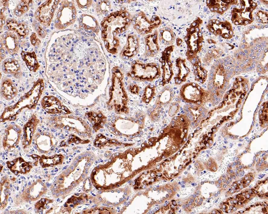
Immunohistochemical analysis of paraffin-embedded human kidney tissue with Rabbit anti-LAMP2a antibody at 1/500 dilution. The section was pre-treated using heat mediated antigen retrieval with Tris-EDTA buffer (pH 9.0) for 20 minutes. The tissues were blocked in 1% BSA for 20 minutes at room temperature, washed with ddH2O and PBS, and then probed with the primary antibody at 1/500 dilution for 1 hour at room temperature. The detection was performed using an HRP conjugated compact polymer system. DAB was used as the chromogen. Tissues were counterstained with hematoxylin and mounted with DPX.IHC
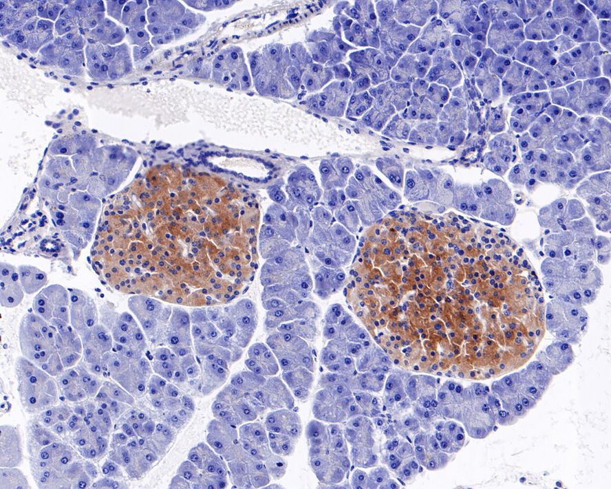
Immunohistochemical analysis of paraffin-embedded mouse pancreas tissue with Rabbit anti-LAMP2a antibody at 1/500 dilution. The section was pre-treated using heat mediated antigen retrieval with Tris-EDTA buffer (pH 9.0) for 20 minutes. The tissues were blocked in 1% BSA for 20 minutes at room temperature, washed with ddH2O and PBS, and then probed with the primary antibody at 1/500 dilution for 1 hour at room temperature. The detection was performed using an HRP conjugated compact polymer system. DAB was used as the chromogen. Tissues were counterstained with hematoxylin and mounted with DPX.IHC
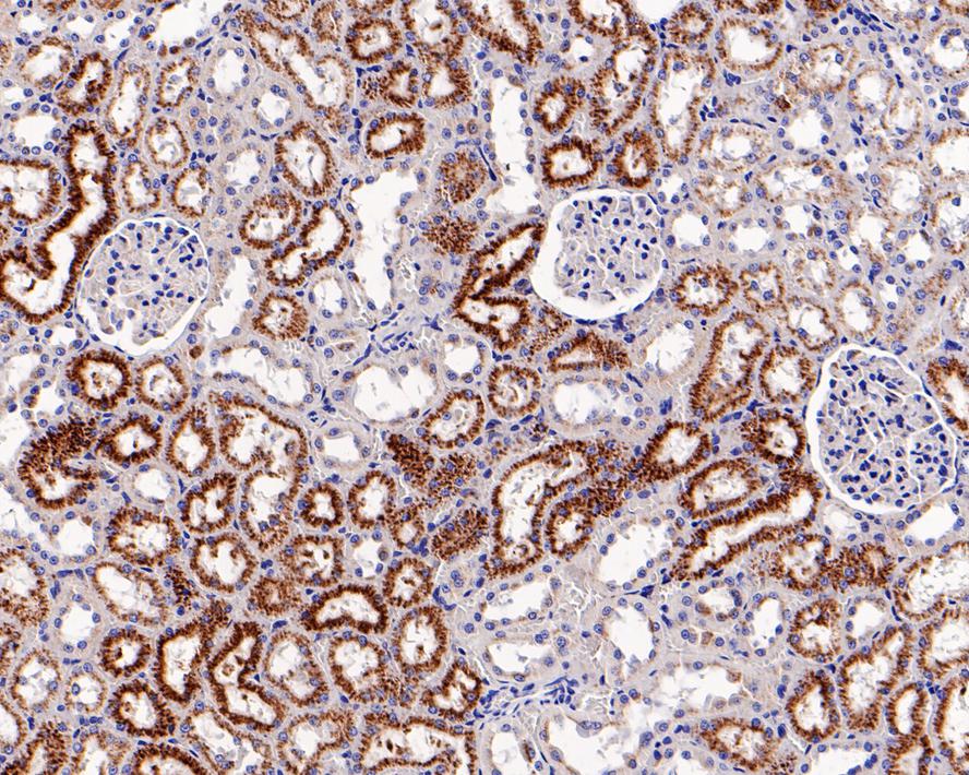
Immunohistochemical analysis of paraffin-embedded rat kidney tissue with Rabbit anti-LAMP2a antibody at 1/500 dilution. The section was pre-treated using heat mediated antigen retrieval with Tris-EDTA buffer (pH 9.0) for 20 minutes. The tissues were blocked in 1% BSA for 20 minutes at room temperature, washed with ddH2O and PBS, and then probed with the primary antibody at 1/500 dilution for 1 hour at room temperature. The detection was performed using an HRP conjugated compact polymer system. DAB was used as the chromogen. Tissues were counterstained with hematoxylin and mounted with DPX.IP
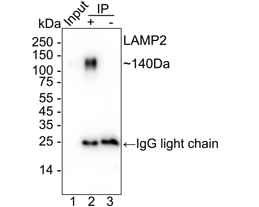
LAMP2a was immunoprecipitated from 0.2 mg SK-MEL-28 cell lysate with Rabbit anti-LAMP2a antibody at 2 µg/25 µl beads. Western blot was performed from the immunoprecipitate using Rabbit anti-LAMP2a antibody at 1/2,000 dilution. HRP Conjugated Rabbit IgG kappa light chain antibody at 1/5,000 dilution was used for 1 hour at room temperature. Lane 1: SK-MEL-28 cell lysate (Input), Lane 2: Rabbit anti-LAMP2a antibody IP in SK-MEL-28 cell lysate, Lane 3: Rabbit IgG instead of Rabbit anti-LAMP2a antibody in SK-MEL-28 cell lysate. Blocking/Dilution buffer: 5% NFDM/TBST Exposure time: 20 seconds.| Product Name | LAMP2a Recombinant Rabbit Monoclonal Antibody |
|---|---|
| Antibody Type | Primary Antibodies |
| Product description | Lysosome-associated membrane proteins (LAMP) are glycosylated type I membrane proteins that play a role in the biogenesis of the pigment melanin. LAMP-1 (also designated CD107A) and LAMP-2 (also designated CD107B) are involved in a variety of functions, including cellular adhesion, and are thought to participate in the process of tumor invasion and metastasis. Newly synthesized LAMP-1 and LAMP-2 proteins are sorted at the trans Golgi network and are transported intracellularly via a pathway that is distinct from the clathrin-coated vesicles used for the mannose-6 phosphate receptor. LAMP-1 is expressed on the surface of thrombin-activated but not resting platelets, and it is thought to be involved in the adhesive, prothrombic properties of these cells. Both LAMP-1 and LAMP-2 are involved in maintaining lysosome acidity and protecting the lysosomal membranes from autodigestion, and their expression is increased in patients with lysosomal storage disorders. |
| Immunogen | Synthetic peptide within Human LAMP2a aa 361-410 / 410. |
| Clonality | Monoclonal |
|---|---|
| Isotype | IgG |
| Host Species | Recombinant rabbit |
| Tested Applications | IHCIPWB |
| WB:1:5000 IHC:1:500 IP:Use at an assay dependent concentration. |
|
| Species Reactivity | HumanMouseRat |
| Concentration | 1mg/ml |
| Purification | Protein A |
| Alternative Names | CD107 antigen-like family member B antibody CD107b antibody LAMP 2 antibody Lamp 2a antibody LAMP-2 antibody LAMP2 antibody LAMP2_HUMAN antibody Lysosome-associated membrane glycoprotein 2 antibody Lysosome-associated membrane protein 2 antibody |
|---|---|
| Molecular Weight(MW) | 45kDa(Observed band size: 70-140kDa) |
| Cellular Localization | Cell membrane, Endosome membrane, Lysosome membrane |
WB
Western blot analysis of LAMP2a on different lysates with Rabbit anti-LAMP2a antibody at 1/5,000 dilution. Lane 1: SK-MEL-28 cell lysate (20 µg/Lane), Lane 2: HeLa cell lysate (20 µg/Lane), Lane 3: JAR cell lysate (20 µg/Lane), Lane 4: U-937 cell lysate (20 µg/Lane), Lane 5: RAW264.7 cell lysate (15 µg/Lane), Lane 6: NIH/3T3 cell lysate (15 µg/Lane), Lane 7: PC-12 cell lysate (15 µg/Lane), Lane 8: Mouse liver tissue lysate (30 µg/Lane), Lane 9: Rat liver tissue lysate (30 µg/Lane), Lane 10: Rat lung tissue lysate (30 µg/Lane), Exposure time: Lane 1-4: 2 minutes 37 seconds; Lane 5-10: 5 minutes; 4-20% SDS-PAGE gel. Proteins were transferred to a PVDF membrane and blocked with 5% NFDM/TBST for 1 hour at room temperature. The primary antibody at 1/5,000 dilution was used in 5% NFDM/TBST at 4℃ overnight. Goat Anti-Rabbit IgG - HRP Secondary Antibody at 1:50,000 dilution was used for 1 hour at room temperature.
IHC
Immunohistochemical analysis of paraffin-embedded human kidney tissue with Rabbit anti-LAMP2a antibody at 1/500 dilution. The section was pre-treated using heat mediated antigen retrieval with Tris-EDTA buffer (pH 9.0) for 20 minutes. The tissues were blocked in 1% BSA for 20 minutes at room temperature, washed with ddH2O and PBS, and then probed with the primary antibody at 1/500 dilution for 1 hour at room temperature. The detection was performed using an HRP conjugated compact polymer system. DAB was used as the chromogen. Tissues were counterstained with hematoxylin and mounted with DPX.
IHC
Immunohistochemical analysis of paraffin-embedded mouse pancreas tissue with Rabbit anti-LAMP2a antibody at 1/500 dilution. The section was pre-treated using heat mediated antigen retrieval with Tris-EDTA buffer (pH 9.0) for 20 minutes. The tissues were blocked in 1% BSA for 20 minutes at room temperature, washed with ddH2O and PBS, and then probed with the primary antibody at 1/500 dilution for 1 hour at room temperature. The detection was performed using an HRP conjugated compact polymer system. DAB was used as the chromogen. Tissues were counterstained with hematoxylin and mounted with DPX.
IHC
Immunohistochemical analysis of paraffin-embedded rat kidney tissue with Rabbit anti-LAMP2a antibody at 1/500 dilution. The section was pre-treated using heat mediated antigen retrieval with Tris-EDTA buffer (pH 9.0) for 20 minutes. The tissues were blocked in 1% BSA for 20 minutes at room temperature, washed with ddH2O and PBS, and then probed with the primary antibody at 1/500 dilution for 1 hour at room temperature. The detection was performed using an HRP conjugated compact polymer system. DAB was used as the chromogen. Tissues were counterstained with hematoxylin and mounted with DPX.
IP
LAMP2a was immunoprecipitated from 0.2 mg SK-MEL-28 cell lysate with Rabbit anti-LAMP2a antibody at 2 µg/25 µl beads. Western blot was performed from the immunoprecipitate using Rabbit anti-LAMP2a antibody at 1/2,000 dilution. HRP Conjugated Rabbit IgG kappa light chain antibody at 1/5,000 dilution was used for 1 hour at room temperature. Lane 1: SK-MEL-28 cell lysate (Input), Lane 2: Rabbit anti-LAMP2a antibody IP in SK-MEL-28 cell lysate, Lane 3: Rabbit IgG instead of Rabbit anti-LAMP2a antibody in SK-MEL-28 cell lysate. Blocking/Dilution buffer: 5% NFDM/TBST Exposure time: 20 seconds.| Positive Control | JAR, human kidney tissue, human liver tissue, human pancreas tissue, mouse placenta tissue, mouse kidney tissue, mouse brain tissue. |
|---|---|
| Application Notes | WB:1:5000 IHC:1:500 IP:Use at an assay dependent concentration. |
| Form | Liquid |
|---|---|
| Storage Instructions | Store at +4℃ after thawing. Aliquot store at -20℃ or -80℃. Avoid repeated freeze / thaw cycles. |
| Storage Buffer | 1*TBS (pH7.4), 1%BSA, 40%Glycerol. Preservative: 0.05% Sodium Azide. |


