Application
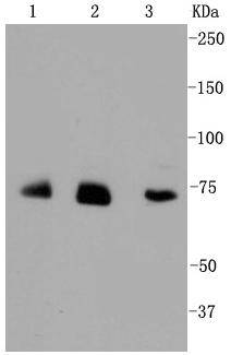
Fig1: Western blot analysis of Alas1 on different cell lysates using anti-Alas1 antibody at 1/1,000 dilution. Positive control: Lane 1: JAR Lane 2: HUVEC Lane 3: HelaApplication
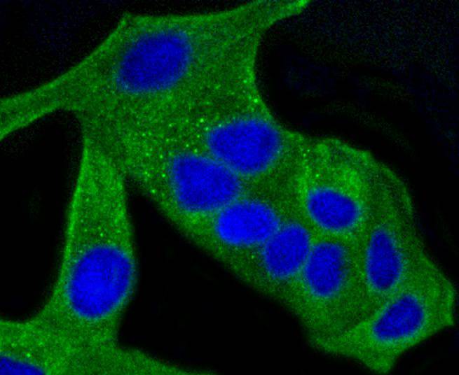
Fig2: ICC staining Alas1 in Hela cells (green). The nuclear counter stain is DAPI (blue). Cells were fixed in paraformaldehyde, permeabilised with 0.25% Triton X100/PBS.Application
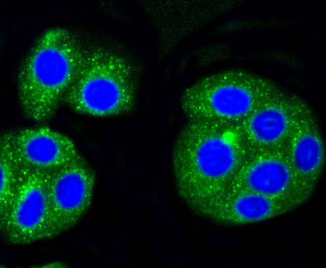
Fig3: ICC staining Alas1 in HepG2 cells (green). The nuclear counter stain is DAPI (blue). Cells were fixed in paraformaldehyde, permeabilised with 0.25% Triton X100/PBS.Application
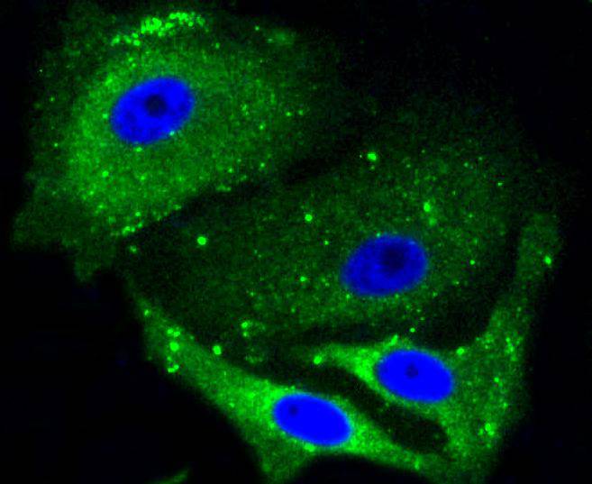
Fig4: ICC staining Alas1 in MCF-7 cells (green). Cells were fixed in paraformaldehyde, permeabilised with 0.25% Triton X100/PBS.| Product Name | Alas1 [SA70-04] |
|---|---|
| Antibody Type | Primary Antibodies |
| Antigen Alias | 5 aminolevulinate synthase antibody 5 aminolevulinate synthase nonspecific mitochondrial antibody 5 aminolevulinic acid synthase antibody 5-aminolevulinate synthase antibody 5-aminolevulinic acid synthase 1 antibody Alas 1 antibody ALAS 3 antibody ALAS antibody ALAS H antibody ALAS HOUSEKEEPING TYPE antibody ALAS N antibody ALAS-H antibody alaS1 antibody ALAS3 antibody ALASH antibody Aminolevulinate delta synthase 1 antibody Aminolevulinic acid synthase 1 antibody Delta ALA synthetase antibody Delta aminolevulinate synthase antibody Delta-ALA synthase 1 antibody Delta-aminolevulinate synthase 1 antibody HEM1_HUMAN antibody MIG 4 antibody MIG4 antibody Migration inducing protein 4 antibody mitochondrial antibody nonspecific antibody |
| Product description | 5-aminolevulinate synthase 1 (ALAS-H) and 2 (ALAS-E) are two isoforms of ALAS, an enzyme catalyzing the first step of the heme biosynthetic pathway in mammals. The erythroid-specific isoenzyme, ALAS-E, regulates the first step of hematopoietic cell differentation and iron metabolism in the liver. ALAS-H is a housekeeping protein which mediates synthesis of early heme in the mitochondria of most cells. Succinyl CoA associates with ALAS-E in protein conformation change and translocation of ALAS-E into the mitochondria and does not interact with ALAS-H. The ALAS-E 5'-flanking region contains binding sites for nuclear activators such as GATA-1, NF-E2 and EKLF. Since the ALAS gene maps to the X chromosome, mutation of the gene leads to the pyridoxine-refractory X-linked sideroblastic anemia. |
| Immunogen | recombinant protein |
| Clonality | Monoclonal |
|---|---|
| Isotype | IgG |
| Host Species | Recombinant rabbit |
| Tested Applications | WBICC/IF |
| WB:1:1,000-1:5,000 ICC:1:50-1:200 |
|
| Species Reactivity | Human |
| Concentration | 1mg/ml |
| Alternative Names | 5 aminolevulinate synthase antibody 5 aminolevulinate synthase nonspecific mitochondrial antibody 5 aminolevulinic acid synthase antibody 5-aminolevulinate synthase antibody 5-aminolevulinic acid synthase 1 antibody Alas 1 antibody ALAS 3 antibody ALAS antibody ALAS H antibody ALAS HOUSEKEEPING TYPE antibody ALAS N antibody ALAS-H antibody alaS1 antibody ALAS3 antibody ALASH antibody Aminolevulinate delta synthase 1 antibody Aminolevulinic acid synthase 1 antibody Delta ALA synthetase antibody Delta aminolevulinate synthase antibody Delta-ALA synthase 1 antibody Delta-aminolevulinate synthase 1 antibody HEM1_HUMAN antibody MIG 4 antibody MIG4 antibody Migration inducing protein 4 antibody mitochondrial antibody nonspecific antibody |
|---|---|
| Molecular Weight(MW) | 71 kDa |
| Cellular Localization | Mitochondrion matrix |
| SwissProt ID | P13196 |
|---|
Application
Fig1: Western blot analysis of Alas1 on different cell lysates using anti-Alas1 antibody at 1/1,000 dilution. Positive control: Lane 1: JAR Lane 2: HUVEC Lane 3: Hela
Application
Fig2: ICC staining Alas1 in Hela cells (green). The nuclear counter stain is DAPI (blue). Cells were fixed in paraformaldehyde, permeabilised with 0.25% Triton X100/PBS.
Application
Fig3: ICC staining Alas1 in HepG2 cells (green). The nuclear counter stain is DAPI (blue). Cells were fixed in paraformaldehyde, permeabilised with 0.25% Triton X100/PBS.
Application
Fig4: ICC staining Alas1 in MCF-7 cells (green). Cells were fixed in paraformaldehyde, permeabilised with 0.25% Triton X100/PBS.| Positive Control | HUVEC, MCF-7, Hela, HepG2, JAR. |
|---|---|
| Application Notes | WB:1:1,000-1:5,000 ICC:1:50-1:200 |
| Form | Liquid |
|---|---|
| Storage Instructions | Store at +4℃ after thawing. Aliquot store at -20℃ or -80℃. Avoid repeated freeze / thaw cycles. |
| Storage Buffer | 1*TBS (pH7.4), 1%BSA, 40%Glycerol. Preservative: 0.05% Sodium Azide. |


