Application
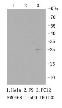
Fig1: Western blot analysis of RPS3 on PC12 cell lysates using anti-RPS3 antibody at 1/500 dilution.Application
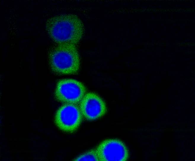
Fig2: ICC staining RPS3 in N2A cells (green). The nuclear counter stain is DAPI (blue). Cells were fixed in paraformaldehyde, permeabilised with 0.25% Triton X100/PBS.Application
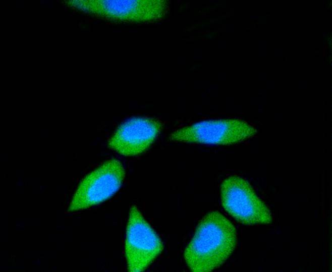
Fig3: ICC staining RPS3 in SH-SY-5Y cells (green). The nuclear counter stain is DAPI (blue). Cells were fixed in paraformaldehyde, permeabilised with 0.25% Triton X100/PBS.Application
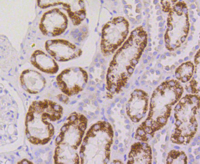
Fig4: Immunohistochemical analysis of paraffin-embedded human kidney tissue using anti-RPS3 antibody. Counter stained with hematoxylin.Application
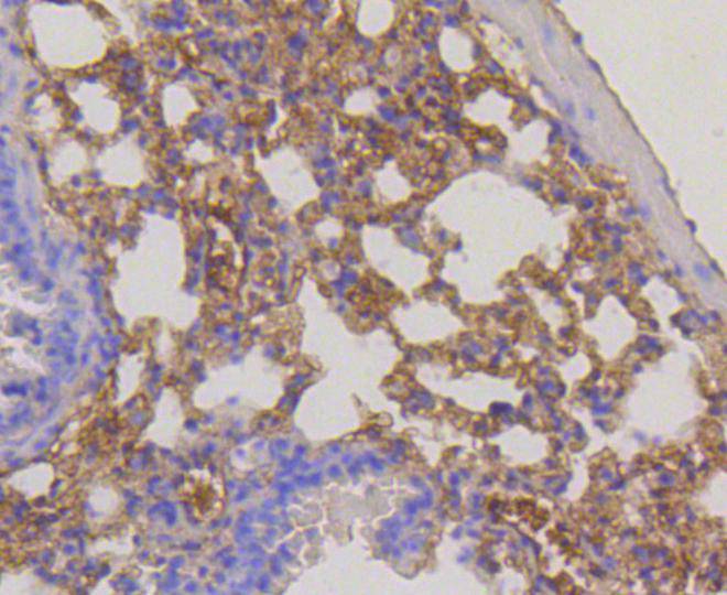
Fig5: Immunohistochemical analysis of paraffin-embedded mouse lung tissue using anti-RPS3 antibody. Counter stained with hematoxylin.| Product Name | RPS3 [SA46-08] |
|---|---|
| Antibody Type | Primary Antibodies |
| Antigen Alias | 40S ribosomal protein S3 antibody fb13d09 antibody FLJ26283 antibody FLJ27450 antibody IMR 90 ribosomal protein S3 antibody MGC56088 antibody MGC87870 antibody OTTHUMP00000229804 antibody OTTHUMP00000229805 antibody OTTHUMP00000229874 antibody OTTHUMP00000229877 antibody OTTHUMP00000229878 antibody OTTHUMP00000229879 antibody OTTHUMP00000229880 antibody OTTHUMP00000229882 antibody OTTHUMP00000229883 antibody OTTHUMP00000229886 antibody Ribosomal protein S3 antibody rps3 antibody RS3_HUMAN antibody S3 antibody wu:fb13d09 antibody zgc:56088 antibody |
| Product description | mal proteins have the ability to pass through the nuclear envelope in the native state, making them the largest of the structures accommodated by the nuclear pore complexes. The nuclear export of ribosomal subunits is a unidirectional, saturable and energy-dependent process. Ribosomal Protein S3 a member of the 40S subunit and plays a role in translation and ribosome maturation. Specifically, Ribosomal Protein S3 mediates the formation of the mRNA binding site 3’ of the codon in the decoding site. In addition, Ribosomal Protein S3 is involved in DNA damage recognition as shown by its affinity for abasic sites and 7,8-dihydro-8-oxoguanine residues and its interaction with human base excision repair (BER) proteins OGG1 and Ref-1. |
| Immunogen | recombinant protein |
| Clonality | Monoclonal |
|---|---|
| Isotype | IgG |
| Host Species | Recombinant rabbit |
| Tested Applications | WBICCIHC |
| WB:1:500-1:1,000 ICC:1:50-1:200 IHC:1:50-1:200 |
|
| Species Reactivity | HumanMouseRat |
| Concentration | 1mg/ml |
| Alternative Names | 40S ribosomal protein S3 antibody fb13d09 antibody FLJ26283 antibody FLJ27450 antibody IMR 90 ribosomal protein S3 antibody MGC56088 antibody MGC87870 antibody OTTHUMP00000229804 antibody OTTHUMP00000229805 antibody OTTHUMP00000229874 antibody OTTHUMP00000229877 antibody OTTHUMP00000229878 antibody OTTHUMP00000229879 antibody OTTHUMP00000229880 antibody OTTHUMP00000229882 antibody OTTHUMP00000229883 antibody OTTHUMP00000229886 antibody Ribosomal protein S3 antibody rps3 antibody RS3_HUMAN antibody S3 antibody wu:fb13d09 antibody zgc:56088 antibody |
|---|---|
| Molecular Weight(MW) | 27 kDa |
| Cellular Localization | Cytoplasm, Nucleus, Mitochondrion inner membrane |
Application
Fig1: Western blot analysis of RPS3 on PC12 cell lysates using anti-RPS3 antibody at 1/500 dilution.
Application
Fig2: ICC staining RPS3 in N2A cells (green). The nuclear counter stain is DAPI (blue). Cells were fixed in paraformaldehyde, permeabilised with 0.25% Triton X100/PBS.
Application
Fig3: ICC staining RPS3 in SH-SY-5Y cells (green). The nuclear counter stain is DAPI (blue). Cells were fixed in paraformaldehyde, permeabilised with 0.25% Triton X100/PBS.
Application
Fig4: Immunohistochemical analysis of paraffin-embedded human kidney tissue using anti-RPS3 antibody. Counter stained with hematoxylin.
Application
Fig5: Immunohistochemical analysis of paraffin-embedded mouse lung tissue using anti-RPS3 antibody. Counter stained with hematoxylin.| Positive Control | PC12, N2A, SH-SY-5Y, human kidney tissue, mouse lung tissue. |
|---|---|
| Application Notes | WB:1:500-1:1,000 ICC:1:50-1:200 IHC:1:50-1:200 |
| Form | Liquid |
|---|---|
| Storage Instructions | Store at +4℃ after thawing. Aliquot store at -20℃ or -80℃. Avoid repeated freeze / thaw cycles. |
| Storage Buffer | 1*TBS (pH7.4), 1%BSA, 40%Glycerol. Preservative: 0.05% Sodium Azide. |


