Application
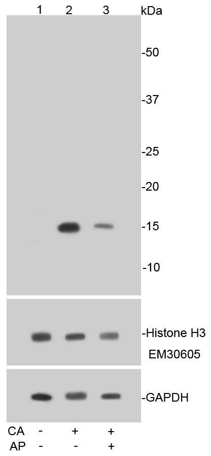
Fig1: Western blot analysis of Phospho-Histone H3(S10) on Hela cell lysates using anti-Phospho-Histone H3(S10) antibody at 1/1,000 dilution. Positive control: Lane 1: Untreated HeLa cell lysate Lane 2: HeLa cell lysate treated with calyculin A Lane 3: HeLa cell lysate treated with calyculin A and alkaline phosphataseApplication
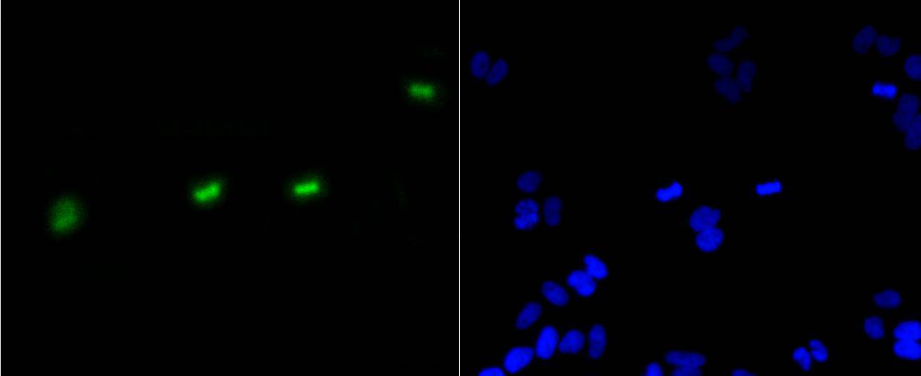
Fig2: ICC staining Phospho-Histone H3(S10) in Hela cells (green). The nuclear counter stain is DAPI (blue). Cells were fixed in paraformaldehyde, permeabilised with 0.25% Triton X100/PBS.Application
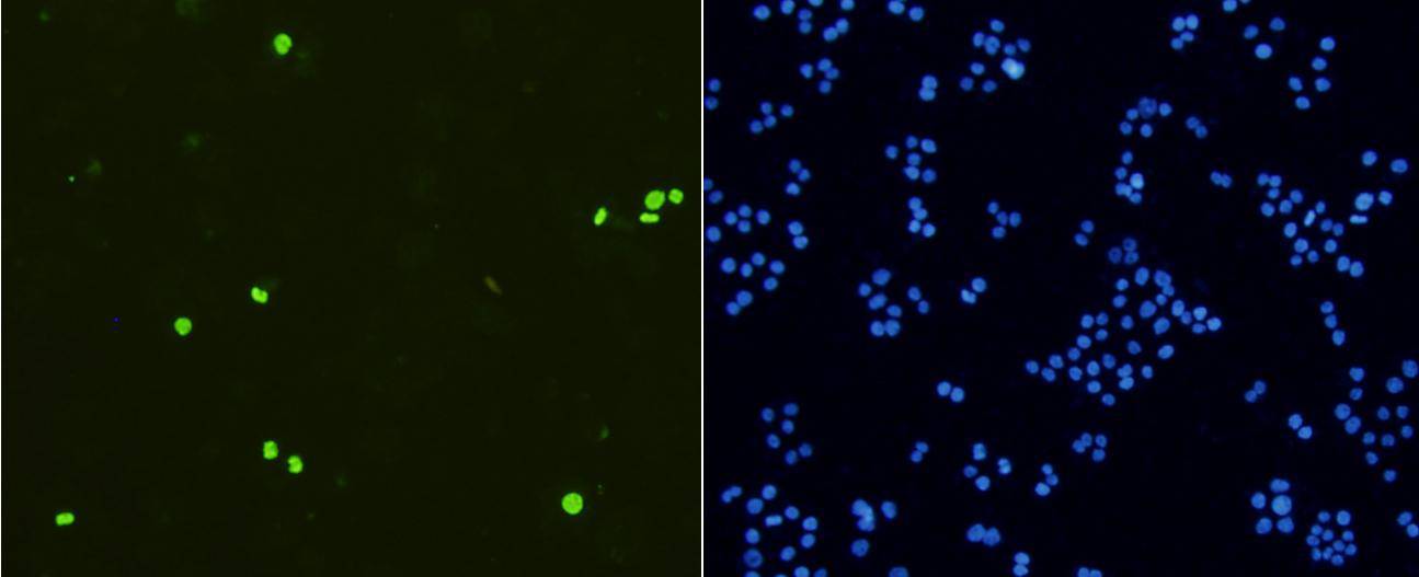
Fig3: ICC staining Phospho-Histone H3(S10) in Ags cells (green). Cells were fixed in paraformaldehyde, permeabilised with 0.25% Triton X100/PBS.Application
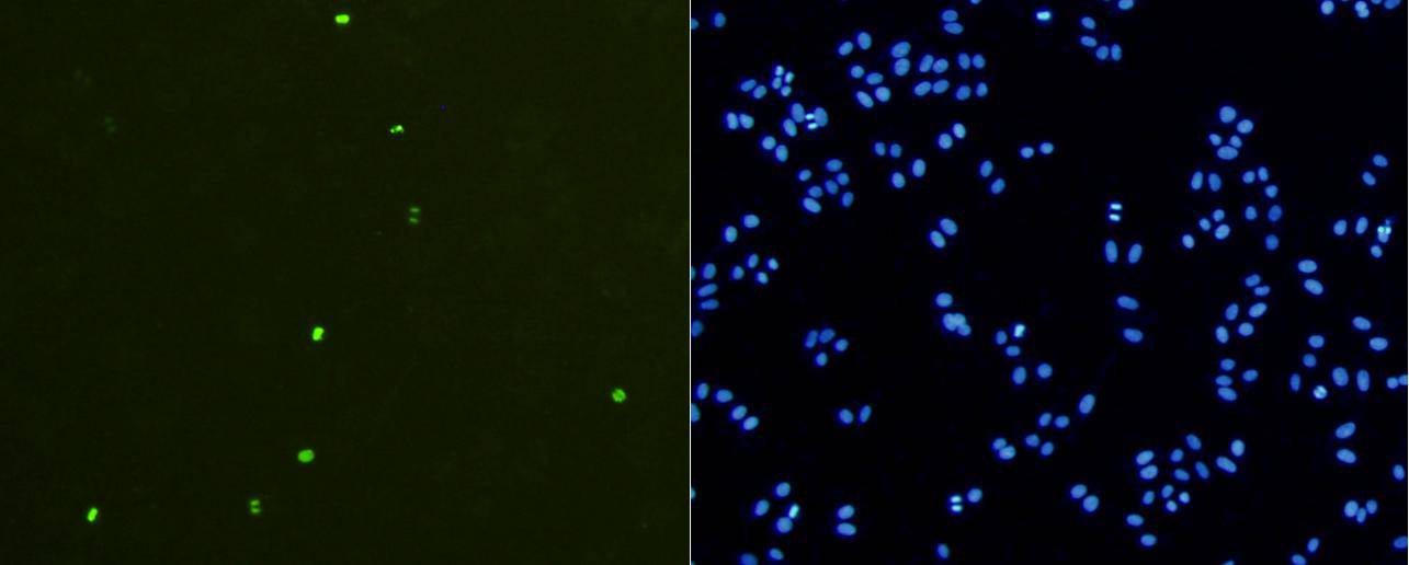
Fig4: ICC staining Phospho-Histone H3(S10) in HepG2 cells (green). Cells were fixed in paraformaldehyde, permeabilised with 0.25% Triton X100/PBS.Application
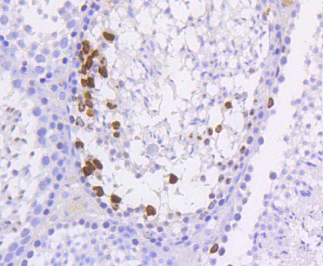
Fig5: Immunohistochemical analysis of paraffin-embedded mouse testis tissue using anti-Phospho-Histone H3(S10) antibody. Counter stained with hematoxylin.| Product Name | Phospho-Histone H3(S10) [SA31-01] |
|---|---|
| Antibody Type | Primary Antibodies |
| Antigen Alias | H3 3 like sequence MH921 antibody H3 3A antibody H3 a antibody H3 b antibody H3 c antibody H3 d antibody H3 f antibody H3 h antibody H3 histone family member E pseudogene antibody H3 i antibody H3 j antibody H3 k antibody H3 l antibody H33_HUMAN antibody H3F3 antibody H3f3b antibody Histone H3 3 pseudogene antibody Histone H3.3 antibody |
| Product description | In eukaryotes, DNA is wrapped around histone octamers to form the basic unit of chromatin structure. The octamer is composed of histones H2A, H2B, H3 and H4, and it associates with approximately 200 base pairs of DNA to form the nucleosome. The association of DNA with histones results in dense packing of chromatin, which restricts proteins involved in gene transcription from binding to DNA. Histone H3, the core protein of the nucleosome, becomes phosphorylated at the end of prophase. The two major sites of phosphorylation are the mitosis-specific site Ser10, and Ser28, both of which are extensively phosphorylated in DNA-bound forms of histone H3 and in nucleosomal histone H3. The nucleosome structure of histone H3 promotes N-terminal phosphorylation in vitro. |
| Immunogen | Synthetic phospho-peptide corresponding to residues surrounding Ser10 of human Histone H3. |
| Modification | p-S10 |
| Clonality | Monoclonal |
|---|---|
| Isotype | IgG |
| Host Species | Recombinant rabbit |
| Tested Applications | WBICC/IFIHCIP |
| WB:1:1,000-1:5,000 ICC:1:50-1:200 IHC:1:50-1:200 |
|
| Species Reactivity | HumanMouseRat |
| Concentration | 1mg/ml |
| Alternative Names | H3 3 like sequence MH921 antibody H3 3A antibody H3 a antibody H3 b antibody H3 c antibody H3 d antibody H3 f antibody H3 h antibody H3 histone family member E pseudogene antibody H3 i antibody H3 j antibody H3 k antibody H3 l antibody H33_HUMAN antibody H3F3 antibody H3f3b antibody Histone H3 3 pseudogene antibody Histone H3.3 antibody |
|---|---|
| Molecular Weight(MW) | 15 kDa |
| Cellular Localization | Nucleus, Chromosome |
Application
Fig1: Western blot analysis of Phospho-Histone H3(S10) on Hela cell lysates using anti-Phospho-Histone H3(S10) antibody at 1/1,000 dilution. Positive control: Lane 1: Untreated HeLa cell lysate Lane 2: HeLa cell lysate treated with calyculin A Lane 3: HeLa cell lysate treated with calyculin A and alkaline phosphatase
Application
Fig2: ICC staining Phospho-Histone H3(S10) in Hela cells (green). The nuclear counter stain is DAPI (blue). Cells were fixed in paraformaldehyde, permeabilised with 0.25% Triton X100/PBS.
Application
Fig3: ICC staining Phospho-Histone H3(S10) in Ags cells (green). Cells were fixed in paraformaldehyde, permeabilised with 0.25% Triton X100/PBS.
Application
Fig4: ICC staining Phospho-Histone H3(S10) in HepG2 cells (green). Cells were fixed in paraformaldehyde, permeabilised with 0.25% Triton X100/PBS.
Application
Fig5: Immunohistochemical analysis of paraffin-embedded mouse testis tissue using anti-Phospho-Histone H3(S10) antibody. Counter stained with hematoxylin.| Positive Control | Ags, HepG2, Hela, mouse testis tissue. |
|---|---|
| Application Notes | WB:1:1,000-1:5,000 ICC:1:50-1:200 IHC:1:50-1:200 |
| Form | Liquid |
|---|---|
| Storage Instructions | Store at +4℃ after thawing. Aliquot store at -20℃ or -80℃. Avoid repeated freeze / thaw cycles. |
| Storage Buffer | 1*TBS (pH7.4), 1%BSA, 40%Glycerol. Preservative: 0.05% Sodium Azide. |


