Application
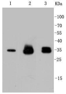
Fig1: Western blot analysis of Cyclin D1 on different lysates using anti-Cyclin D1 antibody at 1/1,000 dilution. Positive control: Lane 1: Hela Lane 2: PC-12 Lane 3: SH-SY-5YApplication
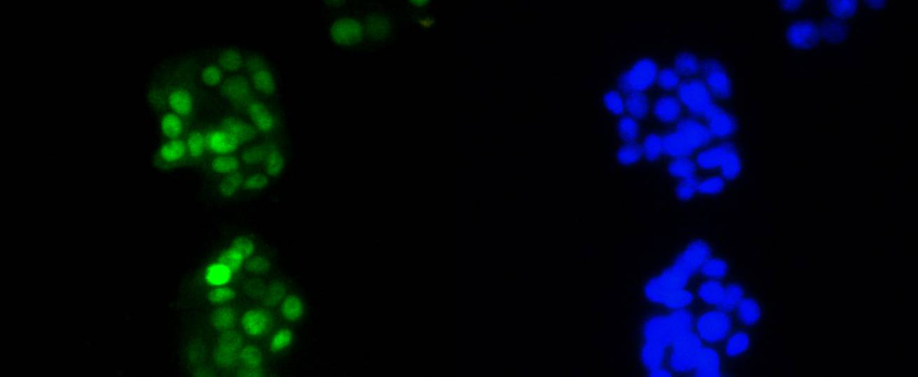
Fig2: ICC staining Cyclin D1 in PC-12 cells (green). The nuclear counter stain is DAPI (blue). Cells were fixed in paraformaldehyde, permeabilised with 0.25% Triton X100/PBS.Application
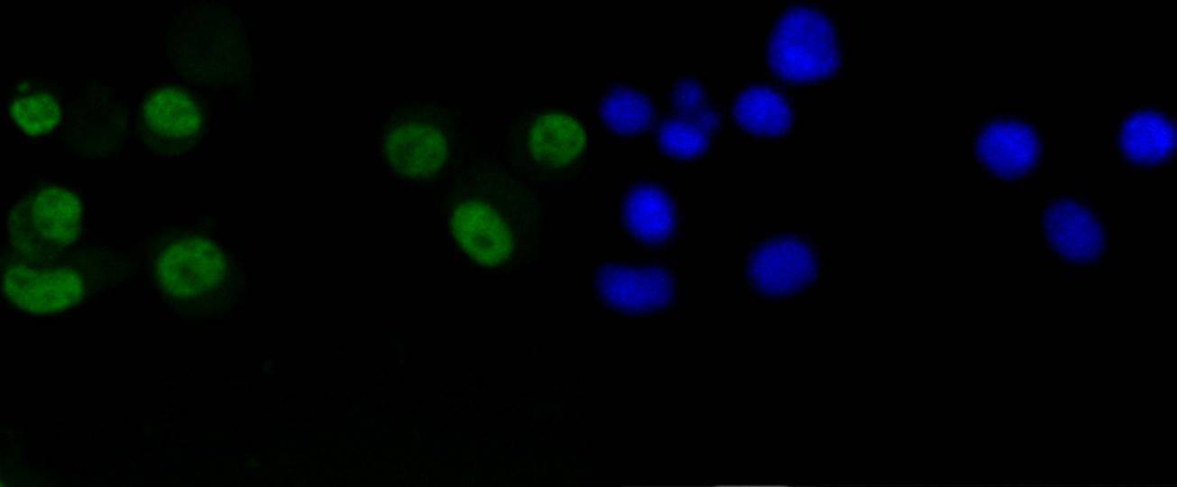
Fig3: ICC staining Cyclin D1 in N2A cells (green). The nuclear counter stain is DAPI (blue). Cells were fixed in paraformaldehyde, permeabilised with 0.25% Triton X100/PBS.Application
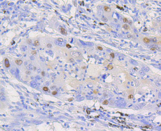
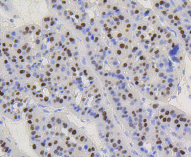
Fig4: Immunohistochemical analysis of paraffin-embedded human lung carcinoma tissue using anti-Cyclin D1 antibody. Counter stained with hematoxylin.Application
Fig5: Immunohistochemical analysis of paraffin-embedded human liver carcinoma tissue using anti-Cyclin D1 antibody. Counter stained with hematoxylin.Application
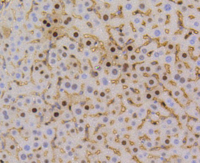
Fig6: Immunohistochemical analysis of paraffin-embedded mouse liver tissue using anti-Cyclin D1 antibody. Counter stained with hematoxylin.Application
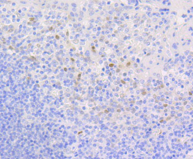
Fig7: Immunohistochemical analysis of paraffin-embedded human tonsil tissue using anti-Cyclin D1 antibody. Counter stained with hematoxylin.| Product Name | Cyclin D1 [SA38-08] |
|---|---|
| Antibody Type | Primary Antibodies |
| Antigen Alias | AI327039 antibody B cell CLL/lymphoma 1 antibody B cell leukemia 1 antibody B cell lymphoma 1 protein antibody B-cell lymphoma 1 protein antibody BCL 1 antibody BCL-1 antibody BCL-1 oncogene antibody BCL1 antibody BCL1 oncogene antibody ccnd1 antibody CCND1/FSTL3 fusion gene, included antibody CCND1/IGHG1 fusion gene, included antibody CCND1/IGLC1 fusion gene, included antibody CCND1/PTH fusion gene, included antibody CCND1_HUMAN antibody cD1 antibody Cyl 1 antibody D11S287E antibody G1/S specific cyclin D1 antibody G1/S-specific cyclin-D1 antibody Parathyroid adenomatosis 1 antibody PRAD1 antibody PRAD1 oncogene antibody U21B31 antibody |
| Product description | The proliferation of eukaryotic cells is controlled at specific points in the cell cycle, particularly at the G1 to S and the G2 to M transitions. It is well established that the Cdc2 p34-cyclin B protein kinase plays a critical role in the G2 to M transition while cyclin A associates with Cdk2 p33 and functions in S phase. Considerable effort directed towards the identification of G1 cyclins has led to the isolation of cyclin D, cyclin C and cyclin E. Of these, cyclin D corresponds to a putative human oncogene, designated PRAD1, which maps at the site of the Bcl1 rearrangement in certain lymphomas and leukemias. Two additional human type D cyclins, as well as their mouse homologs, have been identified. Evidence has established that members of the cyclin D family function to regulate phosphorylation of the retinoblastoma gene product, thereby activating E2F transcription factors. |
| Immunogen | recombinant protein |
| Clonality | Monoclonal |
|---|---|
| Isotype | IgG |
| Host Species | Recombinant rabbit |
| Tested Applications | WBICC/IFIHCIP |
| WB:1:1000-1:5,000 ICC:1:50-1:200 IHC:1:50-1:200 |
|
| Species Reactivity | HumanMouseRat |
| Concentration | 1mg/ml |
| Alternative Names | AI327039 antibody
B cell CLL/lymphoma 1 antibody
B cell leukemia 1 antibody
B cell lymphoma 1 protein antibody
B-cell lymphoma 1 protein antibody
BCL 1 antibody
BCL-1 antibody
BCL-1 oncogene antibody
BCL1 antibody
BCL1 oncogene antibody
ccnd1 antibody
CCND1/FSTL3 fusion gene included antibody CCND1/IGHG1 fusion gene included antibody CCND1/IGLC1 fusion gene included antibody CCND1/PTH fusion gene included antibody CCND1_HUMAN antibody cD1 antibody Cyl 1 antibody D11S287E antibody G1/S specific cyclin D1 antibody G1/S-specific cyclin-D1 antibody Parathyroid adenomatosis 1 antibody PRAD1 antibody PRAD1 oncogene antibody U21B31 antibody |
|---|---|
| Molecular Weight(MW) | 34 kDa |
| Cellular Localization | Cytoplasm, Nucleus, Membrane, Mitochondrion |
Application
Fig1: Western blot analysis of Cyclin D1 on different lysates using anti-Cyclin D1 antibody at 1/1,000 dilution. Positive control: Lane 1: Hela Lane 2: PC-12 Lane 3: SH-SY-5Y
Application
Fig2: ICC staining Cyclin D1 in PC-12 cells (green). The nuclear counter stain is DAPI (blue). Cells were fixed in paraformaldehyde, permeabilised with 0.25% Triton X100/PBS.
Application
Fig3: ICC staining Cyclin D1 in N2A cells (green). The nuclear counter stain is DAPI (blue). Cells were fixed in paraformaldehyde, permeabilised with 0.25% Triton X100/PBS.
Application
Fig4: Immunohistochemical analysis of paraffin-embedded human lung carcinoma tissue using anti-Cyclin D1 antibody. Counter stained with hematoxylin.
Application
Fig5: Immunohistochemical analysis of paraffin-embedded human liver carcinoma tissue using anti-Cyclin D1 antibody. Counter stained with hematoxylin.
Application
Fig6: Immunohistochemical analysis of paraffin-embedded mouse liver tissue using anti-Cyclin D1 antibody. Counter stained with hematoxylin.
Application
Fig7: Immunohistochemical analysis of paraffin-embedded human tonsil tissue using anti-Cyclin D1 antibody. Counter stained with hematoxylin.| Positive Control | Hela, SH-SY-5Y, N2A, PC-12, human tonsil tissue, human lung carcinoma tissue, mouse liver tissue, human liver carcinoma tissue. |
|---|---|
| Application Notes | WB:1:1000-1:5,000 ICC:1:50-1:200 IHC:1:50-1:200 |
| Form | Liquid |
|---|---|
| Storage Instructions | Store at +4℃ after thawing. Aliquot store at -20℃ or -80℃. Avoid repeated freeze / thaw cycles. |
| Storage Buffer | 1*TBS (pH7.4), 1%BSA, 40%Glycerol. Preservative: 0.05% Sodium Azide. |


