WB
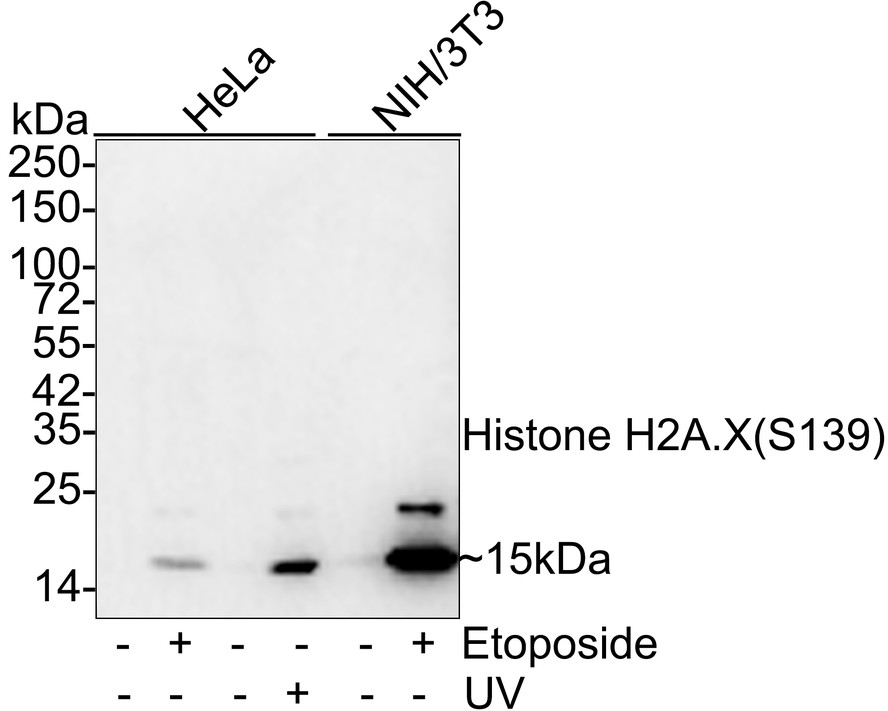
Western blot analysis of Phospho-Histone H2A.X (S139) on different lysates with Rabbit anti-Phospho-Histone H2A.X (S139) antibody at 1/2,000 dilution. Lane 1: HeLa whole cell lysate, Lane 2: HeLa treated with 20μM Etoposide for 2 hours whole cell lysate, Lane 3: HeLa whole cell lysate, Lane 4: HeLa treated with UV for 2 hours whole cell lysate, Lane 5: NIH/3T3 whole cell lysate, Lane 6: NIH/3T3 treated with 25μM Etoposide for 5 hours whole cell lysate, Lysates/proteins at 20 µg/Lane. Exposure time: 53 seconds; 4-20% SDS-PAGE gel. Proteins were transferred to a PVDF membrane and blocked with 5% NFDM/TBST for 1 hour at room temperature. The primary antibody at 1/2,000 dilution was used in 5% NFDM/TBST at room temperature for 2 hours. Goat Anti-Rabbit IgG - HRP Secondary Antibody at 1/100,000 dilution was used for 1 hour at room temperature.IHC
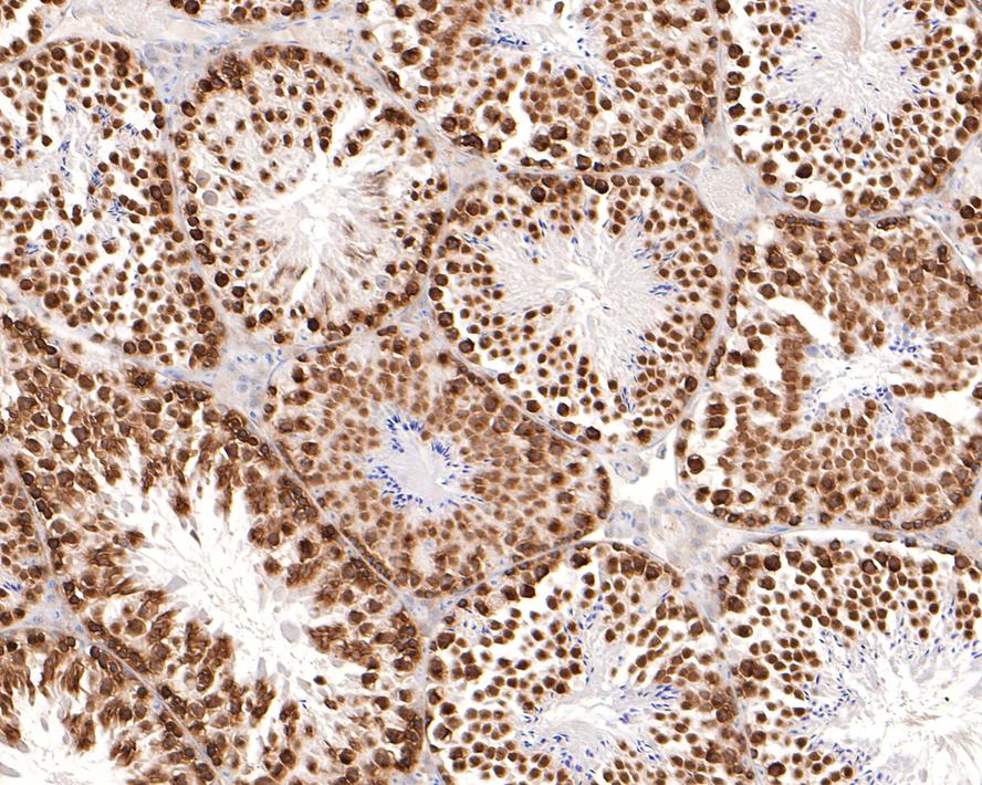
Immunohistochemical analysis of paraffin-embedded mouse testis tissue with Rabbit anti-Phospho-Histone H2A.X (S139) antibody at 1/500 dilution. The section was pre-treated using heat mediated antigen retrieval with sodium citrate buffer (pH 6.0) for 2 minutes. The tissues were blocked in 1% BSA for 20 minutes at room temperature, washed with ddH2O and PBS, and then probed with the primary antibody at 1/500 dilution for 1 hour at room temperature. The detection was performed using an HRP conjugated compact polymer system. DAB was used as the chromogen. Tissues were counterstained with hematoxylin and mounted with DPX.IHC
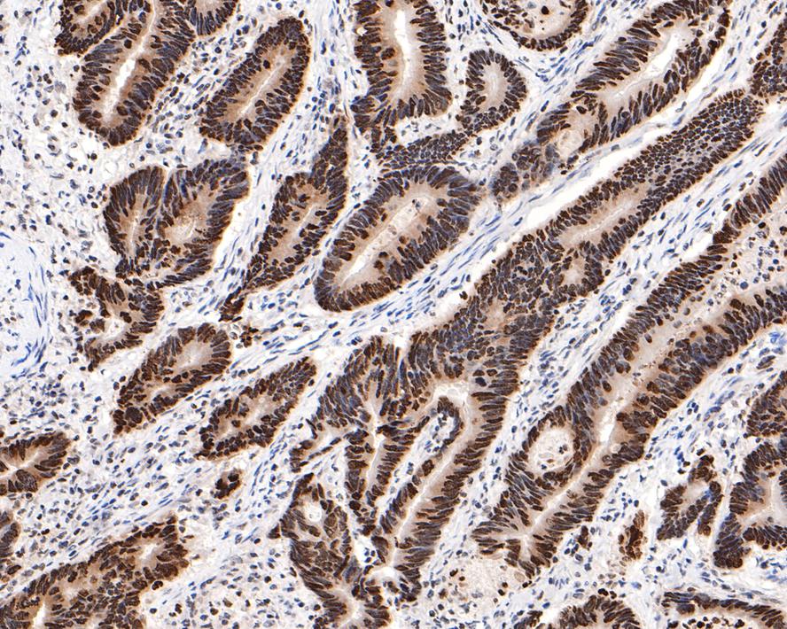
Immunohistochemical analysis of paraffin-embedded human colon carcinoma tissue with Rabbit anti-Phospho-Histone H2A.X (S139) antibody at 1/500 dilution. The section was pre-treated using heat mediated antigen retrieval with sodium citrate buffer (pH 6.0) for 2 minutes. The tissues were blocked in 1% BSA for 20 minutes at room temperature, washed with ddH2O and PBS, and then probed with the primary antibody at 1/500 dilution for 1 hour at room temperature. The detection was performed using an HRP conjugated compact polymer system. DAB was used as the chromogen. Tissues were counterstained with hematoxylin and mounted with DPX.ICC/IF
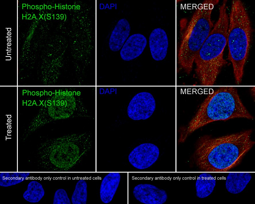
Immunocytochemistry analysis of HeLa cells untreated / treated with 20μM etoposide for 2 hours labeling Phospho-Histone H2A.X (S139) with Rabbit anti-Phospho-Histone H2A.X (S139) antibody at 1/2,000 dilution. Cells were fixed in 4% paraformaldehyde for 15 minutes at room temperature, permeabilized with 0.1% Triton X-100 in PBS for 15 minutes at room temperature, then blocked with 1% BSA in 10% negative goat serum for 1 hour at room temperature. Cells were then incubated with Rabbit anti-Phospho-Histone H2A.X (S139) antibody at 1/2,000 dilution in 1% BSA in PBST overnight at 4 ℃. Goat Anti-Rabbit IgG H&L (488) was used as the secondary antibody at 1/1,000 dilution. PBS instead of the primary antibody was used as the secondary antibody only control. Nuclear DNA was labelled in blue with DAPI. Beta tubulin (red) was stained at 1/100 dilution overnight at +4℃. Goat Anti-Mouse IgG H&L (594) was used as the secondary antibody at 1/1,000 dilution.IF-P
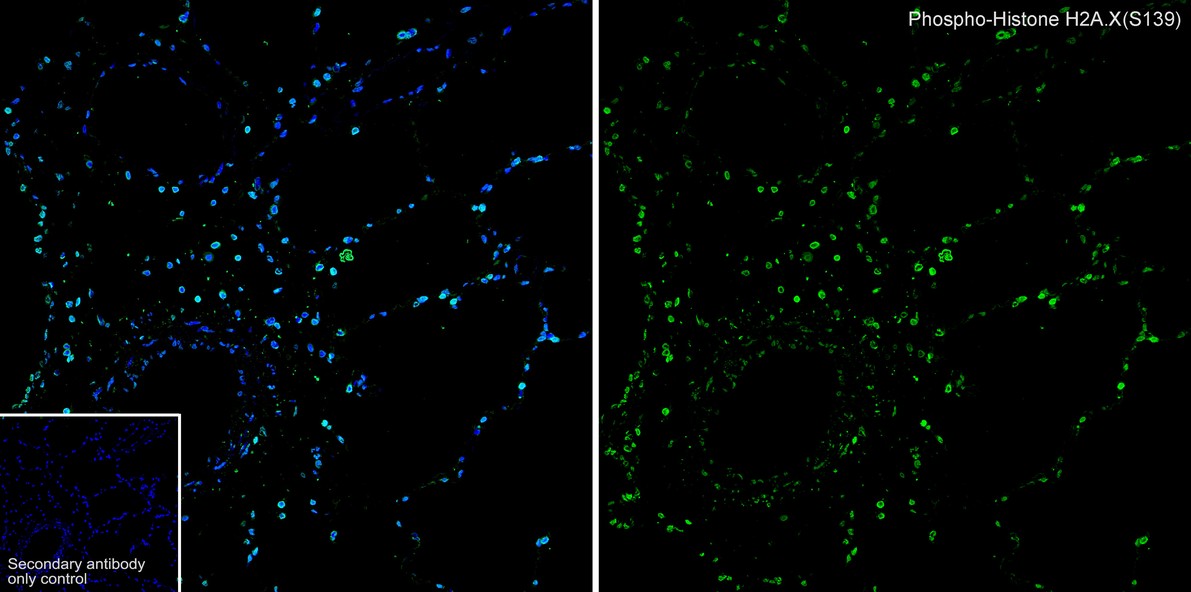
Species: Cynomolgus monkey, Site: Lung, Sample: Paraffin-embedded section, Antibody concentration: 1/500.| Product Name | Phospho-Histone H2A.X (S139) Recombinant Rabbit Monoclonal Antibody |
|---|---|
| Antibody Type | Primary Antibodies |
| Product description | Histone H2A.X is a member of the Histone H2A family, which is involved in nucleosomal organization of chromatin. The H2AFX gene is located in close proximity to the Porphobilinogen deaminase (PBG-D) gene in both mouse and human, and maps to chromosome 9 and 11q23, respectively. H2A.X differs from the other members of the H2A family by the presence of a highly conserved C-terminal motif. It is rapidly phosphorylated in response to ionizing radiation and plays an important role in the recognition and repair of DNA double stranded breaks. The phosphorylated form of H2A.X, designated ?-H2A.X, forms nuclear foci at the heavy chain constant region of cells involved in class switch recombination (CSR), a region-specific DNA reaction that replaces one immunoglobulin heavy chain constant region gene with another. The phosphorylated ?-H2A.X is also thought to initiate subsequent repair factors, including Rad50, Rad51 and BRCA1. |
| Immunogen | Synthetic phospho-peptide corresponding to residues surrounding Ser139 of Human Histone H2AX. |
| Modification | p-S139 |
| Clonality | monoclonal |
|---|---|
| Isotype | IgG |
| Host Species | Recombinant rabbit |
| Tested Applications | ICC/IFIF-PIHCWB |
| WB:1:2000-1:5000 IHC:1:500-1:5000 ICC/IF:1:2000 IF-P:1:500 |
|
| Species Reactivity | HumanMouseRat |
| Concentration | 1mg/ml |
| Purification | Protein A |
| Alternative Names | AW228881 antibody H2A histone family member X antibody H2A.FX antibody H2A.X antibody H2a/x antibody H2AFX antibody H2AX antibody H2AX histone antibody H2AX_HUMAN antibody Hist5.2ax antibody Histone 2A antibody Histone 2AX antibody Histone H2A.X antibody Histone H2AX antibody RGD1566119 antibody |
|---|---|
| Molecular Weight(MW) | 15kDa(Observed band size: 15/20kDa) |
| Cellular Localization | Nucleus, Chromosome |
WB
Western blot analysis of Phospho-Histone H2A.X (S139) on different lysates with Rabbit anti-Phospho-Histone H2A.X (S139) antibody at 1/2,000 dilution. Lane 1: HeLa whole cell lysate, Lane 2: HeLa treated with 20μM Etoposide for 2 hours whole cell lysate, Lane 3: HeLa whole cell lysate, Lane 4: HeLa treated with UV for 2 hours whole cell lysate, Lane 5: NIH/3T3 whole cell lysate, Lane 6: NIH/3T3 treated with 25μM Etoposide for 5 hours whole cell lysate, Lysates/proteins at 20 µg/Lane. Exposure time: 53 seconds; 4-20% SDS-PAGE gel. Proteins were transferred to a PVDF membrane and blocked with 5% NFDM/TBST for 1 hour at room temperature. The primary antibody at 1/2,000 dilution was used in 5% NFDM/TBST at room temperature for 2 hours. Goat Anti-Rabbit IgG - HRP Secondary Antibody at 1/100,000 dilution was used for 1 hour at room temperature.
IHC
Immunohistochemical analysis of paraffin-embedded mouse testis tissue with Rabbit anti-Phospho-Histone H2A.X (S139) antibody at 1/500 dilution. The section was pre-treated using heat mediated antigen retrieval with sodium citrate buffer (pH 6.0) for 2 minutes. The tissues were blocked in 1% BSA for 20 minutes at room temperature, washed with ddH2O and PBS, and then probed with the primary antibody at 1/500 dilution for 1 hour at room temperature. The detection was performed using an HRP conjugated compact polymer system. DAB was used as the chromogen. Tissues were counterstained with hematoxylin and mounted with DPX.
IHC
Immunohistochemical analysis of paraffin-embedded human colon carcinoma tissue with Rabbit anti-Phospho-Histone H2A.X (S139) antibody at 1/500 dilution. The section was pre-treated using heat mediated antigen retrieval with sodium citrate buffer (pH 6.0) for 2 minutes. The tissues were blocked in 1% BSA for 20 minutes at room temperature, washed with ddH2O and PBS, and then probed with the primary antibody at 1/500 dilution for 1 hour at room temperature. The detection was performed using an HRP conjugated compact polymer system. DAB was used as the chromogen. Tissues were counterstained with hematoxylin and mounted with DPX.
ICC/IF
Immunocytochemistry analysis of HeLa cells untreated / treated with 20μM etoposide for 2 hours labeling Phospho-Histone H2A.X (S139) with Rabbit anti-Phospho-Histone H2A.X (S139) antibody at 1/2,000 dilution. Cells were fixed in 4% paraformaldehyde for 15 minutes at room temperature, permeabilized with 0.1% Triton X-100 in PBS for 15 minutes at room temperature, then blocked with 1% BSA in 10% negative goat serum for 1 hour at room temperature. Cells were then incubated with Rabbit anti-Phospho-Histone H2A.X (S139) antibody at 1/2,000 dilution in 1% BSA in PBST overnight at 4 ℃. Goat Anti-Rabbit IgG H&L (488) was used as the secondary antibody at 1/1,000 dilution. PBS instead of the primary antibody was used as the secondary antibody only control. Nuclear DNA was labelled in blue with DAPI. Beta tubulin (red) was stained at 1/100 dilution overnight at +4℃. Goat Anti-Mouse IgG H&L (594) was used as the secondary antibody at 1/1,000 dilution.
IF-P
Species: Cynomolgus monkey, Site: Lung, Sample: Paraffin-embedded section, Antibody concentration: 1/500.| Application Notes | WB:1:2000-1:5000 IHC:1:500-1:5000 ICC/IF:1:2000 IF-P:1:500 |
|---|
| Form | Liquid |
|---|---|
| Storage Instructions | Store at +4℃ after thawing. Aliquot store at -20℃ or -80℃. Avoid repeated freeze / thaw cycles. |
| Storage Buffer | 1*TBS (pH7.4), 1%BSA, 40%Glycerol. Preservative: 0.05% Sodium Azide. |


