WB
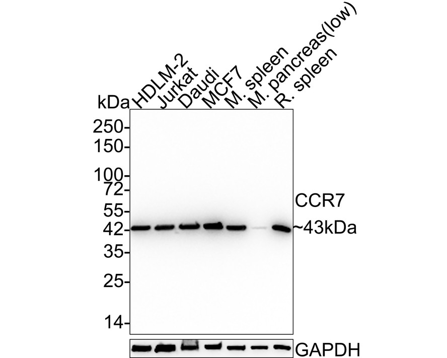
Western blot analysis of CCR7 on different lysates with Rabbit anti-CCR7 antibody at 1/5,000 dilution. Lane 1: HDLM-2 cell lysate, Lane 2: Jurkat cell lysate, Lane 3: Daudi cell lysate, Lane 4: MCF7 cell lysate, Lane 5: Mouse spleen tissue lysate, Lane 6: Mouse pancreas tissue lysate (low expression), Lane 7: Rat spleen tissue lysate, Lysates/proteins at 20 µg/Lane. Exposure time: 24 seconds; 4-20% SDS-PAGE gel. Proteins were transferred to a PVDF membrane and blocked with 5% NFDM/TBST for 1 hour at room temperature. The primary antibody at 1/5,000 dilution was used in 5% NFDM/TBST at 4℃ overnight. Goat Anti-Rabbit IgG - HRP Secondary Antibody at 1:50,000 dilution was used for 1 hour at room temperature.WB
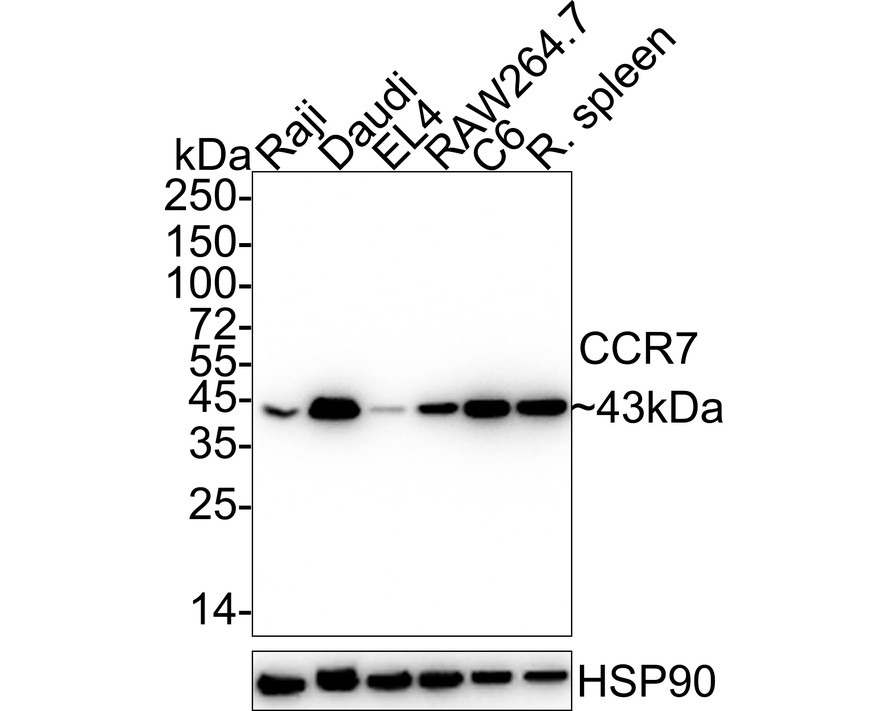
Western blot analysis of CCR7 on different lysates with Rabbit anti-CCR7 antibody at 1/2,000 dilution. Lane 1: Raji cell lysate, Lane 2: Daudi cell lysate, Lane 3: EL4 cell lysate, Lane 4: RAW264.7 cell lysate, Lane 5: C6 cell lysate, Lane 6: Rat spleen tissue lysate, Lysates/proteins at 15 µg/Lane. Exposure time: 10 seconds; 4-20% SDS-PAGE gel. Proteins were transferred to a PVDF membrane and blocked with 5% NFDM/TBST for 1 hour at room temperature. The primary antibody at 1/2,000 dilution was used in 5% NFDM/TBST at 4℃ overnight. Goat Anti-Rabbit IgG - HRP Secondary Antibody at 1:50,000 dilution was used for 1 hour at room temperature.IHC
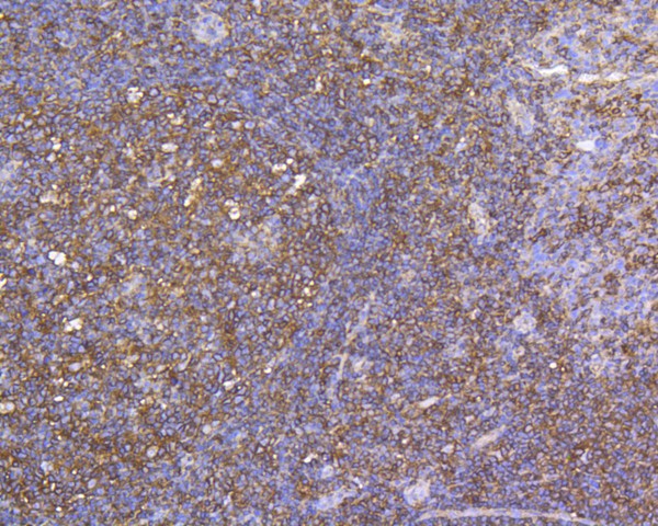
Immunohistochemical analysis of paraffin-embedded human tonsil tissue using anti-CCR7 antibody. The section was pre-treated using heat mediated antigen retrieval with Tris-EDTA buffer (pH 8.0-8.4) for 20 minutes.The tissues were blocked in 5% BSA for 30 minutes at room temperature, washed with ddH2O and PBS, and then probed with the primary antibody (1/200) for 30 minutes at room temperature. The detection was performed using an HRP conjugated compact polymer system. DAB was used as the chromogen. Tissues were counterstained with hematoxylin and mounted with DPX.IHC
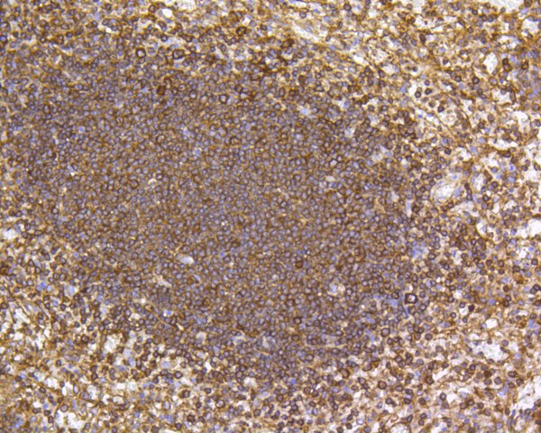
Immunohistochemical analysis of paraffin-embedded human spleen tissue using anti-CCR7 antibody. The section was pre-treated using heat mediated antigen retrieval with Tris-EDTA buffer (pH 8.0-8.4) for 20 minutes.The tissues were blocked in 5% BSA for 30 minutes at room temperature, washed with ddH2O and PBS, and then probed with the primary antibody (1/200) for 30 minutes at room temperature. The detection was performed using an HRP conjugated compact polymer system. DAB was used as the chromogen. Tissues were counterstained with hematoxylin and mounted with DPX.ICC/IF
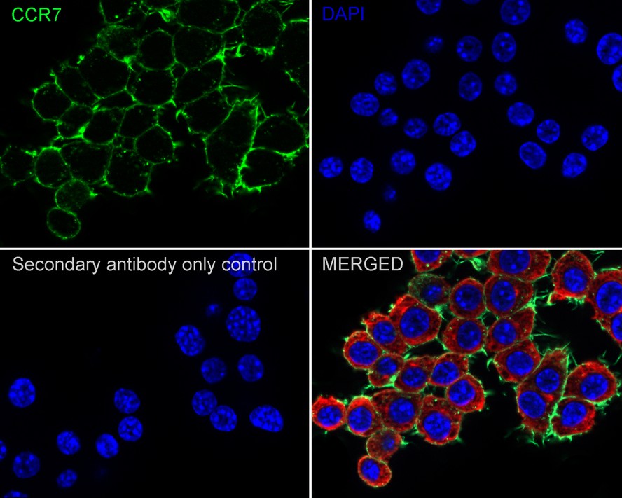
Immunocytochemistry analysis of RAW264.7 cells labeling CCR7 with Rabbit anti-CCR7 antibody at 1/500 dilution. Cells were fixed in 4% paraformaldehyde for 20 minutes at room temperature, permeabilized with 0.1% Triton X-100 in PBS for 5 minutes at room temperature, then blocked with 1% BSA in 10% negative goat serum for 1 hour at room temperature. Cells were then incubated with Rabbit anti-CCR7 antibody at 1/500 dilution in 1% BSA in PBST overnight at 4 ℃. Goat Anti-Rabbit IgG H&L (488) was used as the secondary antibody at 1/1,000 dilution. PBS instead of the primary antibody was used as the secondary antibody only control. Nuclear DNA was labelled in blue with DAPI. Beta tubulin (red) was stained at 1/100 dilution overnight at +4℃. Goat Anti-Mouse IgG H&L (594) was used as the secondary antibody at 1/1,000 dilution.ICC/IF
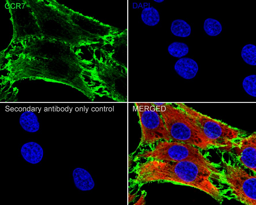
Immunocytochemistry analysis of C6 cells labeling CCR7 with Rabbit anti-CCR7 antibody at 1/200 dilution. Cells were fixed in 4% paraformaldehyde for 20 minutes at room temperature, permeabilized with 0.1% Triton X-100 in PBS for 5 minutes at room temperature, then blocked with 1% BSA in 10% negative goat serum for 1 hour at room temperature. Cells were then incubated with Rabbit anti-CCR7 antibody at 1/200 dilution in 1% BSA in PBST overnight at 4 ℃. Goat Anti-Rabbit IgG H&L (488) was used as the secondary antibody at 1/1,000 dilution. PBS instead of the primary antibody was used as the secondary antibody only control. Nuclear DNA was labelled in blue with DAPI. Beta tubulin (red) was stained at 1/100 dilution overnight at +4℃. Goat Anti-Mouse IgG H&L (594) was used as the secondary antibody at 1/1,000 dilution.IP
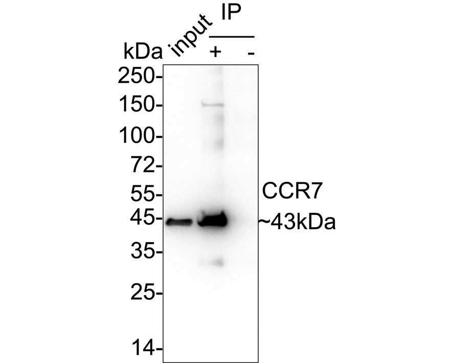
CCR7 was immunoprecipitated from 0.2 mg Daudi cell lysate with Rabbit anti-CCR7 antibody at 2 µg/10 µl beads. Western blot was performed from the immunoprecipitate using Rabbit anti-CCR7 antibody at 1/2,000 dilution. Anti-Rabbit IgG for IP Nano-secondary antibody at 1/5,000 dilution was used for 1 hour at room temperature. Lane 1: Daudi cell lysate (input), Lane 2: Rabbit anti-CCR7 antibody IP in Daudi cell lysate, Lane 3: Rabbit IgG instead of Rabbit anti-CCR7 antibody in Daudi cell lysate, Blocking/Dilution buffer: 5% NFDM/TBST, Exposure time: 8 seconds.| Product Name | CCR7 Recombinant Rabbit Monoclonal Antibody |
|---|---|
| Antibody Type | Primary Antibodies |
| Product description | C-C or ∫ chemokine family members are characterized by a pair of adjacent cysteine residues and serve as potent chemoattractants and activators of monocytes and T cells. C-C chemokine receptor family members include CKR-1, CKR-2A, CKR-2B, CKR-3, CKR-4, CKR-5, CKR-6, CKR-7, CKR-8, CKR-9, CKR-10 and the Duffy blood group antigen. Each of these receptors are G protein-coupled, seven pass transmembrane domain proteins whose major physiological role is to function in the chemotaxis of T cells and phagocytic cells to areas of inflammation. However, this receptor family has also been shown to facilitate viral infection. CKR-7 (C-C chemokine receptor type 7), also known as CCR7, CMKBR7, EBI1 or EVI1, is a 378 amino acid multi-pass membrane protein that belongs to the CC-chemokine receptor family. Expressed in activated B- and T-lymphocytes, as well as in various lymphoid tissues, CKR-7 functions as a receptor for MIP-3∫ and is thought to be involved in mediating normal lymphocyte function. |
| Immunogen | Synthetic peptide within Human CCR7 aa 13-62 / 378. |
| Clonality | Monoclonal |
|---|---|
| Isotype | IgG |
| Host Species | Recombinant rabbit |
| Tested Applications | ICC/IFIHCIPWB |
| WB:1:2000-1:5000 IHC:1:200 ICC/IF:1:200-1:500 IP: 1-2μg/sample |
|
| Species Reactivity | HumanMouseRat |
| Concentration | 1mg/ml |
| Purification | Protein A |
| Alternative Names | BLR 2 antibody BLR2 antibody C C chemokine receptor type 7 antibody C C CKR 7 antibody C-C chemokine receptor type 7 antibody C-C CKR-7 antibody CC chemokine receptor 7 antibody CC chemokine receptor type 7 antibody CC CKR 7 antibody CC-CKR-7 antibody CCCKR7 antibody CCR 7 antibody CCR-7 antibody Ccr7 antibody CCR7_HUMAN antibody CD 197 antibody CD197 antibody CD197 antigen antibody CDw197 antibody Chemokine (C C motif) receptor 7 antibody Chemokine (C C) receptor 7 antibody Chemokine C C motif receptor 7 antibody Chemokine C C receptor 7 antibody Chemokine receptor 7 antibody Chemokine receptor 7-like protein antibody CMKBR7 antibody EBI 1 antibody EBI1 antibody Ebi1h antibody EBV Induced G Protein Coupled Receptor 1 antibody EBV-induced G-protein coupled receptor 1 antibody Epstein Barr virus induced G protein coupled receptor antibody Epstein Barr virus induced gene 1 antibody Epstein-Barr virus-induced G-protein coupled receptor 1 antibody EVI 1 antibody EVI1 antibody Lymphocyte Specific G Protein Coupled Peptide Receptor antibody MGC108519 antibody MIP 3 beta receptor antibody MIP-3 beta receptor antibody MIP3 Beta Receptor antibody |
|---|---|
| Molecular Weight(MW) | 43 kDa |
| Cellular Localization | Cell membrane. |
WB
Western blot analysis of CCR7 on different lysates with Rabbit anti-CCR7 antibody at 1/5,000 dilution. Lane 1: HDLM-2 cell lysate, Lane 2: Jurkat cell lysate, Lane 3: Daudi cell lysate, Lane 4: MCF7 cell lysate, Lane 5: Mouse spleen tissue lysate, Lane 6: Mouse pancreas tissue lysate (low expression), Lane 7: Rat spleen tissue lysate, Lysates/proteins at 20 µg/Lane. Exposure time: 24 seconds; 4-20% SDS-PAGE gel. Proteins were transferred to a PVDF membrane and blocked with 5% NFDM/TBST for 1 hour at room temperature. The primary antibody at 1/5,000 dilution was used in 5% NFDM/TBST at 4℃ overnight. Goat Anti-Rabbit IgG - HRP Secondary Antibody at 1:50,000 dilution was used for 1 hour at room temperature.
WB
Western blot analysis of CCR7 on different lysates with Rabbit anti-CCR7 antibody at 1/2,000 dilution. Lane 1: Raji cell lysate, Lane 2: Daudi cell lysate, Lane 3: EL4 cell lysate, Lane 4: RAW264.7 cell lysate, Lane 5: C6 cell lysate, Lane 6: Rat spleen tissue lysate, Lysates/proteins at 15 µg/Lane. Exposure time: 10 seconds; 4-20% SDS-PAGE gel. Proteins were transferred to a PVDF membrane and blocked with 5% NFDM/TBST for 1 hour at room temperature. The primary antibody at 1/2,000 dilution was used in 5% NFDM/TBST at 4℃ overnight. Goat Anti-Rabbit IgG - HRP Secondary Antibody at 1:50,000 dilution was used for 1 hour at room temperature.
IHC
Immunohistochemical analysis of paraffin-embedded human tonsil tissue using anti-CCR7 antibody. The section was pre-treated using heat mediated antigen retrieval with Tris-EDTA buffer (pH 8.0-8.4) for 20 minutes.The tissues were blocked in 5% BSA for 30 minutes at room temperature, washed with ddH2O and PBS, and then probed with the primary antibody (1/200) for 30 minutes at room temperature. The detection was performed using an HRP conjugated compact polymer system. DAB was used as the chromogen. Tissues were counterstained with hematoxylin and mounted with DPX.
IHC
Immunohistochemical analysis of paraffin-embedded human spleen tissue using anti-CCR7 antibody. The section was pre-treated using heat mediated antigen retrieval with Tris-EDTA buffer (pH 8.0-8.4) for 20 minutes.The tissues were blocked in 5% BSA for 30 minutes at room temperature, washed with ddH2O and PBS, and then probed with the primary antibody (1/200) for 30 minutes at room temperature. The detection was performed using an HRP conjugated compact polymer system. DAB was used as the chromogen. Tissues were counterstained with hematoxylin and mounted with DPX.
ICC/IF
Immunocytochemistry analysis of RAW264.7 cells labeling CCR7 with Rabbit anti-CCR7 antibody at 1/500 dilution. Cells were fixed in 4% paraformaldehyde for 20 minutes at room temperature, permeabilized with 0.1% Triton X-100 in PBS for 5 minutes at room temperature, then blocked with 1% BSA in 10% negative goat serum for 1 hour at room temperature. Cells were then incubated with Rabbit anti-CCR7 antibody at 1/500 dilution in 1% BSA in PBST overnight at 4 ℃. Goat Anti-Rabbit IgG H&L (488) was used as the secondary antibody at 1/1,000 dilution. PBS instead of the primary antibody was used as the secondary antibody only control. Nuclear DNA was labelled in blue with DAPI. Beta tubulin (red) was stained at 1/100 dilution overnight at +4℃. Goat Anti-Mouse IgG H&L (594) was used as the secondary antibody at 1/1,000 dilution.
ICC/IF
Immunocytochemistry analysis of C6 cells labeling CCR7 with Rabbit anti-CCR7 antibody at 1/200 dilution. Cells were fixed in 4% paraformaldehyde for 20 minutes at room temperature, permeabilized with 0.1% Triton X-100 in PBS for 5 minutes at room temperature, then blocked with 1% BSA in 10% negative goat serum for 1 hour at room temperature. Cells were then incubated with Rabbit anti-CCR7 antibody at 1/200 dilution in 1% BSA in PBST overnight at 4 ℃. Goat Anti-Rabbit IgG H&L (488) was used as the secondary antibody at 1/1,000 dilution. PBS instead of the primary antibody was used as the secondary antibody only control. Nuclear DNA was labelled in blue with DAPI. Beta tubulin (red) was stained at 1/100 dilution overnight at +4℃. Goat Anti-Mouse IgG H&L (594) was used as the secondary antibody at 1/1,000 dilution.
IP
CCR7 was immunoprecipitated from 0.2 mg Daudi cell lysate with Rabbit anti-CCR7 antibody at 2 µg/10 µl beads. Western blot was performed from the immunoprecipitate using Rabbit anti-CCR7 antibody at 1/2,000 dilution. Anti-Rabbit IgG for IP Nano-secondary antibody at 1/5,000 dilution was used for 1 hour at room temperature. Lane 1: Daudi cell lysate (input), Lane 2: Rabbit anti-CCR7 antibody IP in Daudi cell lysate, Lane 3: Rabbit IgG instead of Rabbit anti-CCR7 antibody in Daudi cell lysate, Blocking/Dilution buffer: 5% NFDM/TBST, Exposure time: 8 seconds.| Application Notes | WB:1:2000-1:5000 IHC:1:200 ICC/IF:1:200-1:500 IP: 1-2μg/sample |
|---|
| Form | Liquid |
|---|---|
| Storage Instructions | Store at +4℃ after thawing. Aliquot store at -20℃ or -80℃. Avoid repeated freeze / thaw cycles. |
| Storage Buffer | 1*TBS (pH7.4), 1%BSA, 40%Glycerol. Preservative: 0.05% Sodium Azide. |


