Application
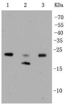
Fig1: Western blot analysis of Rho A+B+C on different lysates using anti-Rho A+B+C antibody at 1/1,000 dilution. Positive control: Lane 1: HepG2 Lane 2: Hela Lane 3: JurkatApplication
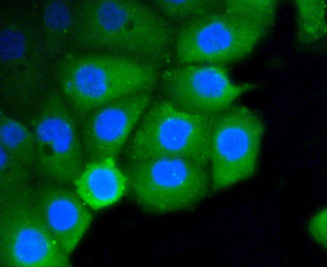
Fig2: ICC staining Rho A+B+C in Ags cells (green). The nuclear counter stain is DAPI (blue). Cells were fixed in paraformaldehyde, permeabilised with 0.25% Triton X100/PBS.Application
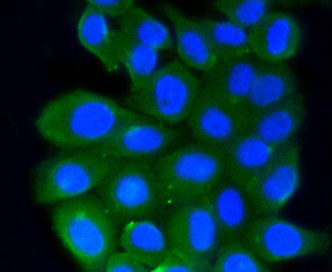
Fig3: ICC staining Rho A+B+C in HepG2 cells (green). The nuclear counter stain is DAPI (blue). Cells were fixed in paraformaldehyde, permeabilised with 0.25% Triton X100/PBS.Application
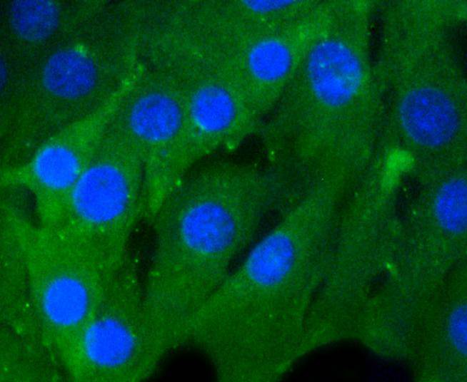
Fig4: ICC staining Rho A+B+C in NIH/3T3 cells (green). Cells were fixed in paraformaldehyde, permeabilised with 0.25% Triton X100/PBS.Application
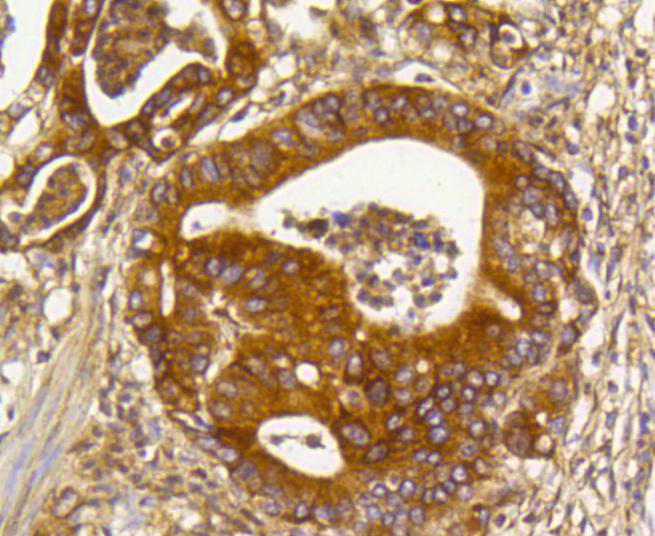
Fig5: Immunohistochemical analysis of paraffin-embedded human gastric carcinoma tissue using anti-Rho A+B+C antibody. Counter stained with hematoxylin.Application
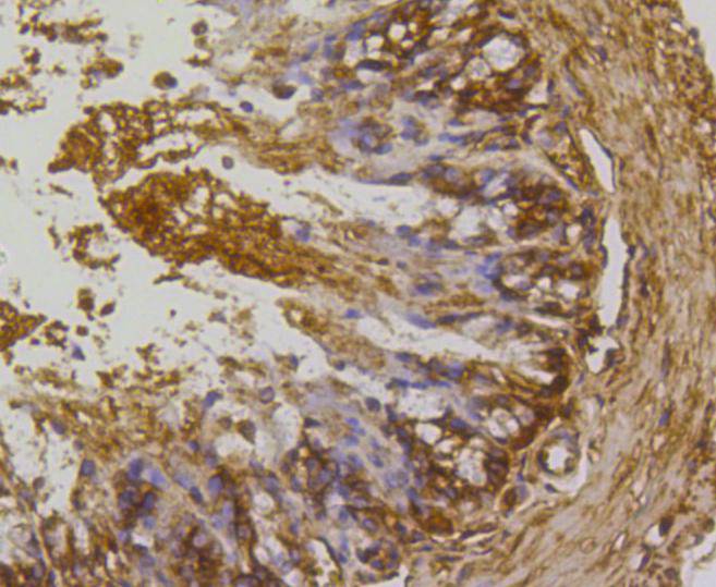
Fig6: Immunohistochemical analysis of paraffin-embedded mouse colon tissue using anti-Rho A+B+C antibody. Counter stained with hematoxylin.Application
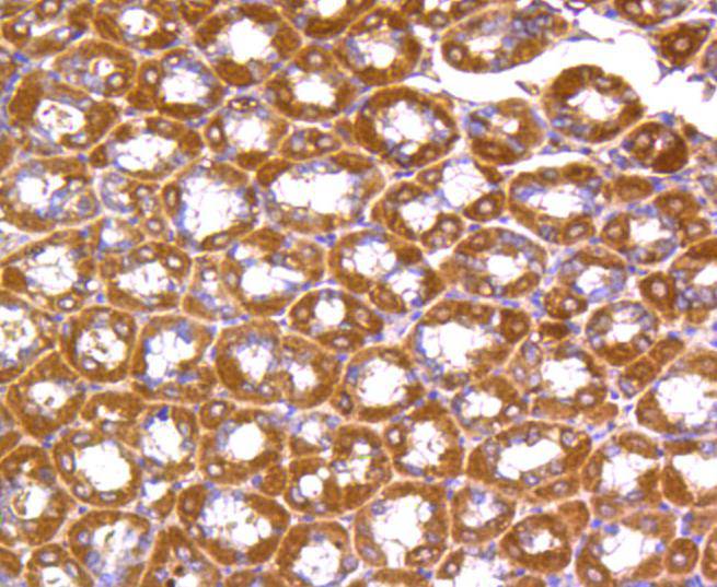
Fig7: Immunohistochemical analysis of paraffin-embedded mouse stomach tissue using anti-Rho A+B+C antibody. Counter stained with hematoxylin.Application
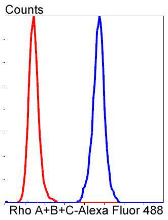
Fig8: Flow cytometric analysis of HepG2 cells with Rho A+B+C antibody at 1/50 dilution (blue) compared with an unlabelled control (cells without incubation with primary antibody; red). Alexa Fluor 488-conjugated goat anti rabbit IgG was used as the secondary antibody.| Product Name | Rho A+B+C [SR38-00] |
|---|---|
| Antibody Type | Primary Antibodies |
| Antigen Alias | AA017882 antibody AI324259, antibody Aplysia ras related homolog 9 (RhoC) antibody Aplysia ras-related homolog 12 antibody Aplysia RAS-related homolog 6 antibody Aplysia RAS-related homolog antibody ARH12 antibody ARH6 antibody ARH9 antibody ARHA antibody ARHB antibody ARHC antibody H12 antibody H6 antibody H9 antibody MGC117867 antibody MGC1448 antibody MGC61427 antibody MST081 antibody MSTP081 antibody Oncogene RHO H12 antibody Oncogene RHO H6 antibody Oncogene RHO H9 antibody OTTHUMP00000013675 antibody OTTHUMP00000013676 antibody OTTHUMP00000013802 antibody OTTHUMP00000013805 antibody OTTHUMP00000013807 antibody OTTHUMP00000013809 antibody OTTMUSP00000025086 antibody OTTMUSP00000025087 antibody Ras homolog 9 (RhoC) antibody Ras homolog gene family, member A antibody Ras homolog gene family, member B antibody Ras homolog gene family, member C antibody RAS-related homolog 9 antibody Rh -related GTP binding protein RhoC antibody Rho cDNA clone 12 antibody rho cDNA clone 6 antibody rho cDNA clone 9 antibody Rho related GTP binding protein RhoB antibody Rho related GTP binding protein RhoC antibody RHO12 antibody RHOA antibody RHOB antibody RHOC antibody rhoC GTPase antibody RHOH12 antibody RHOH6 antibody RHOH9 antibody RP11-426L16.4 antibody small GTP binding protein RhoA antibody Small GTP binding protein RhoC antibody Transforming protein RhoA antibody |
| Product description | The Ras p21 family of guanine nucleotide proteins has been widely studied in view of its apparent role in signal transduction pathways and high frequency of mutations in human malignancies. It is now clear, however, that the Ras proteins (H-, K- and N-Ras p21) are members of a much larger superfamily of related proteins. Six members of this family, Rap 1A, Rap 1B, Rap 2, R-Ras, Ral A and Ral B, exhibit approximately 50% amino acid homology to Ras. The six mammalian Rho proteins (Rho A, B, C, G, 7 and 8) are approximately 30% homologous to Ras and are expressed in a wide range of cell types. Both Ras p21 and Rho p21, as well as other members of the Ras superfamily, contain a carboxy-terminal CAAX sequence (C, cysteine; A, aliphatic amino acid; X, any amino acid) which in the case of Ras has been shown to be essential for correct localization and function. |
| Immunogen | recombinant protein |
| Clonality | Monoclonal |
|---|---|
| Isotype | IgG |
| Host Species | Recombinant rabbit |
| Tested Applications | WBICC/IFIHCFC |
| WB:1:1,000-1:2,000 ICC:1:50-1:200 IHC:1:200-1:500 FC:1:50-1:100 |
|
| Species Reactivity | HumanMouseRat |
| Concentration | 1mg/ml |
| Alternative Names | AA017882 antibody
AI324259 antibody Aplysia ras related homolog 9 (RhoC) antibody Aplysia ras-related homolog 12 antibody Aplysia RAS-related homolog 6 antibody Aplysia RAS-related homolog antibody ARH12 antibody ARH6 antibody ARH9 antibody ARHA antibody ARHB antibody ARHC antibody H12 antibody H6 antibody H9 antibody MGC117867 antibody MGC1448 antibody MGC61427 antibody MST081 antibody MSTP081 antibody Oncogene RHO H12 antibody Oncogene RHO H6 antibody Oncogene RHO H9 antibody OTTHUMP00000013675 antibody OTTHUMP00000013676 antibody OTTHUMP00000013802 antibody OTTHUMP00000013805 antibody OTTHUMP00000013807 antibody OTTHUMP00000013809 antibody OTTMUSP00000025086 antibody OTTMUSP00000025087 antibody Ras homolog 9 (RhoC) antibody Ras homolog gene family member A antibody Ras homolog gene family member B antibody Ras homolog gene family member C antibody RAS-related homolog 9 antibody Rh -related GTP binding protein RhoC antibody Rho cDNA clone 12 antibody rho cDNA clone 6 antibody rho cDNA clone 9 antibody Rho related GTP binding protein RhoB antibody Rho related GTP binding protein RhoC antibody RHO12 antibody RHOA antibody RHOB antibody RHOC antibody rhoC GTPase antibody RHOH12 antibody RHOH6 antibody RHOH9 antibody RP11-426L16.4 antibody small GTP binding protein RhoA antibody Small GTP binding protein RhoC antibody Transforming protein RhoA antibody |
|---|---|
| Molecular Weight(MW) | 24 kDa |
| Cellular Localization | Cytoplasm, Nucleus, Cell membrane, Cleavage furrow. |
Application
Fig1: Western blot analysis of Rho A+B+C on different lysates using anti-Rho A+B+C antibody at 1/1,000 dilution. Positive control: Lane 1: HepG2 Lane 2: Hela Lane 3: Jurkat
Application
Fig2: ICC staining Rho A+B+C in Ags cells (green). The nuclear counter stain is DAPI (blue). Cells were fixed in paraformaldehyde, permeabilised with 0.25% Triton X100/PBS.
Application
Fig3: ICC staining Rho A+B+C in HepG2 cells (green). The nuclear counter stain is DAPI (blue). Cells were fixed in paraformaldehyde, permeabilised with 0.25% Triton X100/PBS.
Application
Fig4: ICC staining Rho A+B+C in NIH/3T3 cells (green). Cells were fixed in paraformaldehyde, permeabilised with 0.25% Triton X100/PBS.
Application
Fig5: Immunohistochemical analysis of paraffin-embedded human gastric carcinoma tissue using anti-Rho A+B+C antibody. Counter stained with hematoxylin.
Application
Fig6: Immunohistochemical analysis of paraffin-embedded mouse colon tissue using anti-Rho A+B+C antibody. Counter stained with hematoxylin.
Application
Fig7: Immunohistochemical analysis of paraffin-embedded mouse stomach tissue using anti-Rho A+B+C antibody. Counter stained with hematoxylin.
Application
Fig8: Flow cytometric analysis of HepG2 cells with Rho A+B+C antibody at 1/50 dilution (blue) compared with an unlabelled control (cells without incubation with primary antibody; red). Alexa Fluor 488-conjugated goat anti rabbit IgG was used as the secondary antibody.| Positive Control | Jurkat, NIH/3T3, Hela, HepG2, mouse stomach tissue, human gastric carcinoma tissue, mouse colon tissue. |
|---|---|
| Application Notes | WB:1:1,000-1:2,000 ICC:1:50-1:200 IHC:1:200-1:500 FC:1:50-1:100 |
| Form | Liquid |
|---|---|
| Storage Instructions | Store at +4℃ after thawing. Aliquot store at -20℃ or -80℃. Avoid repeated freeze / thaw cycles. |
| Storage Buffer | 1*TBS (pH7.4), 1%BSA, 40%Glycerol. Preservative: 0.05% Sodium Azide. |


