WB
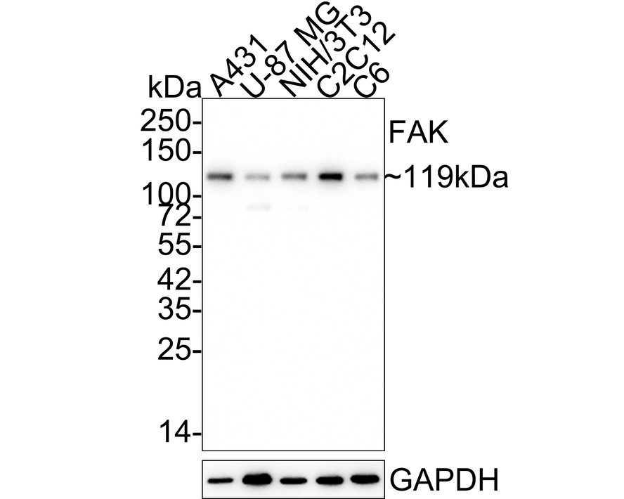
Western blot analysis of FAK on different lysates with Rabbit anti-FAK antibody at 1/5,000 dilution. Lane 1: A431 cell lysate (15 µg/Lane), Lane 2: U-87 MG cell lysate (15 µg/Lane), Lane 3: NIH/3T3 cell lysate (15 µg/Lane), Lane 4: C2C12 cell lysate (15 µg/Lane), Lane 5: C6 cell lysate (15 µg/Lane), Exposure time: 3 minutes; 4-20% SDS-PAGE gel. Proteins were transferred to a PVDF membrane and blocked with 5% NFDM/TBST for 1 hour at room temperature. The primary antibody at 1/5,000 dilution was used in 5% NFDM/TBST at 4℃ overnight. Goat Anti-Rabbit IgG - HRP Secondary Antibody at 1:50,000 dilution was used for 1 hour at room temperature.IHC
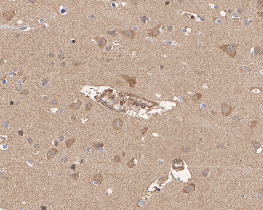
Immunohistochemical analysis of paraffin-embedded human brain tissue with Rabbit anti-FAK antibody at 1/200 dilution. The section was pre-treated using heat mediated antigen retrieval with Tris-EDTA buffer (pH 9.0) for 20 minutes. The tissues were blocked in 1% BSA for 20 minutes at room temperature, washed with ddH2O and PBS, and then probed with the primary antibody at 1/200 dilution for 1 hour at room temperature. The detection was performed using an HRP conjugated compact polymer system. DAB was used as the chromogen. Tissues were counterstained with hematoxylin and mounted with DPX.IHC
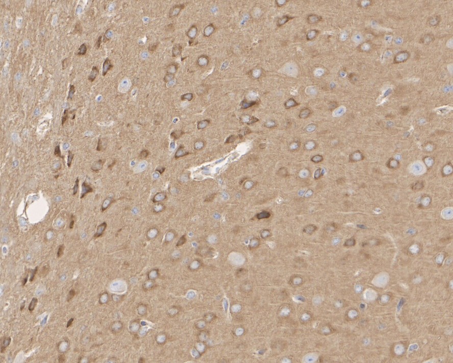
Immunohistochemical analysis of paraffin-embedded mouse brain tissue with Rabbit anti-FAK antibody at 1/1,000 dilution. The section was pre-treated using heat mediated antigen retrieval with Tris-EDTA buffer (pH 9.0) for 20 minutes. The tissues were blocked in 1% BSA for 20 minutes at room temperature, washed with ddH2O and PBS, and then probed with the primary antibody at 1/1,000 dilution for 1 hour at room temperature. The detection was performed using an HRP conjugated compact polymer system. DAB was used as the chromogen. Tissues were counterstained with hematoxylin and mounted with DPX.IHC
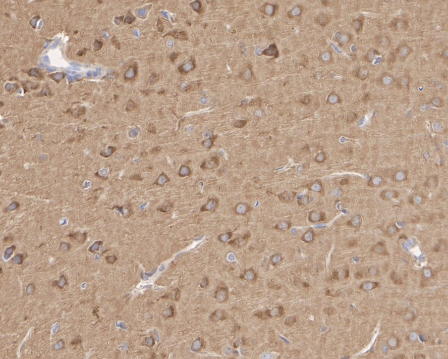
Immunohistochemical analysis of paraffin-embedded rat brain tissue with Rabbit anti-FAK antibody at 1/1,000 dilution. The section was pre-treated using heat mediated antigen retrieval with Tris-EDTA buffer (pH 9.0) for 20 minutes. The tissues were blocked in 1% BSA for 20 minutes at room temperature, washed with ddH2O and PBS, and then probed with the primary antibody at 1/1,000 dilution for 1 hour at room temperature. The detection was performed using an HRP conjugated compact polymer system. DAB was used as the chromogen. Tissues were counterstained with hematoxylin and mounted with DPX.IHC
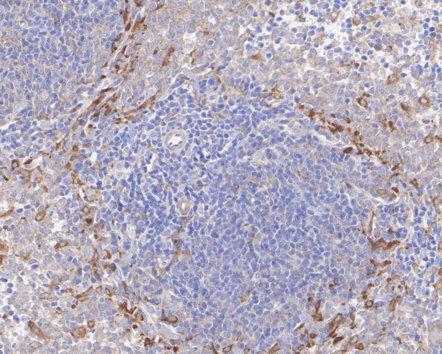
Immunohistochemical analysis of paraffin-embedded rat spleen tissue with Rabbit anti-FAK antibody at 1/1,000 dilution. The section was pre-treated using heat mediated antigen retrieval with Tris-EDTA buffer (pH 9.0) for 20 minutes. The tissues were blocked in 1% BSA for 20 minutes at room temperature, washed with ddH2O and PBS, and then probed with the primary antibody at 1/1,000 dilution for 1 hour at room temperature. The detection was performed using an HRP conjugated compact polymer system. DAB was used as the chromogen. Tissues were counterstained with hematoxylin and mounted with DPX.IHC
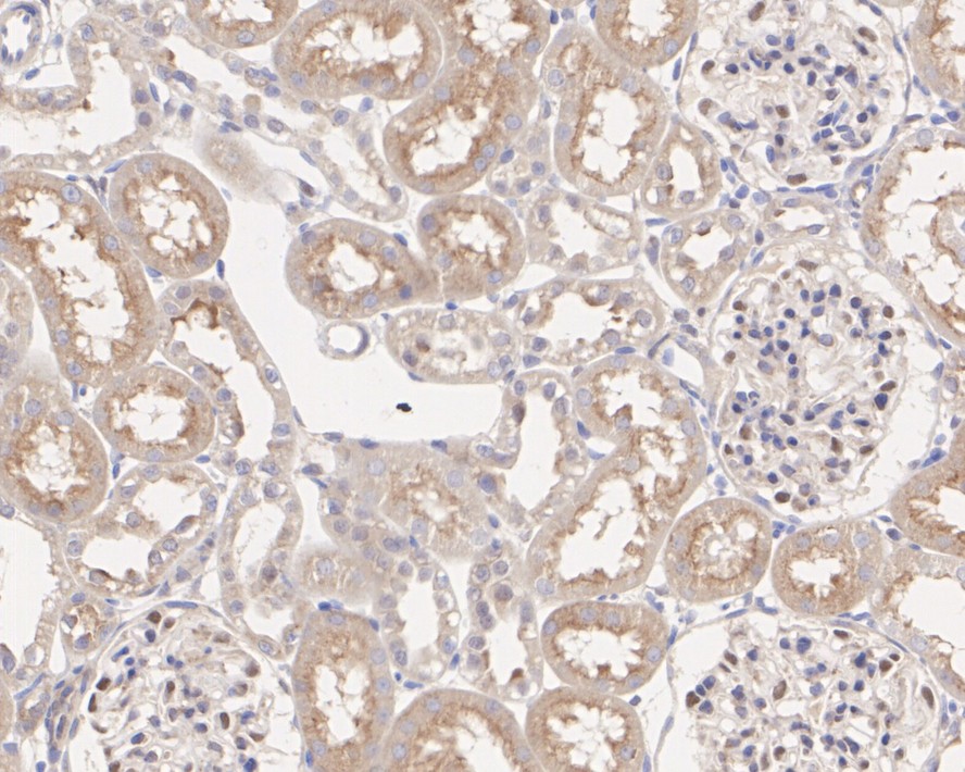
Immunohistochemical analysis of paraffin-embedded rat kidney tissue with Rabbit anti-FAK antibody at 1/1,000 dilution. The section was pre-treated using heat mediated antigen retrieval with Tris-EDTA buffer (pH 9.0) for 20 minutes. The tissues were blocked in 1% BSA for 20 minutes at room temperature, washed with ddH2O and PBS, and then probed with the primary antibody at 1/1,000 dilution for 1 hour at room temperature. The detection was performed using an HRP conjugated compact polymer system. DAB was used as the chromogen. Tissues were counterstained with hematoxylin and mounted with DPX.ICC/IF
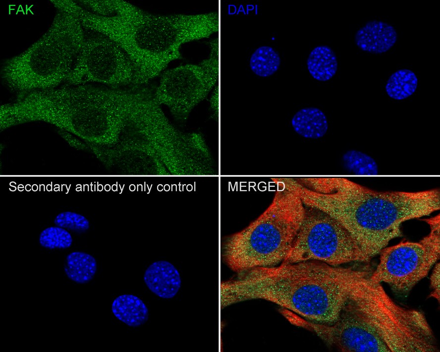
Immunocytochemistry analysis of C2C12 cells labeling FAK with Rabbit anti-FAK antibody at 1/100 dilution. Cells were fixed in 4% paraformaldehyde for 20 minutes at room temperature, permeabilized with 0.1% Triton X-100 in PBS for 5 minutes at room temperature, then blocked with 1% BSA in 10% negative goat serum for 1 hour at room temperature. Cells were then incubated with Rabbit anti-FAK antibody at 1/100 dilution in 1% BSA in PBST overnight at 4 ℃. Goat Anti-Rabbit IgG H&L (488) was used as the secondary antibody at 1/1,000 dilution. PBS instead of the primary antibody was used as the secondary antibody only control. Nuclear DNA was labelled in blue with DAPI. Beta tubulin (red) was stained at 1/100 dilution overnight at +4℃. Goat Anti-Mouse IgG H&L (594) was used as the secondary antibody at 1/1,000 dilution.FC
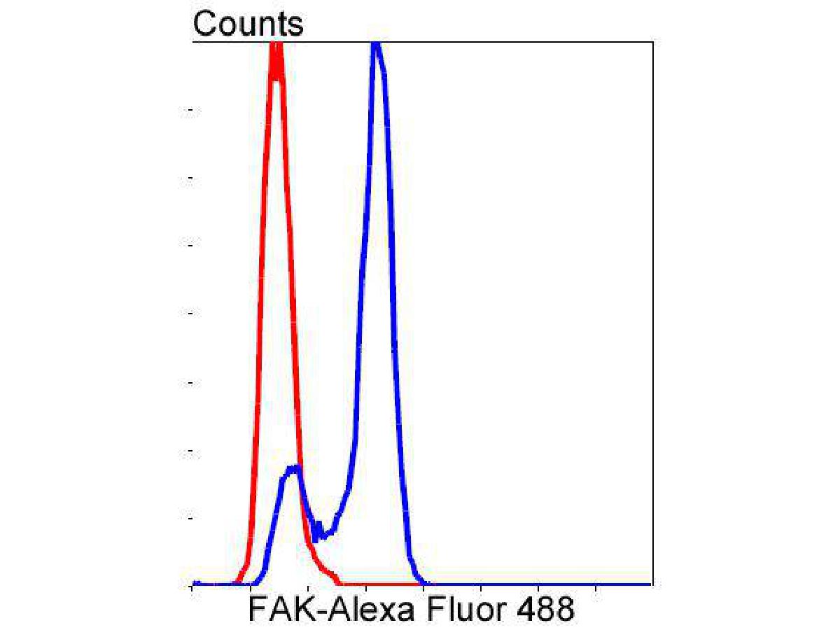
Flow cytometric analysis of FAK was done on Hela cells. The cells were fixed, permeabilized and stained with the primary antibody (1/50) (blue). After incubation of the primary antibody at room temperature for an hour, the cells were stained with a Alexa Fluor 488-conjugated Goat anti-Rabbit IgG Secondary antibody at 1/1000 dilution for 30 minutes.Unlabelled sample was used as a control (cells without incubation with primary antibody; red).IP
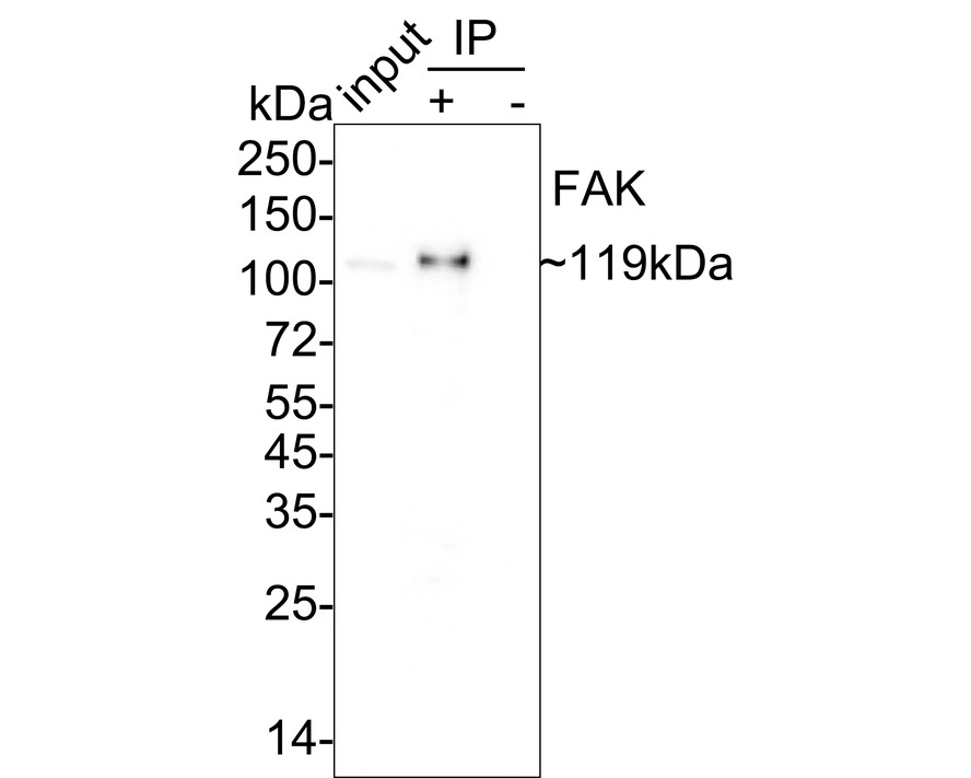
FAK was immunoprecipitated in 0.2mg A431 cell lysate with Rabbit anti-FAK antibody at 2 µg/25 µl agarose. Western blot was performed from the immunoprecipitate using Rabbit anti-FAK antibody at 1/2,000 dilution. Anti-Rabbit IgG for IP Nano-secondary antibody at 1/5,000 dilution was used for 1 hour at room temperature. Lane 1: A431 cell lysate (input), Lane 2: Rabbit anti-FAK antibody IP in A431 cell lysate, Lane 3: Rabbit IgG instead of Rabbit anti-FAK antibody in A431 cell lysate, Blocking/Dilution buffer: 5% NFDM/TBST, Exposure time: 3 minutes.| Product Name | FAK Recombinant Rabbit Monoclonal Antibody |
|---|---|
| Antibody Type | Primary Antibodies |
| Product description | Focal adhesion kinase was initially identified as a major substrate for the intrinsic protein tyrosine kinase activity of Src encoded pp60. The deduced amino acid sequence of FAK p125 has shown it to be a cytoplasmic protein tyrosine kinase whose sequence and structural organization are unique as compared to other proteins described to date. Localization of p125 by immunofluorescence suggests that it is primarily found in cellular focal adhesions leading to its designation as focal adhesion kinase (FAK). FAK is concentrated at the basal edge of only those basal keratinocytes that are actively migrating and rapidly proliferating in repairing burn wounds and is activated and localized to the focal adhesions of spreading keratinocytes in culture. Thus, it has been postulated that FAK may have an important in vivo role in the reepithelialization of human wounds. FAK protein tyrosine kinase activity has also been shown to increase in cells stimulated to grow by use of mitogenic neuropeptides or neurotransmitters acting through G protein coupled receptors. |
| Immunogen | Synthetic peptide within human FAK aa 700-740. |
| Clonality | Monoclonal |
|---|---|
| Isotype | IgG |
| Host Species | Recombinant rabbit |
| Tested Applications | FCICC/IFIHCIPWB |
| WB:1:5000 IHC:1:200-1:1000 ICC/IF:1:50-1:100 FC:1:50 IP:Use at an assay dependent concentration. |
|
| Species Reactivity | HumanMouseRat |
| Concentration | 1mg/ml |
| Purification | Protein A |
| Alternative Names | FADK 1 antibody FADK antibody FAK related non kinase polypeptide antibody FAK1 antibody FAK1_HUMAN antibody Focal adhesion kinase 1 antibody Focal adhesion Kinase antibody Focal adhesion kinase isoform FAK Del33 antibody Focal adhesion kinase related nonkinase antibody FRNK antibody p125FAK antibody pp125FAK antibody PPP1R71 antibody Protein phosphatase 1 regulatory subunit 71 antibody Protein tyrosine kinase 2 antibody Protein-tyrosine kinase 2 antibody Ptk2 antibody PTK2 protein tyrosine kinase 2 antibody |
|---|---|
| Molecular Weight(MW) | 119kDa |
| Cellular Localization | Cytoplasm, Nucleus, Cell membrane, Cell junction. |
WB
Western blot analysis of FAK on different lysates with Rabbit anti-FAK antibody at 1/5,000 dilution. Lane 1: A431 cell lysate (15 µg/Lane), Lane 2: U-87 MG cell lysate (15 µg/Lane), Lane 3: NIH/3T3 cell lysate (15 µg/Lane), Lane 4: C2C12 cell lysate (15 µg/Lane), Lane 5: C6 cell lysate (15 µg/Lane), Exposure time: 3 minutes; 4-20% SDS-PAGE gel. Proteins were transferred to a PVDF membrane and blocked with 5% NFDM/TBST for 1 hour at room temperature. The primary antibody at 1/5,000 dilution was used in 5% NFDM/TBST at 4℃ overnight. Goat Anti-Rabbit IgG - HRP Secondary Antibody at 1:50,000 dilution was used for 1 hour at room temperature.
IHC
Immunohistochemical analysis of paraffin-embedded human brain tissue with Rabbit anti-FAK antibody at 1/200 dilution. The section was pre-treated using heat mediated antigen retrieval with Tris-EDTA buffer (pH 9.0) for 20 minutes. The tissues were blocked in 1% BSA for 20 minutes at room temperature, washed with ddH2O and PBS, and then probed with the primary antibody at 1/200 dilution for 1 hour at room temperature. The detection was performed using an HRP conjugated compact polymer system. DAB was used as the chromogen. Tissues were counterstained with hematoxylin and mounted with DPX.
IHC
Immunohistochemical analysis of paraffin-embedded mouse brain tissue with Rabbit anti-FAK antibody at 1/1,000 dilution. The section was pre-treated using heat mediated antigen retrieval with Tris-EDTA buffer (pH 9.0) for 20 minutes. The tissues were blocked in 1% BSA for 20 minutes at room temperature, washed with ddH2O and PBS, and then probed with the primary antibody at 1/1,000 dilution for 1 hour at room temperature. The detection was performed using an HRP conjugated compact polymer system. DAB was used as the chromogen. Tissues were counterstained with hematoxylin and mounted with DPX.
IHC
Immunohistochemical analysis of paraffin-embedded rat brain tissue with Rabbit anti-FAK antibody at 1/1,000 dilution. The section was pre-treated using heat mediated antigen retrieval with Tris-EDTA buffer (pH 9.0) for 20 minutes. The tissues were blocked in 1% BSA for 20 minutes at room temperature, washed with ddH2O and PBS, and then probed with the primary antibody at 1/1,000 dilution for 1 hour at room temperature. The detection was performed using an HRP conjugated compact polymer system. DAB was used as the chromogen. Tissues were counterstained with hematoxylin and mounted with DPX.
IHC
Immunohistochemical analysis of paraffin-embedded rat spleen tissue with Rabbit anti-FAK antibody at 1/1,000 dilution. The section was pre-treated using heat mediated antigen retrieval with Tris-EDTA buffer (pH 9.0) for 20 minutes. The tissues were blocked in 1% BSA for 20 minutes at room temperature, washed with ddH2O and PBS, and then probed with the primary antibody at 1/1,000 dilution for 1 hour at room temperature. The detection was performed using an HRP conjugated compact polymer system. DAB was used as the chromogen. Tissues were counterstained with hematoxylin and mounted with DPX.
IHC
Immunohistochemical analysis of paraffin-embedded rat kidney tissue with Rabbit anti-FAK antibody at 1/1,000 dilution. The section was pre-treated using heat mediated antigen retrieval with Tris-EDTA buffer (pH 9.0) for 20 minutes. The tissues were blocked in 1% BSA for 20 minutes at room temperature, washed with ddH2O and PBS, and then probed with the primary antibody at 1/1,000 dilution for 1 hour at room temperature. The detection was performed using an HRP conjugated compact polymer system. DAB was used as the chromogen. Tissues were counterstained with hematoxylin and mounted with DPX.
ICC/IF
Immunocytochemistry analysis of C2C12 cells labeling FAK with Rabbit anti-FAK antibody at 1/100 dilution. Cells were fixed in 4% paraformaldehyde for 20 minutes at room temperature, permeabilized with 0.1% Triton X-100 in PBS for 5 minutes at room temperature, then blocked with 1% BSA in 10% negative goat serum for 1 hour at room temperature. Cells were then incubated with Rabbit anti-FAK antibody at 1/100 dilution in 1% BSA in PBST overnight at 4 ℃. Goat Anti-Rabbit IgG H&L (488) was used as the secondary antibody at 1/1,000 dilution. PBS instead of the primary antibody was used as the secondary antibody only control. Nuclear DNA was labelled in blue with DAPI. Beta tubulin (red) was stained at 1/100 dilution overnight at +4℃. Goat Anti-Mouse IgG H&L (594) was used as the secondary antibody at 1/1,000 dilution.
FC
Flow cytometric analysis of FAK was done on Hela cells. The cells were fixed, permeabilized and stained with the primary antibody (1/50) (blue). After incubation of the primary antibody at room temperature for an hour, the cells were stained with a Alexa Fluor 488-conjugated Goat anti-Rabbit IgG Secondary antibody at 1/1000 dilution for 30 minutes.Unlabelled sample was used as a control (cells without incubation with primary antibody; red).
IP
FAK was immunoprecipitated in 0.2mg A431 cell lysate with Rabbit anti-FAK antibody at 2 µg/25 µl agarose. Western blot was performed from the immunoprecipitate using Rabbit anti-FAK antibody at 1/2,000 dilution. Anti-Rabbit IgG for IP Nano-secondary antibody at 1/5,000 dilution was used for 1 hour at room temperature. Lane 1: A431 cell lysate (input), Lane 2: Rabbit anti-FAK antibody IP in A431 cell lysate, Lane 3: Rabbit IgG instead of Rabbit anti-FAK antibody in A431 cell lysate, Blocking/Dilution buffer: 5% NFDM/TBST, Exposure time: 3 minutes.| Application Notes | WB:1:5000 IHC:1:200-1:1000 ICC/IF:1:50-1:100 FC:1:50 IP:Use at an assay dependent concentration. |
|---|
| Form | Liquid |
|---|---|
| Storage Instructions | Store at +4℃ after thawing. Aliquot store at -20℃ or -80℃. Avoid repeated freeze / thaw cycles. |
| Storage Buffer | 1*TBS (pH7.4), 1%BSA, 40%Glycerol. Preservative: 0.05% Sodium Azide. |


