Application
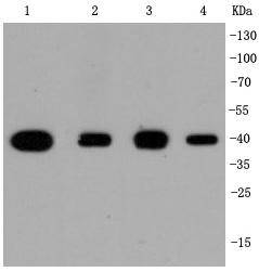
Fig1: Western blot analysis of p38 on different cell lysates using anti-p38 antibody at 1/1,000 dilution. Positive control: Lane 1: Hela Lane 2: NIH/3T3 Lane 3: PC-12 Lane 4: JurkatApplication
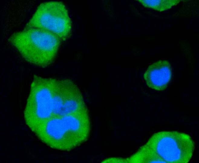
Fig2: ICC staining p38 in Hela cells (green). The nuclear counter stain is DAPI (blue). Cells were fixed in paraformaldehyde, permeabilised with 0.25% Triton X100/PBS.Application
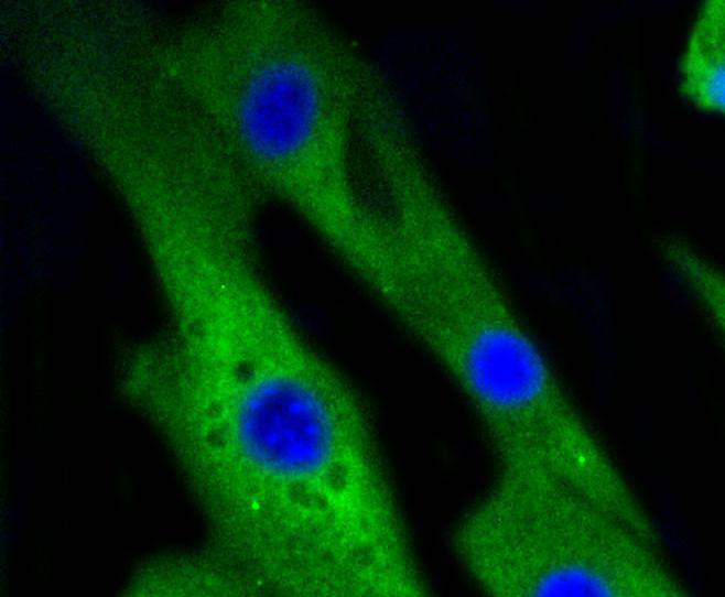
Fig3: ICC staining p38 in NIH/3T3 cells (green). The nuclear counter stain is DAPI (blue). Cells were fixed in paraformaldehyde, permeabilised with 0.25% Triton X100/PBS.| Product Name | p38 [SR43-04] |
|---|---|
| Antibody Type | Primary Antibodies |
| Antigen Alias | CSAID Binding Protein 1 antibody CSAID binding protein antibody CSAID-binding protein antibody Csaids binding protein antibody CSBP 1 antibody CSBP 2 antibody CSBP antibody CSBP1 antibody CSBP2 antibody CSPB1 antibody Cytokine suppressive anti-inflammatory drug-binding protein antibody EXIP antibody MAP kinase 14 antibody MAP kinase MXI2 antibody MAP kinase p38 alpha antibody MAPK 14 antibody MAPK14 antibody MAX interacting protein 2 antibody MAX-interacting protein 2 antibody Mitogen Activated Protein Kinase 14 antibody Mitogen activated protein kinase p38 alpha antibody Mitogen-activated protein kinase 14 antibody Mitogen-activated protein kinase p38 alpha antibody MK14_HUMAN antibody Mxi 2 antibody MXI2 antibody p38 ALPHA antibody p38 antibody p38 MAP kinase antibody p38 MAPK antibody p38 mitogen activated protein kinase antibody p38ALPHA antibody p38alpha Exip antibody PRKM14 antibody PRKM15 antibody RK antibody SAPK2A antibody |
| Product description | MAP (mitogen-activated protein) kinases play a significant role in many biological processes, including cell adhesion and spreading, cell differentiation and apoptosis. p38α, p38β and p38γ, also known as MAPK14, MAPK11 and MAPK12, respectively, each contain one protein kinase domain and belong to the MAP kinase family. Expressed in different areas throughout the body with common expression patterns in heart, p38 proteins use magnesium as a cofactor to catalyze the ATP-dependent phosphorylation of target proteins. Via their catalytic activity, p38α, p38β and p38γ are involved in a variety of events throughout the cell, including signal transduction pathways, cytokine production and cell proliferation and differentiation. The p38 proteins are subject to phosphoryation on Thr and Tyr residues, an event which is thought to activate the phosphorylated protein. |
| Immunogen | recombinant protein |
| Clonality | Monoclonal |
|---|---|
| Isotype | IgG |
| Host Species | Recombinant rabbit |
| Tested Applications | WBICC/IF |
| WB:1:1,000-1:5,000 ICC:1:50-1:200 |
|
| Species Reactivity | HumanMouseRat |
| Concentration | 1mg/ml |
| Alternative Names | CSAID Binding Protein 1 antibody CSAID binding protein antibody CSAID-binding protein antibody Csaids binding protein antibody CSBP 1 antibody CSBP 2 antibody CSBP antibody CSBP1 antibody CSBP2 antibody CSPB1 antibody Cytokine suppressive anti-inflammatory drug-binding protein antibody EXIP antibody MAP kinase 14 antibody MAP kinase MXI2 antibody MAP kinase p38 alpha antibody MAPK 14 antibody MAPK14 antibody MAX interacting protein 2 antibody MAX-interacting protein 2 antibody Mitogen Activated Protein Kinase 14 antibody Mitogen activated protein kinase p38 alpha antibody Mitogen-activated protein kinase 14 antibody Mitogen-activated protein kinase p38 alpha antibody MK14_HUMAN antibody Mxi 2 antibody MXI2 antibody p38 ALPHA antibody p38 antibody p38 MAP kinase antibody p38 MAPK antibody p38 mitogen activated protein kinase antibody p38ALPHA antibody p38alpha Exip antibody PRKM14 antibody PRKM15 antibody RK antibody SAPK2A antibody |
|---|---|
| Molecular Weight(MW) | 38 kDa |
| Cellular Localization | Cytoplasm, Nucleus. |
Application
Fig1: Western blot analysis of p38 on different cell lysates using anti-p38 antibody at 1/1,000 dilution. Positive control: Lane 1: Hela Lane 2: NIH/3T3 Lane 3: PC-12 Lane 4: Jurkat
Application
Fig2: ICC staining p38 in Hela cells (green). The nuclear counter stain is DAPI (blue). Cells were fixed in paraformaldehyde, permeabilised with 0.25% Triton X100/PBS.
Application
Fig3: ICC staining p38 in NIH/3T3 cells (green). The nuclear counter stain is DAPI (blue). Cells were fixed in paraformaldehyde, permeabilised with 0.25% Triton X100/PBS.| Positive Control | NIH/3T3, PC-12, Hela, Jurkat. |
|---|---|
| Application Notes | WB:1:1,000-1:5,000 ICC:1:50-1:200 |
| Form | Liquid |
|---|---|
| Storage Instructions | Store at +4℃ after thawing. Aliquot store at -20℃ or -80℃. Avoid repeated freeze / thaw cycles. |
| Storage Buffer | 1*TBS (pH7.4), 1%BSA, 40%Glycerol. Preservative: 0.05% Sodium Azide. |


