Application
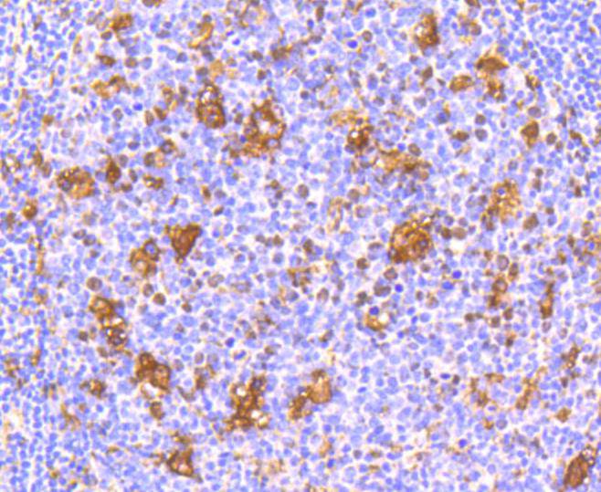
Fig1: Immunohistochemical analysis of paraffin-embedded human tonsil tissue using anti-IFNAR1 antibody. Counter stained with hematoxylin.Application
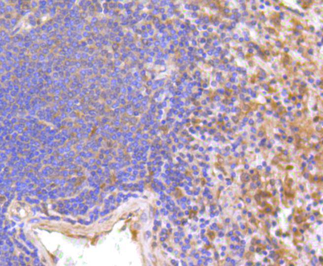
Fig2: Immunohistochemical analysis of paraffin-embedded human spleen tissue using anti-IFNAR1 antibody. Counter stained with hematoxylin.Application
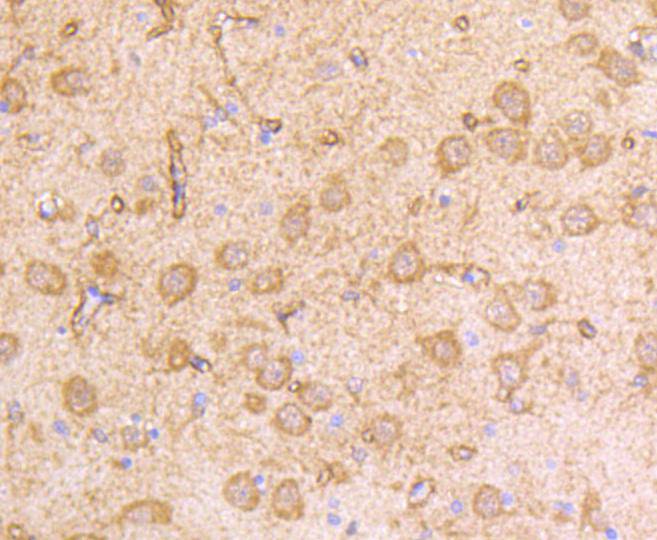
Fig3: Immunohistochemical analysis of paraffin-embedded rat brain tissue using anti-IFNAR1 antibody. Counter stained with hematoxylin.Application
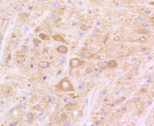
Fig4: Immunohistochemical analysis of paraffin-embedded mouse brain tissue using anti-IFNAR1 antibody. Counter stained with hematoxylin.Application
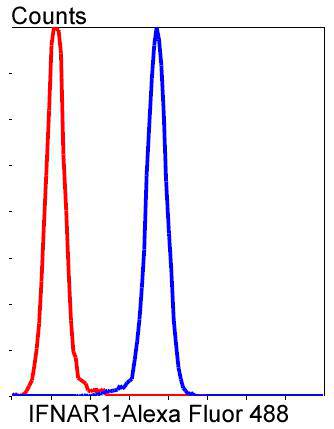
Fig5: Flow cytometric analysis of Jurkat cells with IFNAR1 antibody at 1/50 dilution (blue) compared with an unlabelled control (cells without incubation with primary antibody; red). Alexa Fluor 488-conjugated goat anti rabbit IgG was used as the secondary antibody.Application
Fig6: Western blot analysis of IFNAR1 on Siha cell lysates using anti-IFNAR1 antibody at 1/500 dilution.| Product Name | IFNAR1 [SR45-08] |
|---|---|
| Antibody Type | Primary Antibodies |
| Antigen Alias | Alpha type antiviral protein antibody AVP antibody Beta type antiviral protein antibody CRF2-1 antibody Cytokine receptor class-II member 1 antibody Cytokine receptor family 2 member 1 antibody IFN alpha REC antibody IFN alpha receptor antibody IFN alpha/beta Receptor alpha antibody IFN beta receptor antibody IFN-alpha/beta receptor 1 antibody IFN-R-1 antibody IFNAR antibody Ifnar1 antibody IFNBR antibody IFRC antibody INAR1_HUMAN antibody Interferon (alpha beta and omega) receptor 1 antibody Interferon alpha/beta receptor 1 antibody Interferon alpha/beta receptor alpha chain antibody Interferon beta receptor 1 antibody Type I interferon receptor 1 antibody |
| Product description | The type I interferons (IFNs), α and β, are a group of structurally and functionally related proteins that are induced by either viruses or double stranded RNA and defined by their ability to confer an antiviral state in cells. The α and β IFNs appear to compete with one another for binding to a common cell surface receptor, while immune IFN (IFNγ) binds to a distinct receptor. The latter protein, IFN-αR, is only weakly responsive to type I interferons in contrast to IFN-α/βR, which binds to and responds effectively to IFN-β and to several of the IFN-? subtypes. Moreover, IFN-α/βR is physically associated with the cytoplasmic tyrosine kinase JAK1 and thus, in addition to ligand binding, appears to be functionally involved in signal transduction. IFN-αR1 is a receptor for IFN-α/β and is present as the full chain (IFN-αR1a) and as a splice-variant (IFN-αR1). The IFN-γ receptor complex consists of an alpha subunit (IFN-γRα) and a beta subunit that is 332 amino acids in length (mouse) and 337 amino acids in length (human). |
| Immunogen | recombinant protein |
| Clonality | Monoclonal |
|---|---|
| Isotype | IgG |
| Host Species | Recombinant rabbit |
| Tested Applications | WBIPIHCFC |
| WB:1:1,000-1:2,000 IHC:1:50-1:200: FC:1:50-1:100 |
|
| Species Reactivity | HumanMouseRat |
| Concentration | 1mg/ml |
| Alternative Names | Alpha type antiviral protein antibody AVP antibody Beta type antiviral protein antibody CRF2-1 antibody Cytokine receptor class-II member 1 antibody Cytokine receptor family 2 member 1 antibody IFN alpha REC antibody IFN alpha receptor antibody IFN alpha/beta Receptor alpha antibody IFN beta receptor antibody IFN-alpha/beta receptor 1 antibody IFN-R-1 antibody IFNAR antibody Ifnar1 antibody IFNBR antibody IFRC antibody INAR1_HUMAN antibody Interferon (alpha beta and omega) receptor 1 antibody Interferon alpha/beta receptor 1 antibody Interferon alpha/beta receptor alpha chain antibody Interferon beta receptor 1 antibody Type I interferon receptor 1 antibody |
|---|---|
| Molecular Weight(MW) | 90/130 kDa |
| Cellular Localization | Cell membrane, Late endosome, Lysosome. |
| SwissProt ID | P17181 |
|---|
Application
Fig1: Immunohistochemical analysis of paraffin-embedded human tonsil tissue using anti-IFNAR1 antibody. Counter stained with hematoxylin.
Application
Fig2: Immunohistochemical analysis of paraffin-embedded human spleen tissue using anti-IFNAR1 antibody. Counter stained with hematoxylin.
Application
Fig3: Immunohistochemical analysis of paraffin-embedded rat brain tissue using anti-IFNAR1 antibody. Counter stained with hematoxylin.
Application
Fig4: Immunohistochemical analysis of paraffin-embedded mouse brain tissue using anti-IFNAR1 antibody. Counter stained with hematoxylin.
Application
Fig5: Flow cytometric analysis of Jurkat cells with IFNAR1 antibody at 1/50 dilution (blue) compared with an unlabelled control (cells without incubation with primary antibody; red). Alexa Fluor 488-conjugated goat anti rabbit IgG was used as the secondary antibody.
Application
Fig6: Western blot analysis of IFNAR1 on Siha cell lysates using anti-IFNAR1 antibody at 1/500 dilution.| Positive Control | Human tonsil tissue, human spleen tissue, rat brain tissue, mouse brain tissue. |
|---|---|
| Application Notes | WB:1:1,000-1:2,000 IHC:1:50-1:200: FC:1:50-1:100 |
| Form | Liquid |
|---|---|
| Storage Instructions | Store at +4℃ after thawing. Aliquot store at -20℃ or -80℃. Avoid repeated freeze / thaw cycles. |
| Storage Buffer | 1*TBS (pH7.4), 1%BSA, 40%Glycerol. Preservative: 0.05% Sodium Azide. |


