Application
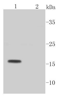
Fig1: Western blot analysis of active Caspase-3 on different cell lysates using anti-active Caspase-3 antibody at 1/1,000 dilution. Positive control: Lane 1: Camptothecin (2 μM) treated Jurkat cells Lane 2: Untreated Jurkat cellsApplication
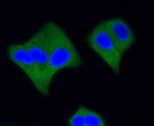
Fig2: ICC staining active Caspase-3 in Hela cells (green). The nuclear counter stain is DAPI (blue). Cells were fixed in paraformaldehyde, permeabilised with 0.25% Triton X100/PBS.Application
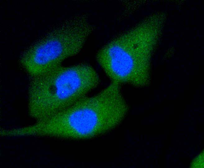
Fig3: ICC staining active Caspase-3 in PC-3M cells (green). The nuclear counter stain is DAPI (blue). Cells were fixed in paraformaldehyde, permeabilised with 0.25% Triton X100/PBS.Application
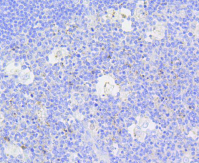
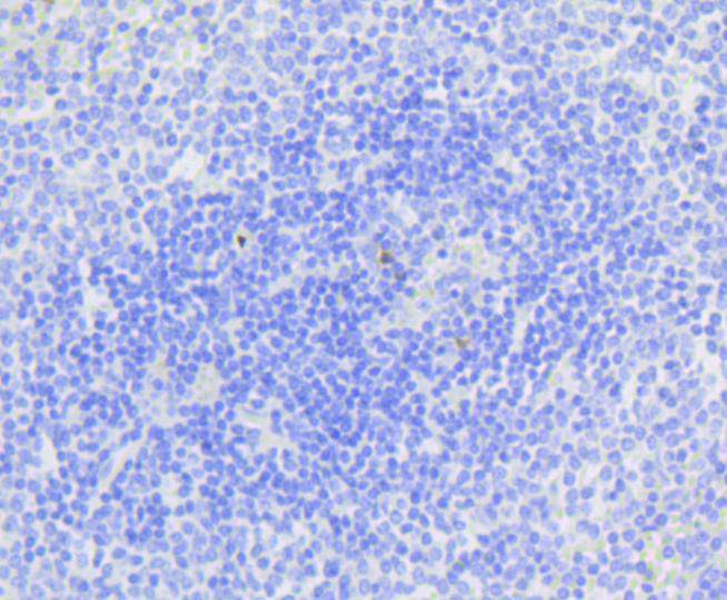
Fig4: Immunohistochemical analysis of paraffin-embedded human tonsil tissue using anti-active Caspase-3 antibody. Counter stained with hematoxylin.Application
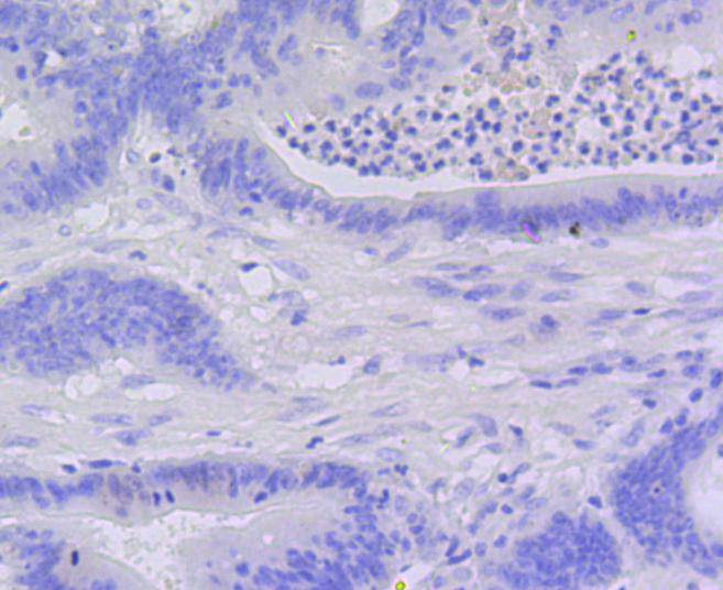
Fig5: Immunohistochemical analysis of paraffin-embedded human colon cancer tissue using anti-active Caspase-3 antibody. Counter stained with hematoxylin.Application
Fig6: Immunohistochemical analysis of paraffin-embedded human spleen tissue using anti-active Caspase-3 antibody. Counter stained with hematoxylin.| Product Name | Active Caspase-3 [SR01-02] |
|---|---|
| Antibody Type | Primary Antibodies |
| Antigen Alias | Apopain antibody CASP 3 antibody CASP-3 antibody CASP3 antibody CASP3_HUMAN antibody Caspase 3 antibody Caspase-3 subunit p12 antibody CPP 32 antibody CPP-32 antibody CPP32B antibody Cysteine protease CPP32 antibody PARP cleavage protease antibody Protein Yama antibody SCA-1 antibody SCA1 antibody SREBP cleavage activity 1 antibody Yama antibody |
| Product description | Caspase-3, also known as apopain, SCA-1, Yama and CPP32, is an aspartate-specific cysteine protease that belongs to the ICE subfamily of caspases. Caspase-3 is expressed in cells as an inactive precursor from which the p17 and p11 subunits of the mature caspase-3 are proteolytically generated during apoptosis. The caspase-3 precursor is first cleaved at Asp175-Ser176 to produce the p11 subunit and the p20 peptide. Subsequently, the p20 peptide is cleaved at Asp28-Ser29 to generate the mature p17 subunit. The active caspase-3 enzyme is a heterodimer composed of two p17 and two p11 subunits. At the onset of apoptosis, caspase-3 proteolytically cleaves PARP at an Asp216-Gly217 bond. During the execution of the apoptotic cascade, activated caspase-3 releases SREBP from the membrane of the ER in a proteolytic reaction that is distinct from their normal sterol-dependent activation. Caspase-3 cleaves and activates SREBPs between the basic helix-loop-helix leucine zipper domain and the membrane attachment domain. Caspase-3 also cleaves and activates caspase-6, -7 and -9. The human caspase-3 gene encodes a cytoplasmic protein that is highly expressed in lung, spleen, heart, liver, kidney and cells of the immune system. |
| Immunogen | recombinant protein |
| Clonality | Monoclonal |
|---|---|
| Isotype | IgG |
| Host Species | Recombinant rabbit |
| Tested Applications | WBICC/IFIHC |
| WB:1:1,000-1:2,000 ICC:1:50-1:200 IHC:1:50-1:200 |
|
| Species Reactivity | Human |
| Concentration | 1mg/ml |
| Alternative Names | Apopain antibody CASP 3 antibody CASP-3 antibody CASP3 antibody CASP3_HUMAN antibody Caspase 3 antibody Caspase-3 subunit p12 antibody CPP 32 antibody CPP-32 antibody CPP32B antibody Cysteine protease CPP32 antibody PARP cleavage protease antibody Protein Yama antibody SCA-1 antibody SCA1 antibody SREBP cleavage activity 1 antibody Yama antibody |
|---|---|
| Molecular Weight(MW) | 17 kDa |
| Cellular Localization | Cytoplasm |
| SwissProt ID | P42574 |
|---|
Application
Fig1: Western blot analysis of active Caspase-3 on different cell lysates using anti-active Caspase-3 antibody at 1/1,000 dilution. Positive control: Lane 1: Camptothecin (2 μM) treated Jurkat cells Lane 2: Untreated Jurkat cells
Application
Fig2: ICC staining active Caspase-3 in Hela cells (green). The nuclear counter stain is DAPI (blue). Cells were fixed in paraformaldehyde, permeabilised with 0.25% Triton X100/PBS.
Application
Fig3: ICC staining active Caspase-3 in PC-3M cells (green). The nuclear counter stain is DAPI (blue). Cells were fixed in paraformaldehyde, permeabilised with 0.25% Triton X100/PBS.
Application
Fig4: Immunohistochemical analysis of paraffin-embedded human tonsil tissue using anti-active Caspase-3 antibody. Counter stained with hematoxylin.
Application
Fig5: Immunohistochemical analysis of paraffin-embedded human colon cancer tissue using anti-active Caspase-3 antibody. Counter stained with hematoxylin.
Application
Fig6: Immunohistochemical analysis of paraffin-embedded human spleen tissue using anti-active Caspase-3 antibody. Counter stained with hematoxylin.| Positive Control | PC-3M, Hela, HepG2, human tonsil tissue, human colon cancer tissue, human spleen tissue. |
|---|---|
| Application Notes | WB:1:1,000-1:2,000 ICC:1:50-1:200 IHC:1:50-1:200 |
| Form | Liquid |
|---|---|
| Storage Instructions | Store at +4℃ after thawing. Aliquot store at -20℃ or -80℃. Avoid repeated freeze / thaw cycles. |
| Storage Buffer | 1*TBS (pH7.4), 1%BSA, 40%Glycerol. Preservative: 0.05% Sodium Azide. |


