Application
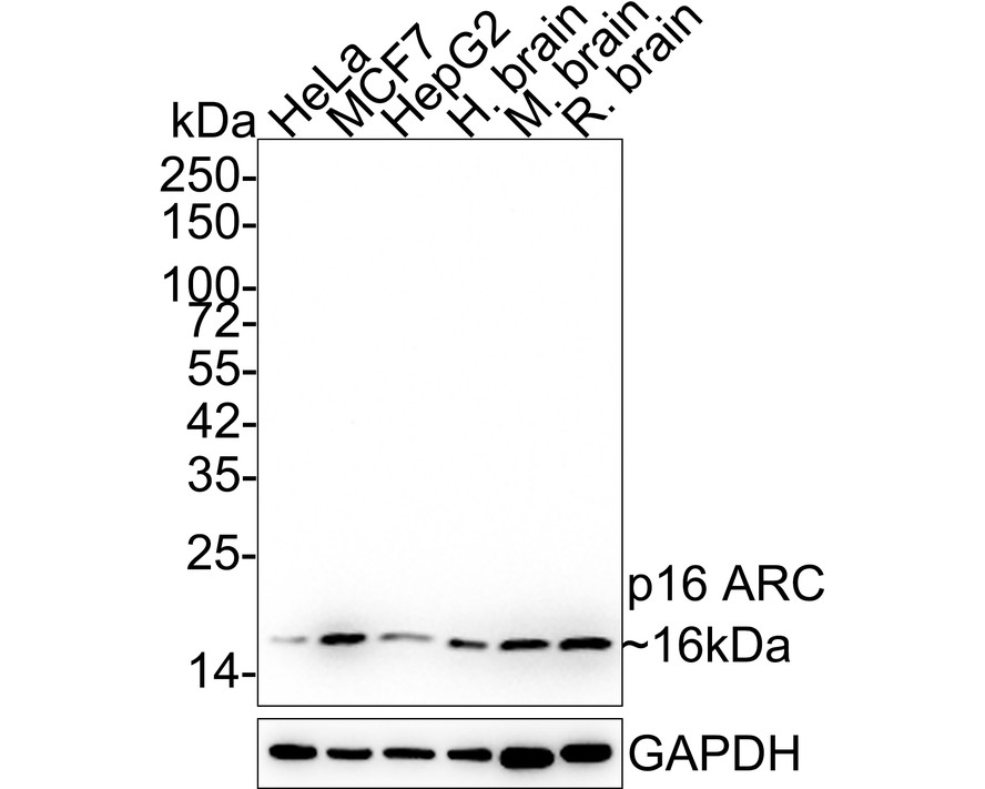
Fig1:Western blot analysis of p16 ARC on different lysates with Rabbit anti-p16 ARC antibody at 1/5,000 dilution. Lane 1: HeLa cell lysate (15 µg/Lane), Lane 2: MCF7 cell lysate (15 µg/Lane), Lane 3: HepG2 cell lysate (15 µg/Lane), Lane 4: Human brain tissue lysate (20 µg/Lane), Lane 5: Mouse brain tissue lysate (20 µg/Lane), Lane 6: Rat brain tissue lysate (20 µg/Lane), Exposure time: 3 minutes; 4-20% SDS-PAGE gel. Proteins were transferred to a PVDF membrane and blocked with 5% NFDM/TBST for 1 hour at room temperature. The primary antibody at 1/5,000 dilution was used in 5% NFDM/TBST at 4℃ overnight. Goat Anti-Rabbit IgG - HRP Secondary Antibody at 1/50,000 dilution was used for 1 hour at room temperature.Application
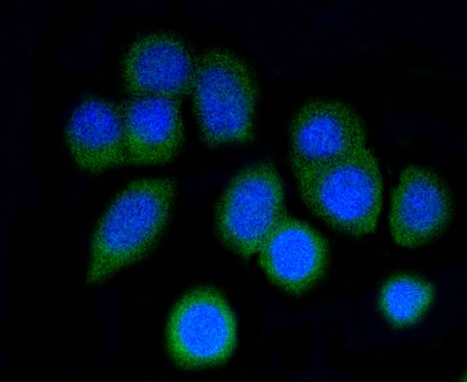
Fig2: ICC staining p16 ARC in N2A cells (green). The nuclear counter stain is DAPI (blue). Cells were fixed in paraformaldehyde, permeabilised with 0.25% Triton X100/PBS.Application
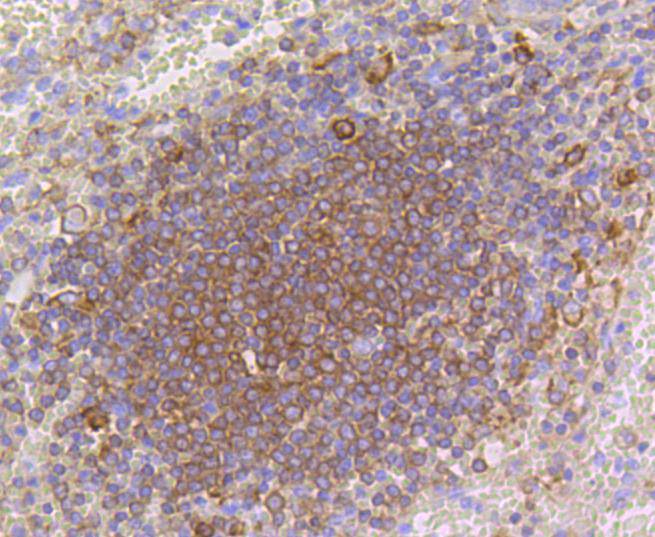
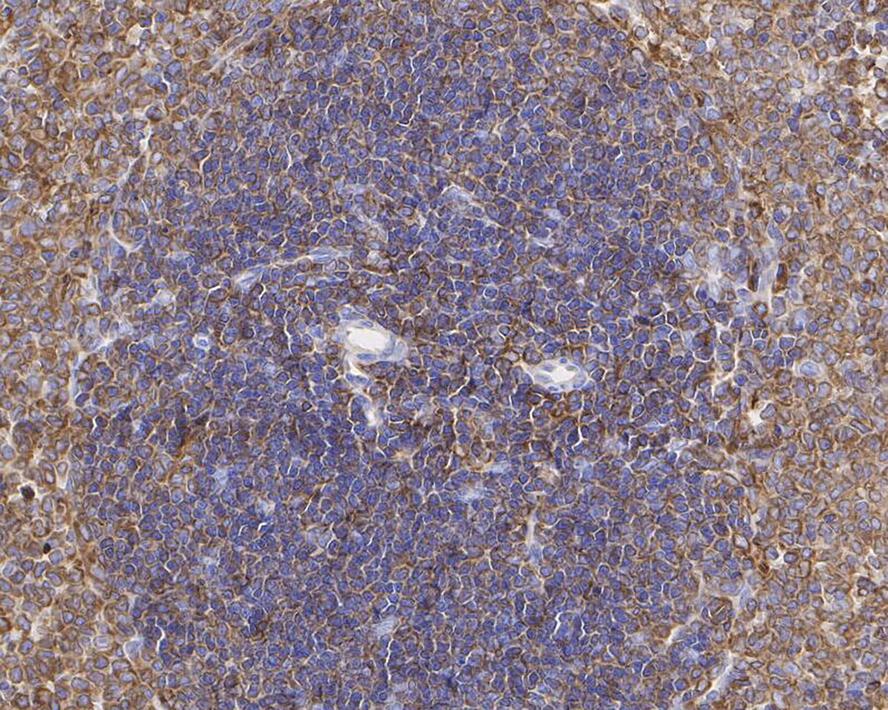
Fig3: Immunohistochemical analysis of paraffin-embedded human spleen tissue using anti-p16 ARC antibody. Counter stained with hematoxylin.Application
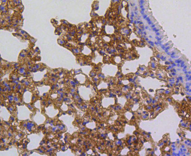
Fig4: Immunohistochemical analysis of paraffin-embedded mouse lung tissue using anti-p16 ARC antibody. Counter stained with hematoxylin.Application
Fig5: Immunohistochemical analysis of paraffin-embedded rat spleen tissue with Rabbit anti-ARPC5 / p16 ARC antibody at 1/1,000 dilution.Application
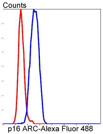
Fig6: Flow cytometric analysis of SH-SY-5Y cells with p16 ARC antibody at 1/50 dilution (blue) compared with an unlabelled control (cells without incubation with primary antibody; red). Alexa Fluor 488-conjugated goat anti rabbit IgG was used as the secondary antibody.| Product Name | ARPC5 / p16 ARC Recombinant Rabbit Monoclonal Antibody |
|---|---|
| Antibody Type | Primary Antibodies |
| Product description | The Arp2/3 (Actin-related protein 2/3) complex consists of seven subunits, all of which are actin-related proteins. The complex is involved in the control of actin polymerization and in mediating the formation of branched actin networks. p16-ARC, also known as ARPC5 (Actin-related protein 2/3 complex subunit 5) or ARC16 (Arp2/3 complex 16 kDa subunit), is a 151 amino acid subunit of the Arp2/3 complex. Thought to play a role in maintaining the integrity of Arp2/3, p16-ARC is a substrate for MAPKAPK-2 which, through phosphorylation of p16-ARC, may participate in Arp2/3 regulatory functions and remodeling of the Actin cytoskeleton. Two isoforms of p16-ARC exist due to alternative splicing events. |
| Immunogen | Synthetic peptide within Human p16 ARC aa 1-50 / 151. |
| Clonality | monoclonal |
|---|---|
| Isotype | IgG |
| Host Species | Recombinant rabbit |
| Tested Applications | FCICC/IFIHCWB |
| WB:1:5000 ICC/IF:1:50 IHC:1:50-1:200 FC:1:50-1:100 |
|
| Species Reactivity | HumanMouseRat |
| Concentration | 1mg/ml |
| Purification | Protein A |
| Alternative Names | Actin related protein 2/3 complex subunit 5 (16 kD) antibody
Actin related protein 2/3 complex subunit 5 antibody
Actin related protein 2/3 complex subunit 5 16kDa antibody Actin-related protein 2/3 complex subunit 5 antibody ARC16 antibody Arp2/3 complex 16 kDa subunit antibody Arp2/3 protein complex subunit p16 antibody ARPC 5 antibody Arpc5 antibody ARPC5_HUMAN antibody dJ127C7.3 antibody MGC88523 antibody p16 Arc antibody p16-ARC antibody RP1 127C7.3 antibody |
|---|---|
| Molecular Weight(MW) | 16kDa |
| Cellular Localization | Cytoplasm |
Application
Fig1:Western blot analysis of p16 ARC on different lysates with Rabbit anti-p16 ARC antibody at 1/5,000 dilution. Lane 1: HeLa cell lysate (15 µg/Lane), Lane 2: MCF7 cell lysate (15 µg/Lane), Lane 3: HepG2 cell lysate (15 µg/Lane), Lane 4: Human brain tissue lysate (20 µg/Lane), Lane 5: Mouse brain tissue lysate (20 µg/Lane), Lane 6: Rat brain tissue lysate (20 µg/Lane), Exposure time: 3 minutes; 4-20% SDS-PAGE gel. Proteins were transferred to a PVDF membrane and blocked with 5% NFDM/TBST for 1 hour at room temperature. The primary antibody at 1/5,000 dilution was used in 5% NFDM/TBST at 4℃ overnight. Goat Anti-Rabbit IgG - HRP Secondary Antibody at 1/50,000 dilution was used for 1 hour at room temperature.
Application
Fig2: ICC staining p16 ARC in N2A cells (green). The nuclear counter stain is DAPI (blue). Cells were fixed in paraformaldehyde, permeabilised with 0.25% Triton X100/PBS.
Application
Fig3: Immunohistochemical analysis of paraffin-embedded human spleen tissue using anti-p16 ARC antibody. Counter stained with hematoxylin.
Application
Fig4: Immunohistochemical analysis of paraffin-embedded mouse lung tissue using anti-p16 ARC antibody. Counter stained with hematoxylin.
Application
Fig5: Immunohistochemical analysis of paraffin-embedded rat spleen tissue with Rabbit anti-ARPC5 / p16 ARC antibody at 1/1,000 dilution.
Application
Fig6: Flow cytometric analysis of SH-SY-5Y cells with p16 ARC antibody at 1/50 dilution (blue) compared with an unlabelled control (cells without incubation with primary antibody; red). Alexa Fluor 488-conjugated goat anti rabbit IgG was used as the secondary antibody.| Application Notes | WB:1:5000 ICC/IF:1:50 IHC:1:50-1:200 FC:1:50-1:100 |
|---|
| Form | Liquid |
|---|---|
| Storage Instructions | Store at +4℃ after thawing. Aliquot store at -20℃ or -80℃. Avoid repeated freeze / thaw cycles. |
| Storage Buffer | 1*TBS (pH7.4), 1%BSA, 40%Glycerol. Preservative: 0.05% Sodium Azide. |


