Application
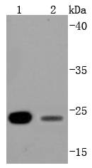
Fig1: Western blot analysis of Bcl-2 on different lysates using anti-Bcl-2 antibody at 1/1,000 dilution. Positive control: Lane 1: Hela Lane 2: JurkatApplication
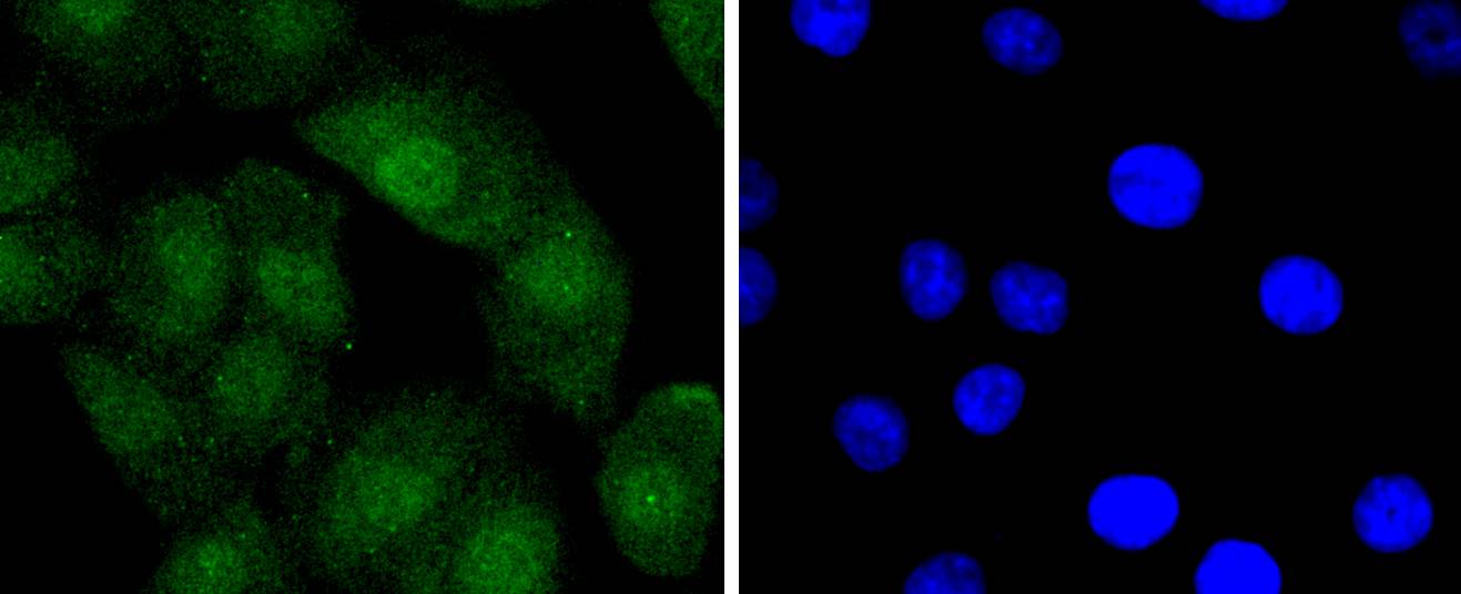
Fig2: ICC staining Bcl-2 in A549 cells (green). The nuclear counter stain is DAPI (blue). Cells were fixed in paraformaldehyde, permeabilised with 0.25% Triton X100/PBS.Application
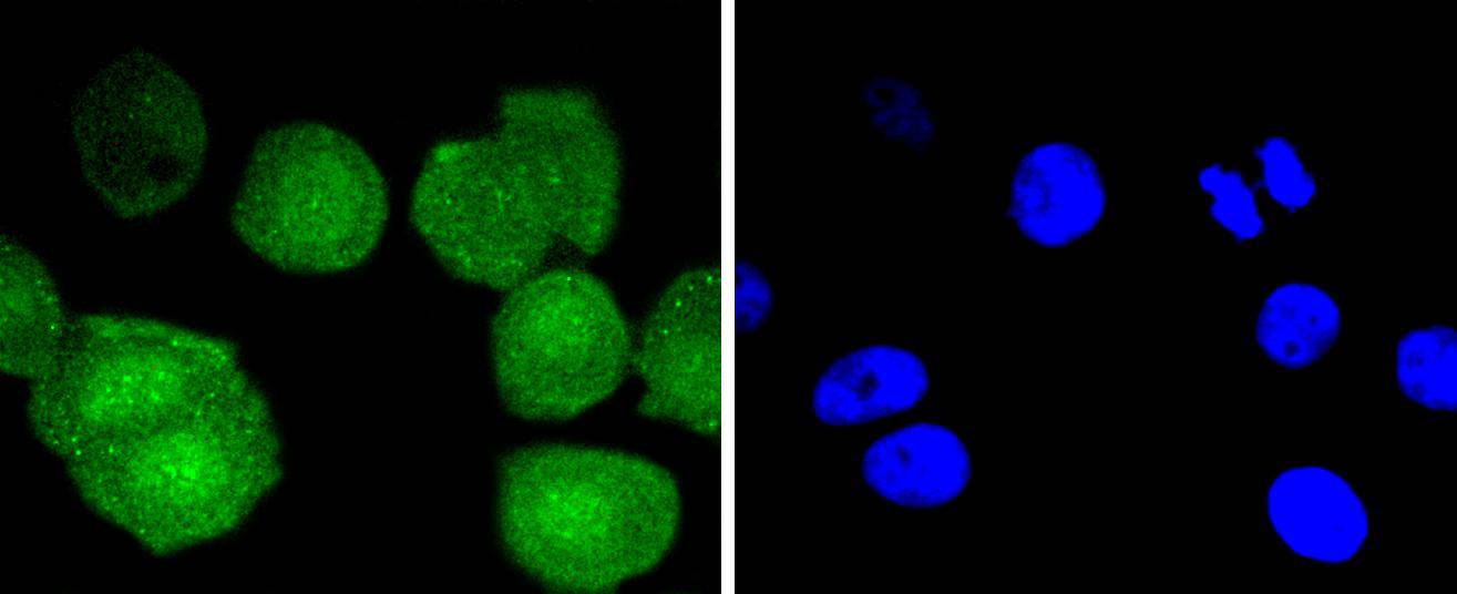
Fig3: ICC staining Bcl-2 in MCF-7 cells (green). The nuclear counter stain is DAPI (blue). Cells were fixed in paraformaldehyde, permeabilised with 0.25% Triton X100/PBS.Application
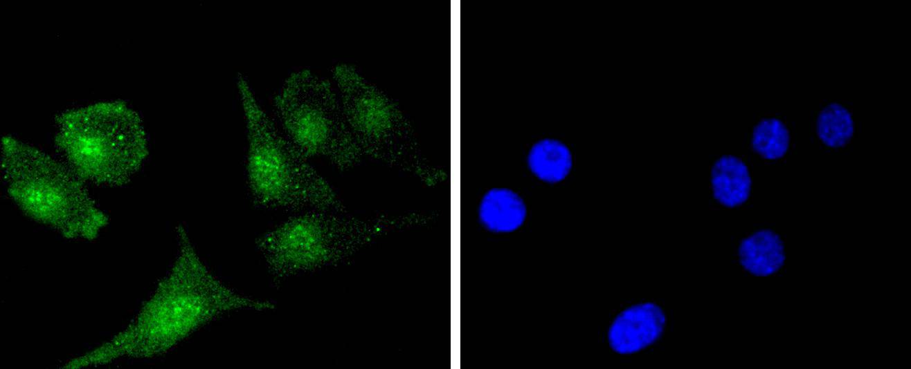
Fig4: ICC staining Bcl-2 in SH-SY-5Y cells (green). The nuclear counter stain is DAPI (blue). Cells were fixed in paraformaldehyde, permeabilised with 0.25% Triton X100/PBS.Application
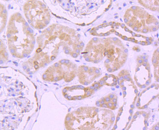
Fig5: Immunohistochemical analysis of paraffin-embedded human kidney tissue using anti-Bcl-2 antibody. Counter stained with hematoxylin.Application
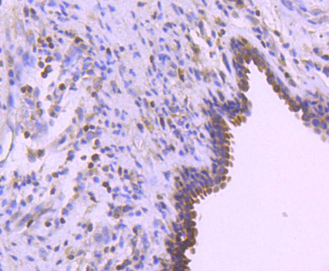
Fig6: Immunohistochemical analysis of paraffin-embedded human breast carcinoma tissue using anti-Bcl-2 antibody. Counter stained with hematoxylin.| Product Name | Bcl-2 [SZ10-03] |
|---|---|
| Antibody Type | Primary Antibodies |
| Antigen Alias | Apoptosis regulator Bcl 2 antibody Apoptosis regulator Bcl-2 antibody Apoptosis regulator Bcl2 antibody AW986256 antibody B cell CLL/lymphoma 2 antibody B cell leukemia/lymphoma 2 antibody Bcl-2 antibody Bcl2 antibody BCL2_HUMAN antibody C430015F12Rik antibody D630044D05Rik antibody D830018M01Rik antibody Leukemia/lymphoma, B-cell, 2 antibody Oncogene B-cell leukemia 2 antibody PPP1R50 antibody Protein phosphatase 1, regulatory subunit 50 antibody |
| Product description | Apoptosis is defined as a set of cascades which, when initiated, programs the cell to undergo lethal changes such as membrane blebbing, mitochondrial break down and DNA fragmentation. Bcl-2 is one among many key regulators of apoptosis, which are essential for proper development, tissue homeostasis, and protection against foreign pathogens. Human Bcl-2 is an anti-apoptotic, membrane-associated oncoprotein that can promote cell survival through protein-protein interactions with other Bcl-2 related family members, such as the death suppressors Bcl-xl, Mcl-1, Bcl-w, and A1 or the death agonists Bax, Bak, Bik, Bad, and Bid. The anti-apoptotic function of Bcl-2 can also be regulated through proteolytic processing and phospho-rylation. Bcl-2 may promote cell survival by interfering with the activation of the cytochrome c/Apaf-1 pathway through stabilization of the mitochondrial membrane. Mutations in the Bcl-2 gene can contribute to cancers where normal physiological cell death mechanisms are compromised by deregulation of the anti-apoptotic influence of Bcl-2. |
| Immunogen | recombinant protein |
| Clonality | Monoclonal |
|---|---|
| Isotype | IgG |
| Host Species | Recombinant rabbit |
| Tested Applications | WBICC/IFIHCIPFC |
| WB:1:1,000-1:2,000 ICC:1:50-1:200 IHC:1:50-1:500 FC:1:50-1:100 |
|
| Species Reactivity | HumanMouse |
| Concentration | 1mg/ml |
| Alternative Names | Apoptosis regulator Bcl 2 antibody
Apoptosis regulator Bcl-2 antibody
Apoptosis regulator Bcl2 antibody
AW986256 antibody
B cell CLL/lymphoma 2 antibody
B cell leukemia/lymphoma 2 antibody
Bcl-2 antibody
Bcl2 antibody
BCL2_HUMAN antibody
C430015F12Rik antibody
D630044D05Rik antibody
D830018M01Rik antibody
Leukemia/lymphoma B-cell 2 antibody Oncogene B-cell leukemia 2 antibody PPP1R50 antibody Protein phosphatase 1 regulatory subunit 50 antibody |
|---|---|
| Molecular Weight(MW) | 26/22 kDa |
| Cellular Localization | Mitochondrion outer membrane, Nucleus membrane, Endoplasmic reticulum membrane. |
Application
Fig1: Western blot analysis of Bcl-2 on different lysates using anti-Bcl-2 antibody at 1/1,000 dilution. Positive control: Lane 1: Hela Lane 2: Jurkat
Application
Fig2: ICC staining Bcl-2 in A549 cells (green). The nuclear counter stain is DAPI (blue). Cells were fixed in paraformaldehyde, permeabilised with 0.25% Triton X100/PBS.
Application
Fig3: ICC staining Bcl-2 in MCF-7 cells (green). The nuclear counter stain is DAPI (blue). Cells were fixed in paraformaldehyde, permeabilised with 0.25% Triton X100/PBS.
Application
Fig4: ICC staining Bcl-2 in SH-SY-5Y cells (green). The nuclear counter stain is DAPI (blue). Cells were fixed in paraformaldehyde, permeabilised with 0.25% Triton X100/PBS.
Application
Fig5: Immunohistochemical analysis of paraffin-embedded human kidney tissue using anti-Bcl-2 antibody. Counter stained with hematoxylin.
Application
Fig6: Immunohistochemical analysis of paraffin-embedded human breast carcinoma tissue using anti-Bcl-2 antibody. Counter stained with hematoxylin.| Positive Control | A549, MCF-7, Hela, Jurkat, SH-SY-5Y, human breast carcinoma tissue, human kidney tissue. |
|---|---|
| Application Notes | WB:1:1,000-1:2,000 ICC:1:50-1:200 IHC:1:50-1:500 FC:1:50-1:100 |
| Form | Liquid |
|---|---|
| Storage Instructions | Store at +4℃ after thawing. Aliquot store at -20℃ or -80℃. Avoid repeated freeze / thaw cycles. |
| Storage Buffer | 1*TBS (pH7.4), 1%BSA, 40%Glycerol. Preservative: 0.05% Sodium Azide. |


