WB
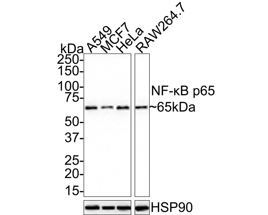
Western blot analysis of NF-kB p65 on different lysates with Rabbit anti-NF-kB p65 antibody at 1/5,000 dilution. Lane 1: A549 cell lysate, Lane 2: MCF7 cell lysate, Lane 3: HeLa cell lysate, Lane 4: RAW264.7 cell lysate, Lysates/proteins at 15 µg/Lane. Exposure time: 49 seconds; 4-20% SDS-PAGE gel. Proteins were transferred to a PVDF membrane and blocked with 5% NFDM/TBST for 1 hour at room temperature. The primary antibody at 1/5,000 dilution was used in 5% NFDM/TBST at room temperature for 2 hours. Goat Anti-Rabbit IgG - HRP Secondary Antibody at 1:100,000 dilution was used for 1 hour at room temperature.IHC
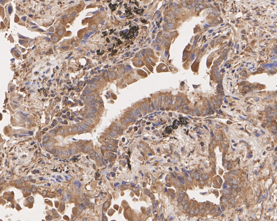
Immunohistochemical analysis of paraffin-embedded human lung cancer tissue with Rabbit anti-NF-kB p65 antibody at 1/200 dilution. The section was pre-treated using heat mediated antigen retrieval with sodium citrate buffer (pH 6.0) for 2 minutes. The tissues were blocked in 1% BSA for 20 minutes at room temperature, washed with ddH2O and PBS, and then probed with the primary antibody at 1/200 dilution for 1 hour at room temperature. The detection was performed using an HRP conjugated compact polymer system. DAB was used as the chromogen. Tissues were counterstained with hematoxylin and mounted with DPX.IHC
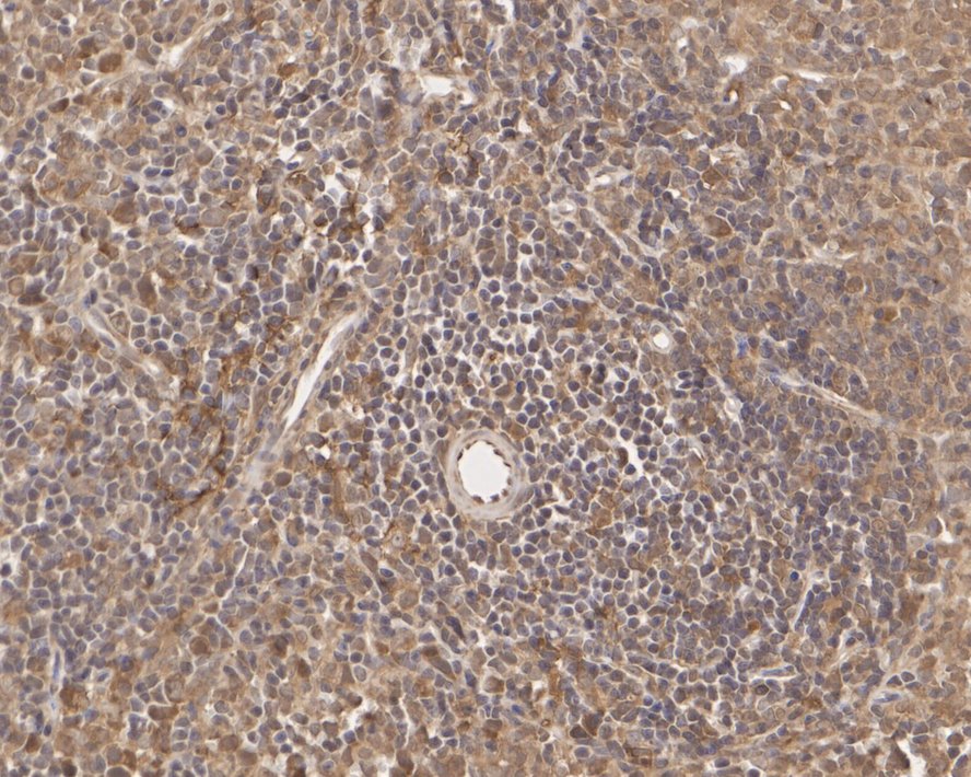
Immunohistochemical analysis of paraffin-embedded rat spleen tissue with Rabbit anti-NF-kB p65 antibody at 1/200 dilution. The section was pre-treated using heat mediated antigen retrieval with sodium citrate buffer (pH 6.0) for 2 minutes. The tissues were blocked in 1% BSA for 20 minutes at room temperature, washed with ddH2O and PBS, and then probed with the primary antibody at 1/200 dilution for 1 hour at room temperature. The detection was performed using an HRP conjugated compact polymer system. DAB was used as the chromogen. Tissues were counterstained with hematoxylin and mounted with DPX.ICC/IF
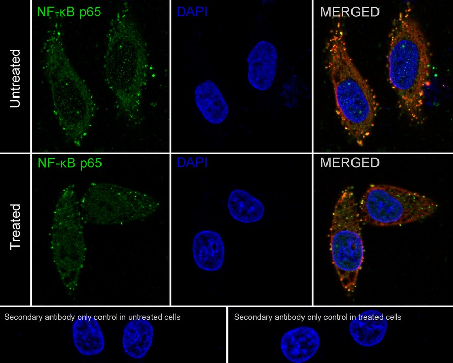
Immunocytochemistry analysis of HeLa cells treated with or without 50 ng/mL TNF-alpha for 20 minutes labeling NF-kB p65 with Rabbit anti-NF-kB p65 antibody at 1/400 dilution. Cells were fixed in 4% paraformaldehyde for 10 minutes at 37 ℃, permeabilized with 0.05% Triton X-100 in PBS for 20 minutes, and then blocked with 2% negative goat serum for 30 minutes at room temperature. Cells were then incubated with Rabbit anti-NF-kB p65 antibody at 1/400 dilution in 2% negative goat serum overnight at 4 ℃. Goat Anti-Rabbit IgG H&L (488) was used as the secondary antibody at 1/1,000 dilution. Nuclear DNA was labelled in blue with DAPI. Beta tubulin (red) was stained at 1/100 dilution overnight at +4℃. Goat Anti-Mouse IgG H&L (594) was used as the secondary antibody at 1/1,000 dilution.FC
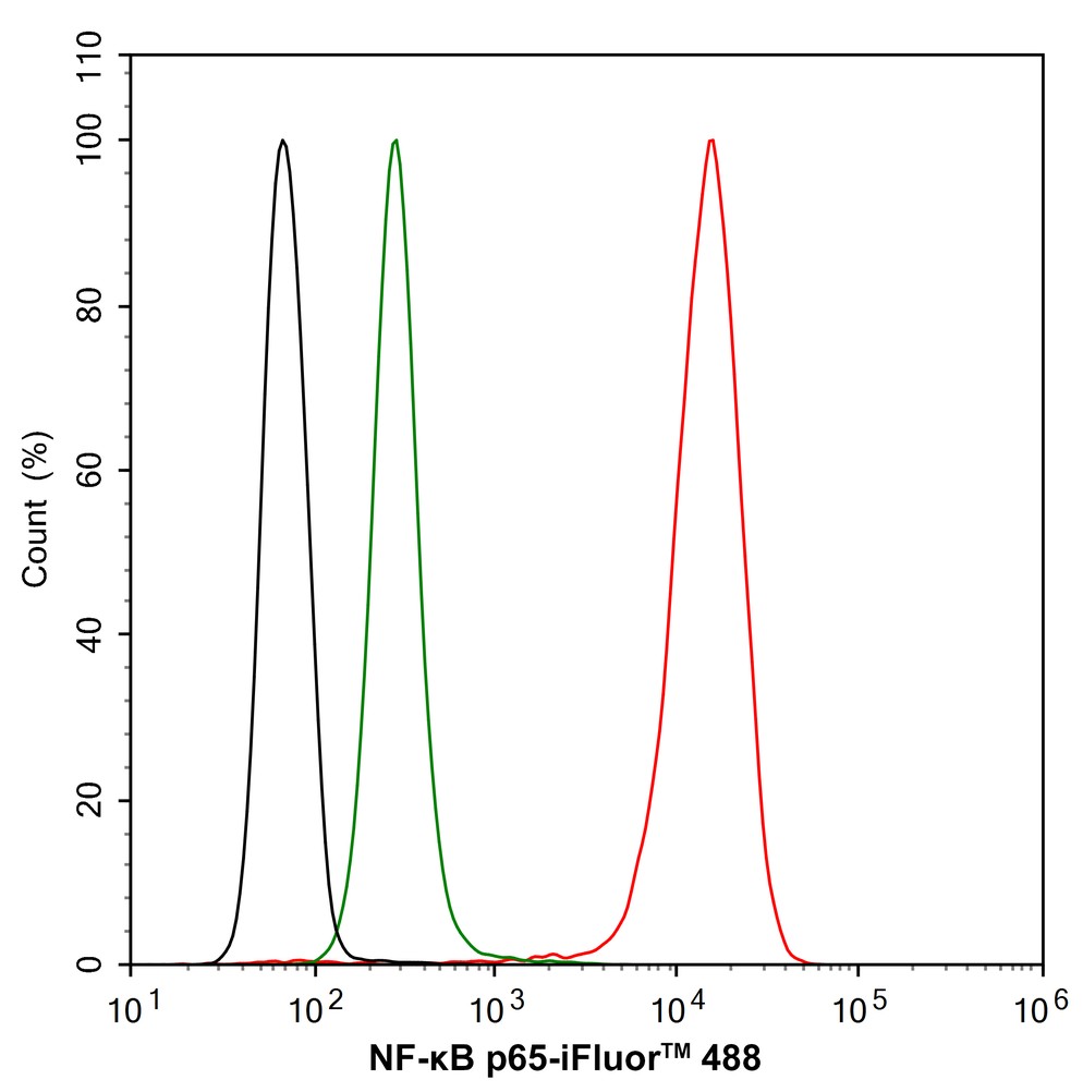
Flow cytometric analysis of HeLa cells labeling NF-kB p65. Cells were fixed and permeabilized. Then stained with the primary antibody (red) at 1/2,000 dilution, compared with Rabbit IgG Isotype Control (green). After incubation of the primary antibody at +4℃ for an hour, the cells were stained with a iFluor™ 488 conjugate-Goat anti-Rabbit IgG Secondary antibody at 1/1,000 dilution for 30 minutes at +4℃. Unlabelled sample was used as a control (cells without incubation with primary antibody; black).IP
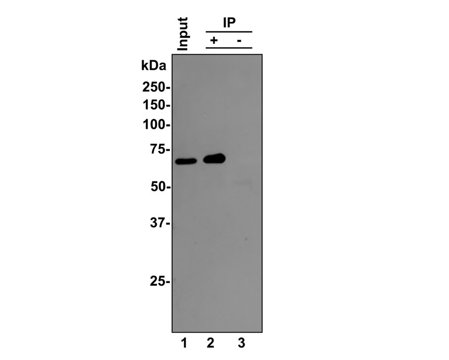
NF-κB p65 was immunoprecipitated from 0.5 mg Hela whole cell lysates with Rabbit anti-NF-kB p65 antibody at 2 μg/mL. Western blot was performed from the immunoprecipitate using Rabbit anti-NF-kB p65 antibody at 1/500 dilution for 45 minutes at room temperature. Goat anti-Rabbit IgG-HRP Secondary Antibody was used at 1:300,000 dilution for 30 minutes at room temperature. Lane 1: Hela whole cell lysates at 10 μg; Lane 2: Rabbit anti-NF-kB p65 antibody IP in Hela whole cell lysates; Lane 3: Rabbit IgG instead of Rabbit anti-NF-kB p65 antibody in Hela whole cell lysates. Exposure time: 10 seconds; 8% SDS-PAGE gel.| Product Name | NF-kB p65 Recombinant Rabbit Monoclonal Antibody |
|---|---|
| Antibody Type | Primary Antibodies |
| Product description | Proteins encoded by the v-Rel viral oncogene and its cellular homolog, c-Rel, are members of a family of transcription factors that include the two subunits of the transcription factor NF kB (p50 and p65) and the Drosophila maternal morphogen, dorsal. Both proteins specifically bind to DNA sequences that are the same or slight variations of the 10 bp kB sequence in the immunoglobulin k light chain enhancer. This same sequence is also present in a number of other cellular and viral enhancers. The DNA binding activity of NFkB is activated and NFkB is subsequently transported from the cytoplasm to the nucleus in cells exposed to mitogens or growth factors. cDNAs encoding precursors for two distinct proteins of the same size have been described, designated p105 and p100. The p105 precursor contains p50 at its N-terminus and a C-terminal region that when expressed as a separate molecule, designated pdI, binds to p50 and regulates its activity. |
| Immunogen | Synthetic peptide within human RELA aa 490-540. |
| Clonality | monoclonal |
|---|---|
| Isotype | IgG |
| Host Species | Recombinant rabbit |
| Tested Applications | FCICC/IFIHCIPWB |
| WB:1:5000-1:10000 IHC:1:200-1:500 ICC/IF:1:100-1:400 FC:1:2000 IP:Use at an assay dependent concentration. |
|
| Species Reactivity | HumanMouseRat |
| Concentration | 1mg/ml |
| Purification | Protein A |
| Alternative Names | Avian reticuloendotheliosis viral (v rel) oncogene homolog A antibody
MGC131774 antibody
NF kappa B p65delta3 antibody
NFKB3 antibody
Nuclear Factor NF Kappa B p65 Subunit antibody
Nuclear factor NF-kappa-B p65 subunit antibody
Nuclear factor of kappa light polypeptide gene enhancer in B cells 3 antibody
Nuclear factor of kappa light polypeptide gene enhancer in B-cells 3 antibody
OTTHUMP00000233473 antibody
OTTHUMP00000233474 antibody
OTTHUMP00000233475 antibody
OTTHUMP00000233476 antibody
OTTHUMP00000233900 antibody
p65 antibody
p65 NF kappaB antibody
p65 NFkB antibody
relA antibody
TF65_HUMAN antibody
Transcription factor p65 antibody
v rel avian reticuloendotheliosis viral oncogene homolog A (nuclear factor of kappa light polypeptide gene enhancer in B cells 3 (p65)) antibody
V rel avian reticuloendotheliosis viral oncogene homolog A antibody
v rel reticuloendotheliosis viral oncogene homolog A (avian) antibody
V rel reticuloendotheliosis viral oncogene homolog A nuclear factor of kappa light polypeptide gene enhancer in B cells 3 p65 antibody |
|---|---|
| Molecular Weight(MW) | 65kDa |
| Cellular Localization | Nucleus, Cytoplasm. |
WB
Western blot analysis of NF-kB p65 on different lysates with Rabbit anti-NF-kB p65 antibody at 1/5,000 dilution. Lane 1: A549 cell lysate, Lane 2: MCF7 cell lysate, Lane 3: HeLa cell lysate, Lane 4: RAW264.7 cell lysate, Lysates/proteins at 15 µg/Lane. Exposure time: 49 seconds; 4-20% SDS-PAGE gel. Proteins were transferred to a PVDF membrane and blocked with 5% NFDM/TBST for 1 hour at room temperature. The primary antibody at 1/5,000 dilution was used in 5% NFDM/TBST at room temperature for 2 hours. Goat Anti-Rabbit IgG - HRP Secondary Antibody at 1:100,000 dilution was used for 1 hour at room temperature.
IHC
Immunohistochemical analysis of paraffin-embedded human lung cancer tissue with Rabbit anti-NF-kB p65 antibody at 1/200 dilution. The section was pre-treated using heat mediated antigen retrieval with sodium citrate buffer (pH 6.0) for 2 minutes. The tissues were blocked in 1% BSA for 20 minutes at room temperature, washed with ddH2O and PBS, and then probed with the primary antibody at 1/200 dilution for 1 hour at room temperature. The detection was performed using an HRP conjugated compact polymer system. DAB was used as the chromogen. Tissues were counterstained with hematoxylin and mounted with DPX.
IHC
Immunohistochemical analysis of paraffin-embedded rat spleen tissue with Rabbit anti-NF-kB p65 antibody at 1/200 dilution. The section was pre-treated using heat mediated antigen retrieval with sodium citrate buffer (pH 6.0) for 2 minutes. The tissues were blocked in 1% BSA for 20 minutes at room temperature, washed with ddH2O and PBS, and then probed with the primary antibody at 1/200 dilution for 1 hour at room temperature. The detection was performed using an HRP conjugated compact polymer system. DAB was used as the chromogen. Tissues were counterstained with hematoxylin and mounted with DPX.
ICC/IF
Immunocytochemistry analysis of HeLa cells treated with or without 50 ng/mL TNF-alpha for 20 minutes labeling NF-kB p65 with Rabbit anti-NF-kB p65 antibody at 1/400 dilution. Cells were fixed in 4% paraformaldehyde for 10 minutes at 37 ℃, permeabilized with 0.05% Triton X-100 in PBS for 20 minutes, and then blocked with 2% negative goat serum for 30 minutes at room temperature. Cells were then incubated with Rabbit anti-NF-kB p65 antibody at 1/400 dilution in 2% negative goat serum overnight at 4 ℃. Goat Anti-Rabbit IgG H&L (488) was used as the secondary antibody at 1/1,000 dilution. Nuclear DNA was labelled in blue with DAPI. Beta tubulin (red) was stained at 1/100 dilution overnight at +4℃. Goat Anti-Mouse IgG H&L (594) was used as the secondary antibody at 1/1,000 dilution.
FC
Flow cytometric analysis of HeLa cells labeling NF-kB p65. Cells were fixed and permeabilized. Then stained with the primary antibody (red) at 1/2,000 dilution, compared with Rabbit IgG Isotype Control (green). After incubation of the primary antibody at +4℃ for an hour, the cells were stained with a iFluor™ 488 conjugate-Goat anti-Rabbit IgG Secondary antibody at 1/1,000 dilution for 30 minutes at +4℃. Unlabelled sample was used as a control (cells without incubation with primary antibody; black).
IP
NF-κB p65 was immunoprecipitated from 0.5 mg Hela whole cell lysates with Rabbit anti-NF-kB p65 antibody at 2 μg/mL. Western blot was performed from the immunoprecipitate using Rabbit anti-NF-kB p65 antibody at 1/500 dilution for 45 minutes at room temperature. Goat anti-Rabbit IgG-HRP Secondary Antibody was used at 1:300,000 dilution for 30 minutes at room temperature. Lane 1: Hela whole cell lysates at 10 μg; Lane 2: Rabbit anti-NF-kB p65 antibody IP in Hela whole cell lysates; Lane 3: Rabbit IgG instead of Rabbit anti-NF-kB p65 antibody in Hela whole cell lysates. Exposure time: 10 seconds; 8% SDS-PAGE gel.| Application Notes | WB:1:5000-1:10000 IHC:1:200-1:500 ICC/IF:1:100-1:400 FC:1:2000 IP:Use at an assay dependent concentration. |
|---|
| Form | Liquid |
|---|---|
| Storage Instructions | Store at +4℃ after thawing. Aliquot store at -20℃ or -80℃. Avoid repeated freeze / thaw cycles. |
| Storage Buffer | 1*TBS (pH7.4), 1%BSA, 40%Glycerol. Preservative: 0.05% Sodium Azide. |


