Application
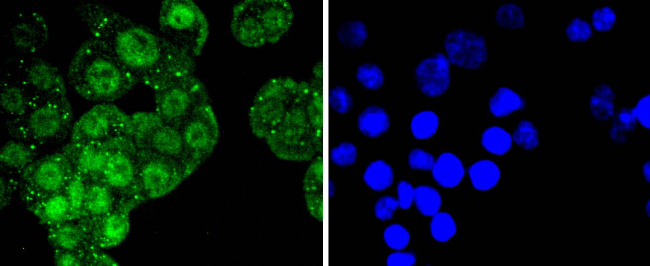
Fig1: ICC staining ATF2 in SW480 cells (green). The nuclear counter stain is DAPI (blue). Cells were fixed in paraformaldehyde, permeabilised with 0.25% Triton X100/PBS.Application
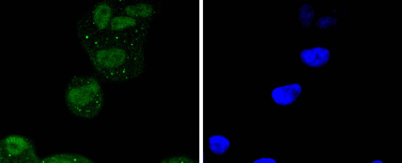
Fig2: ICC staining ATF2 in Hela cells (green). The nuclear counter stain is DAPI (blue). Cells were fixed in paraformaldehyde, permeabilised with 0.25% Triton X100/PBS.Application
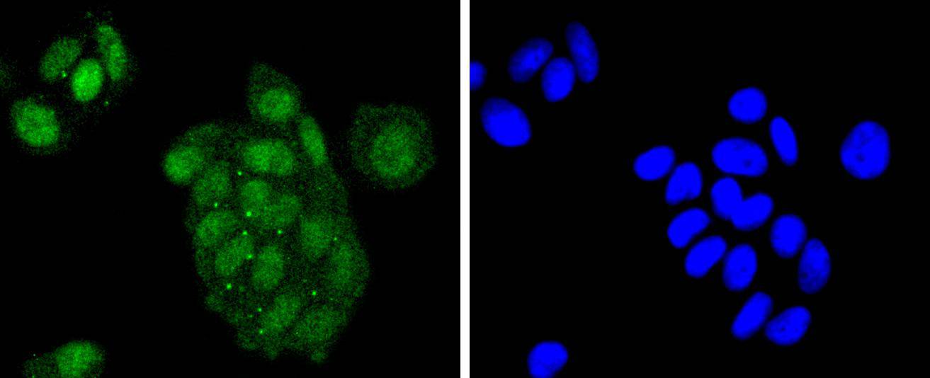
Fig3: ICC staining ATF2 in MCF-7 cells (green). The nuclear counter stain is DAPI (blue). Cells were fixed in paraformaldehyde, permeabilised with 0.25% Triton X100/PBS.Application
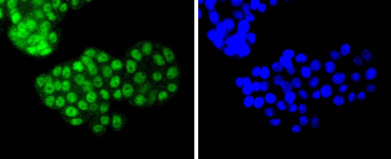
Fig4: ICC staining ATF2 in PC-12 cells (green). The nuclear counter stain is DAPI (blue). Cells were fixed in paraformaldehyde, permeabilised with 0.25% Triton X100/PBS.Application
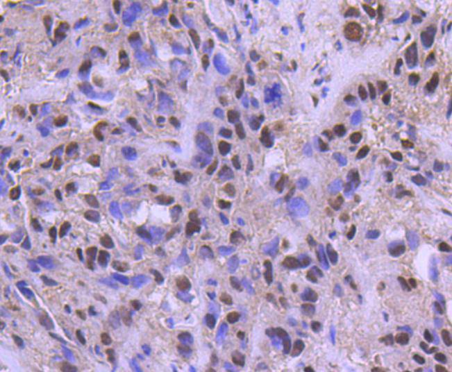
Fig5: Immunohistochemical analysis of paraffin-embedded human breast carcinoma tissue using anti-ATF2 antibody. Counter stained with hematoxylin.| Product Name | ATF2 [SZ17-01] |
|---|---|
| Antibody Type | Primary Antibodies |
| Antigen Alias | Activating transcription factor 2 antibody Activating transcription factor 2 splice variant ATF2 var2 antibody ATF 2 antibody Atf-2 antibody Atf2 antibody ATF2 protein antibody ATF2_HUMAN antibody cAMP Response Element Binding Protein 2 antibody cAMP response element binding protein CRE BP1 antibody cAMP response element-binding protein CRE-BP1 antibody cAMP responsive element binding protein 2, formerly antibody cAMP-dependent transcription factor ATF-2 antibody cAMP-responsive element-binding protein 2 antibody CRE BP1 antibody CRE-BP antibody CREB 2 antibody CREB-2 antibody CREB2 antibody CREBP1 antibody Cyclic AMP dependent transcription factor ATF 2 antibody Cyclic AMP-dependent transcription factor ATF-2 antibody Cyclic AMP-responsive element-binding protein 2 antibody D130078H02Rik antibody D18875 antibody HB 16 antibody HB16 antibody Histone acetyltransferase ATF2 antibody MGC105211 antibody MGC105222 antibody MGC111558 antibody MGC142504 antibody mXBP antibody MXBP protein antibody Tg(Gzma-Klra1)7Wum antibody TREB 7 antibody TREB7 antibody |
| Product description | Eukaryotic gene transcription is regulated by sequence-specific transcription factors which bind modular cis-acting promotor and enhancer elements. The ATF/CREB transcription factor family binds the palindromic cAMP response element (CRE) octanucleotide TGACGTCA. The ATF/CREB family includes CREB-1, CREB-2 (also designated ATF-4), ATF-1, ATF-2 and ATF-3. This family of proteins contain highly divergent N-terminal domains, but share a C-terminal leucine zipper for dimerization and DNA binding. ATF-2 forms homodimers and heterodimers with c-Jun to initiate CRE-dependent transcription. Phosphorylation of ATF-2 at Thr 69 and Thr 71 by stress-activated kinases is necessary for transcriptional activation. Myc also induces phosphorylation of ATF-2 at Thr 69 and Thr 71 to prolong the half-life of ATF-2. ATF-2 also functions as a histone acetyltransferase (HAT) by specifically acetylating histones H2B and H4 in vitro. The gene encoding human ATF-2 maps to chromosome 2q31.1. |
| Immunogen | recombinant protein |
| Clonality | Monoclonal |
|---|---|
| Isotype | IgG |
| Host Species | Recombinant rabbit |
| Tested Applications | WBICC/IFIHCIP |
| WB:1:1,000 ICC:1:50-1:200 IHC:1:50-1:200 |
|
| Species Reactivity | Human |
| Concentration | 1mg/ml |
| Alternative Names | Activating transcription factor 2 antibody
Activating transcription factor 2 splice variant ATF2 var2 antibody
ATF 2 antibody
Atf-2 antibody
Atf2 antibody
ATF2 protein antibody
ATF2_HUMAN antibody
cAMP Response Element Binding Protein 2 antibody
cAMP response element binding protein CRE BP1 antibody
cAMP response element-binding protein CRE-BP1 antibody
cAMP responsive element binding protein 2 formerly antibody cAMP-dependent transcription factor ATF-2 antibody cAMP-responsive element-binding protein 2 antibody CRE BP1 antibody CRE-BP antibody CREB 2 antibody CREB-2 antibody CREB2 antibody CREBP1 antibody Cyclic AMP dependent transcription factor ATF 2 antibody Cyclic AMP-dependent transcription factor ATF-2 antibody Cyclic AMP-responsive element-binding protein 2 antibody D130078H02Rik antibody D18875 antibody HB 16 antibody HB16 antibody Histone acetyltransferase ATF2 antibody MGC105211 antibody MGC105222 antibody MGC111558 antibody MGC142504 antibody mXBP antibody MXBP protein antibody Tg(Gzma-Klra1)7Wum antibody TREB 7 antibody TREB7 antibody |
|---|---|
| Molecular Weight(MW) | 54 kDa |
| Cellular Localization | Nucleus, Cytoplasm, Mitochondrion outer membrane. |
| SwissProt ID | P15336 |
|---|
Application
Fig1: ICC staining ATF2 in SW480 cells (green). The nuclear counter stain is DAPI (blue). Cells were fixed in paraformaldehyde, permeabilised with 0.25% Triton X100/PBS.
Application
Fig2: ICC staining ATF2 in Hela cells (green). The nuclear counter stain is DAPI (blue). Cells were fixed in paraformaldehyde, permeabilised with 0.25% Triton X100/PBS.
Application
Fig3: ICC staining ATF2 in MCF-7 cells (green). The nuclear counter stain is DAPI (blue). Cells were fixed in paraformaldehyde, permeabilised with 0.25% Triton X100/PBS.
Application
Fig4: ICC staining ATF2 in PC-12 cells (green). The nuclear counter stain is DAPI (blue). Cells were fixed in paraformaldehyde, permeabilised with 0.25% Triton X100/PBS.
Application
Fig5: Immunohistochemical analysis of paraffin-embedded human breast carcinoma tissue using anti-ATF2 antibody. Counter stained with hematoxylin.| Positive Control | SW480, MCF-7, Hela, PC-12, human breast carcinoma tissue. |
|---|---|
| Application Notes | WB:1:1,000 ICC:1:50-1:200 IHC:1:50-1:200 |
| Form | Liquid |
|---|---|
| Storage Instructions | Store at +4℃ after thawing. Aliquot store at -20℃ or -80℃. Avoid repeated freeze / thaw cycles. |
| Storage Buffer | 1*TBS (pH7.4), 1%BSA, 40%Glycerol. Preservative: 0.05% Sodium Azide. |


