Application
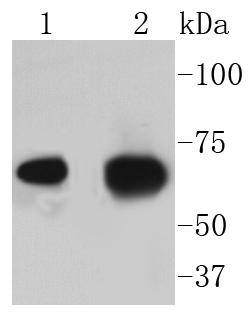
Fig1: Western blot analysis of AIF on different lysates using anti-AIF antibody at 1/1,000 dilution.Positive control:Lane 1: HelaLane 2: SKOV-3Application
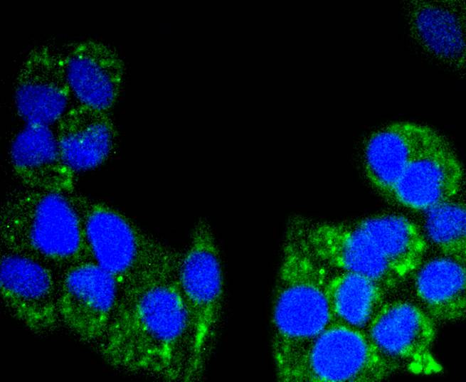
Fig2: ICC staining AIF in Hela cells (green). The nuclear counter stain is DAPI (blue). Cells were fixed in paraformaldehyde, permeabilised with 0.25% Triton X100/PBS.Application
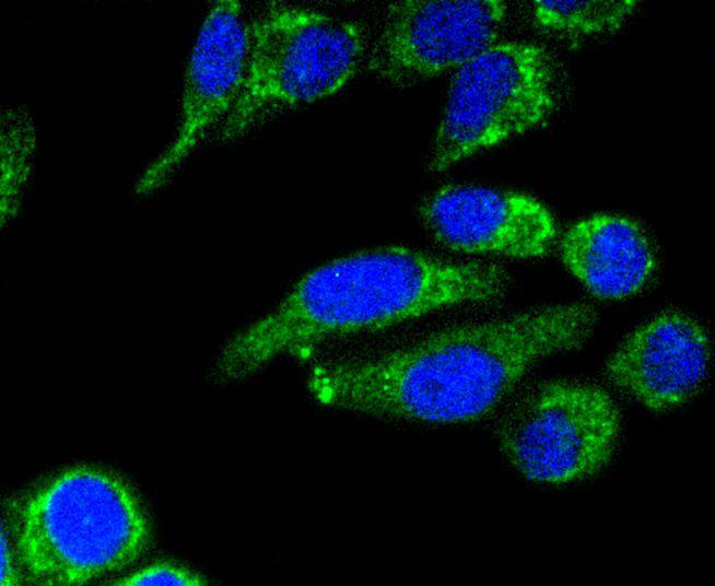
Fig3: ICC staining AIF in HepG2 cells (green). The nuclear counter stain is DAPI (blue). Cells were fixed in paraformaldehyde, permeabilised with 0.25% Triton X100/PBS.Application
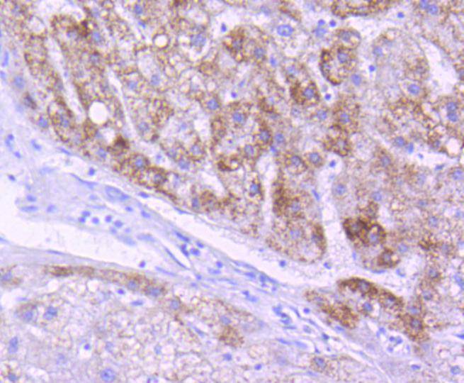
Fig4: Immunohistochemical analysis of paraffin-embedded human liver tissue using anti-AIF antibody. Counter stained with hematoxylin.Application
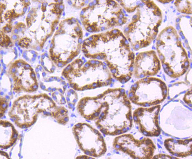
Fig5: Immunohistochemical analysis of paraffin-embedded human kidney tissue using anti-AIF antibody. Counter stained with hematoxylin.Application
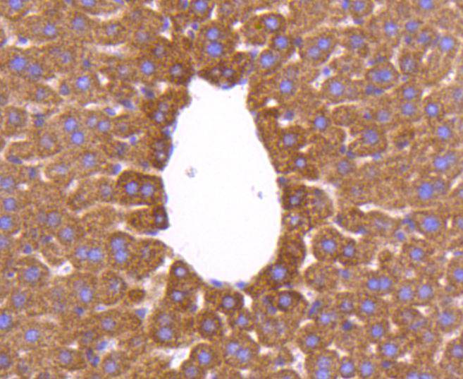
Fig6: Immunohistochemical analysis of paraffin-embedded mouse liver tissue using anti-AIF antibody. Counter stained with hematoxylin.Application
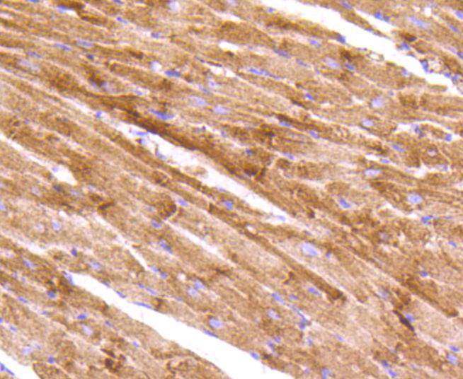
Fig7: Immunohistochemical analysis of paraffin-embedded mouse heart tissue using anti-AIF antibody. Counter stained with hematoxylin.Application
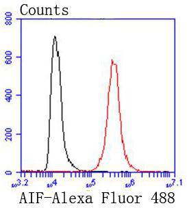
Fig8: Flow cytometric analysis of Hela cells with AIF antibody at 1/50 dilution (red) compared with an unlabelled control (cells without incubation with primary antibody; black). Alexa Fluor 488-conjugated goat anti rabbit IgG was used as the secondary antibody.| Product Name | AIF [SZ05-01] |
|---|---|
| Antibody Type | Primary Antibodies |
| Antigen Alias | AIFM1 antibody AIFM1_HUMAN antibody Apoptosis inducing factor 1, mitochondrial antibody Apoptosis inducing factor antibody Apoptosis inducing factor, mitochondrion associated, 1 antibody Apoptosis-inducing factor 1 antibody CMTX4 antibody COWCK antibody COXPD6 antibody Harlequin antibody Hq antibody mAIF antibody MGC111425 antibody MGC5706 antibody mitochondrial antibody Neuropathy, axonal motor-sensory, with deafness and mental retardation antibody neuropathy, axonal, motor-sensory with deafness and mental retardation (Cowchock syndrome) antibody PDCD 8 antibody PDCD8 antibody Programmed cell death 8 (apoptosis inducing factor) antibody Programmed cell death 8 antibody Programmed cell death 8 isoform 1 antibody Programmed cell death 8 isoform 2 antibody Programmed cell death 8 isoform 3 antibody Programmed cell death protein 8 antibody Programmed cell death protein 8 mitochondrial antibody Programmed cell death protein 8 mitochondrial precursor antibody Programmed cell death protein 8 mitochondrial precursor antibody Striatal apoptosis inducing factor antibody |
| Product description | A key event in the apoptotic process is the opening of the mitochondrial permeability transition pore, an event that is regulated by Bcl-2 family proteins, resulting in the release of several proteins from the mitochondrial intermembrane space. Several of these proteins participate in apoptosis, including cytochrome c, procaspases 2, 3, and 9, and AIF (apoptosis-inducing factor). AIF has been shown to cause DNA fragmentation and chromatin condensation and to induce the release of cytochrome c and caspase-9 from mitochondria. Bcl-2 overexpression has been shown to prevent the release of AIF from mitochondria, but not to block its apoptogenic activity. |
| Immunogen | recombinant protein |
| Clonality | Monoclonal |
|---|---|
| Isotype | IgG |
| Host Species | Recombinant rabbit |
| Tested Applications | WBICCIHCIPFC |
| WB:1:1,000-1:5,000 ICC:1:50-1:200 IHC:1:50-1:200 FC:1:50-1:100 |
|
| Species Reactivity | HumanMouseRat |
| Concentration | 1mg/ml |
| Alternative Names | AIFM1 antibody
AIFM1_HUMAN antibody
Apoptosis inducing factor 1 mitochondrial antibody Apoptosis inducing factor antibody Apoptosis inducing factor mitochondrion associated 1 antibody Apoptosis-inducing factor 1 antibody CMTX4 antibody COWCK antibody COXPD6 antibody Harlequin antibody Hq antibody mAIF antibody MGC111425 antibody MGC5706 antibody mitochondrial antibody Neuropathy axonal motor-sensory with deafness and mental retardation antibody neuropathy axonal motor-sensory with deafness and mental retardation (Cowchock syndrome) antibody PDCD 8 antibody PDCD8 antibody Programmed cell death 8 (apoptosis inducing factor) antibody Programmed cell death 8 antibody Programmed cell death 8 isoform 1 antibody Programmed cell death 8 isoform 2 antibody Programmed cell death 8 isoform 3 antibody Programmed cell death protein 8 antibody Programmed cell death protein 8 mitochondrial antibody Programmed cell death protein 8 mitochondrial precursor antibody Programmed cell death protein 8 mitochondrial precursor antibody Striatal apoptosis inducing factor antibody |
|---|---|
| Molecular Weight(MW) | 67 kDa |
| Cellular Localization | Mitochondrion intermembrane space, Mitochondrion inner membrane, Cytoplasm, Nucleus. |
Application
Fig1: Western blot analysis of AIF on different lysates using anti-AIF antibody at 1/1,000 dilution.Positive control:Lane 1: HelaLane 2: SKOV-3
Application
Fig2: ICC staining AIF in Hela cells (green). The nuclear counter stain is DAPI (blue). Cells were fixed in paraformaldehyde, permeabilised with 0.25% Triton X100/PBS.
Application
Fig3: ICC staining AIF in HepG2 cells (green). The nuclear counter stain is DAPI (blue). Cells were fixed in paraformaldehyde, permeabilised with 0.25% Triton X100/PBS.
Application
Fig4: Immunohistochemical analysis of paraffin-embedded human liver tissue using anti-AIF antibody. Counter stained with hematoxylin.
Application
Fig5: Immunohistochemical analysis of paraffin-embedded human kidney tissue using anti-AIF antibody. Counter stained with hematoxylin.
Application
Fig6: Immunohistochemical analysis of paraffin-embedded mouse liver tissue using anti-AIF antibody. Counter stained with hematoxylin.
Application
Fig7: Immunohistochemical analysis of paraffin-embedded mouse heart tissue using anti-AIF antibody. Counter stained with hematoxylin.
Application
Fig8: Flow cytometric analysis of Hela cells with AIF antibody at 1/50 dilution (red) compared with an unlabelled control (cells without incubation with primary antibody; black). Alexa Fluor 488-conjugated goat anti rabbit IgG was used as the secondary antibody.| Positive Control | SKOV-3, Hela, HepG2, human liver tissue, mouse heart tissue, human kidney tissue, mouse liver tissue. |
|---|---|
| Application Notes | WB:1:1,000-1:5,000 ICC:1:50-1:200 IHC:1:50-1:200 FC:1:50-1:100 |
| Form | Liquid |
|---|---|
| Storage Instructions | Store at +4℃ after thawing. Aliquot store at -20℃ or -80℃. Avoid repeated freeze / thaw cycles. |
| Storage Buffer | 1*TBS (pH7.4), 1%BSA, 40%Glycerol. Preservative: 0.05% Sodium Azide. |


