Application
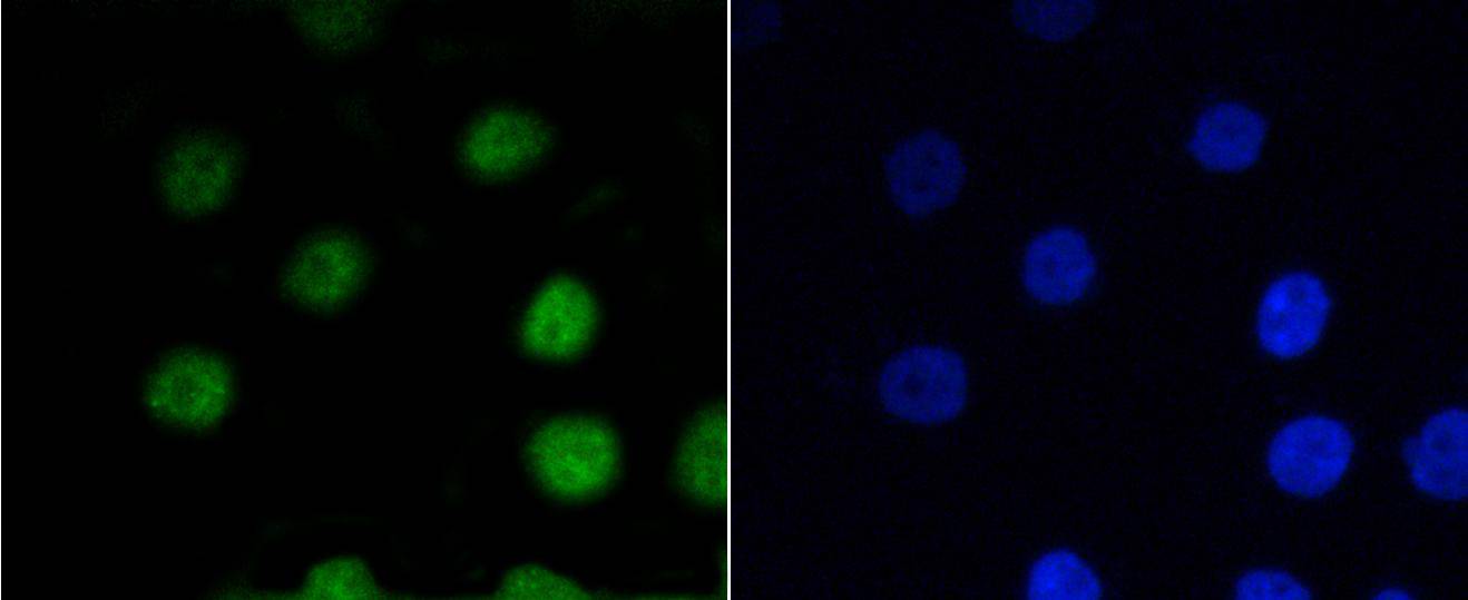
Fig1: ICC staining P-V-Myb+C-Myb(S11) in Ags cells (green). The nuclear counter stain is DAPI (blue). Cells were fixed in paraformaldehyde, permeabilised with 0.25% Triton X100/PBS.Application
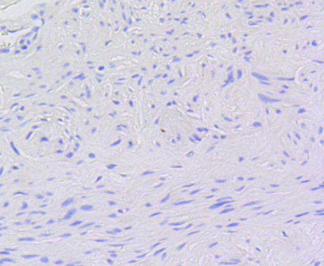
Fig2: Immunohistochemical analysis of paraffin-embedded human uterine neck tissue using anti-P-V-Myb+C-Myb(S11) antibody. Counter stained with hematoxylin.Application
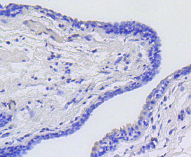
Fig3: Immunohistochemical analysis of paraffin-embedded human breast carcinoma tissue using anti-P-V-Myb+C-Myb(S11) antibody. Counter stained with hematoxylin.Application
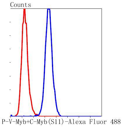
Fig4: Flow cytometric analysis of MCF-7 cells with P-V-Myb+C-Myb(S11) antibody at 1/50 dilution (blue) compared with an unlabelled control (cells without incubation with primary antibody; red). Alexa Fluor 488-conjugated goat anti rabbit IgG was used as antibody| Product Name | P-V-Myb+C-Myb(S11) [SZ04-81] |
|---|---|
| Antibody Type | Primary Antibodies |
| Antigen Alias | C myb antibody C myb protein antibody Cmyb antibody MYB antibody Myb proto oncogene antibody V myb myeloblastosis viral oncogene homolog antibody |
| Product description | The Myb family of transcription factors regulates proliferation, differentiation and apoptosis of hematopoletic cells. The avian myeloblastosis viral (v-Myb) protein is nuclear and binds to specific DNA sequences. The gene encoding for v-Myb is oncogenic, causing monoblastic leukemia and transforming myelomonocytic cells by deregulating the expression of specific target genes. v-Myb functions as a transcriptional activator, and it can repress biologically relevant genes such as Ets-2, which promotes macrophage differentiation. The proto-oncogene c-Myb encodes for a nuclear protein that plays a role in transcriptional regulation and may be essential for hematopoietic cell proliferation. A-Myb is also member of the Myb family that is expressed in proliferating B cell centroblasts. Transgenic mice overexpressing A-Myb posses enhanced hyperplasia of the lymph nodes. |
| Immunogen | Synthetic phospho-peptide corresponding to residues surrounding Ser11 of human V-Myb+C-Myb. |
| Clonality | Monoclonal |
|---|---|
| Isotype | IgG |
| Host Species | Recombinant rabbit |
| Tested Applications | WBICC/IFIHCFC |
| WB:1:1,000-1:2,000 ICC:1:50-1:200 IHC:1:50-1:200 FC:1:50-1:100 |
|
| Species Reactivity | Human |
| Concentration | 1mg/ml |
| Alternative Names | C myb antibody C myb protein antibody Cmyb antibody MYB antibody Myb proto oncogene antibody V myb myeloblastosis viral oncogene homolog antibody |
|---|---|
| Molecular Weight(MW) | 72/75 kDa |
| Cellular Localization | Nucleus. |
| SwissProt ID | P10242 |
|---|
Application
Fig1: ICC staining P-V-Myb+C-Myb(S11) in Ags cells (green). The nuclear counter stain is DAPI (blue). Cells were fixed in paraformaldehyde, permeabilised with 0.25% Triton X100/PBS.
Application
Fig2: Immunohistochemical analysis of paraffin-embedded human uterine neck tissue using anti-P-V-Myb+C-Myb(S11) antibody. Counter stained with hematoxylin.
Application
Fig3: Immunohistochemical analysis of paraffin-embedded human breast carcinoma tissue using anti-P-V-Myb+C-Myb(S11) antibody. Counter stained with hematoxylin.
Application
Fig4: Flow cytometric analysis of MCF-7 cells with P-V-Myb+C-Myb(S11) antibody at 1/50 dilution (blue) compared with an unlabelled control (cells without incubation with primary antibody; red). Alexa Fluor 488-conjugated goat anti rabbit IgG was used as antibody| Positive Control | Ags, human uterine neck tissue, human breast carcinoma tissue. |
|---|---|
| Application Notes | WB:1:1,000-1:2,000 ICC:1:50-1:200 IHC:1:50-1:200 FC:1:50-1:100 |
| Form | Liquid |
|---|---|
| Storage Instructions | Store at +4℃ after thawing. Aliquot store at -20℃ or -80℃. Avoid repeated freeze / thaw cycles. |
| Storage Buffer | 1*TBS (pH7.4), 1%BSA, 40%Glycerol. Preservative: 0.05% Sodium Azide. |


