Application
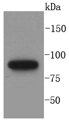
Fig1: Western blot analysis of MLH1 on A431 cells lysates using anti-MLH1 antibody at 1/1,000 dilution.Application
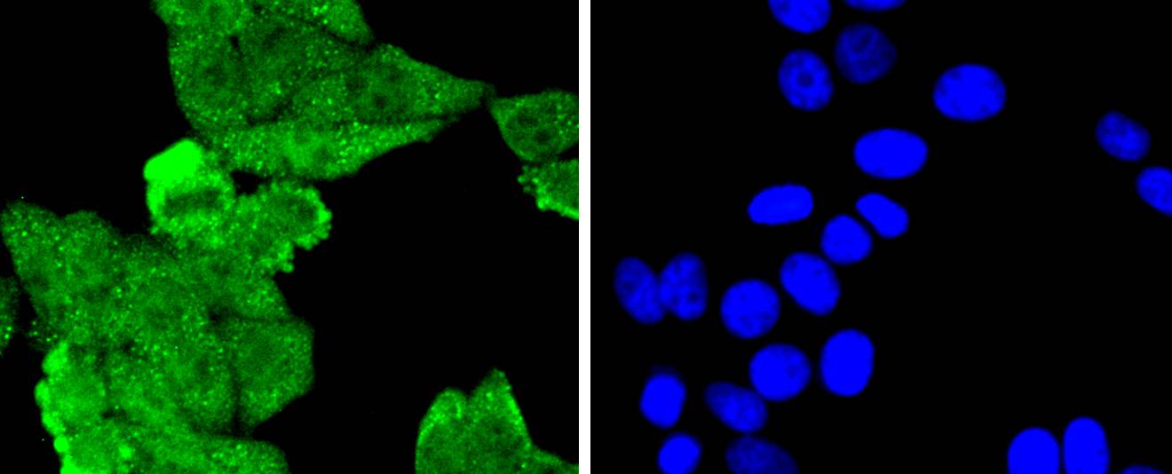
Fig2: ICC staining MLH1 in HepG2 cells (green). The nuclear counter stain is DAPI (blue). Cells were fixed in paraformaldehyde, permeabilised with 0.25% Triton X100/PBS.Application
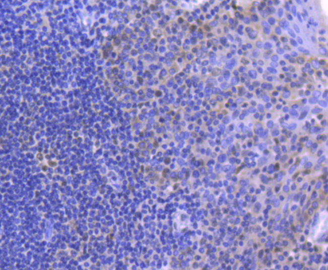
Fig3: Immunohistochemical analysis of paraffin-embedded human tonsil tissue using anti-MLH1 antibody. Counter stained with hematoxylin.Application
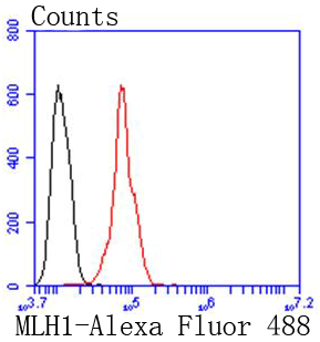
Fig4: Flow cytometric analysis of Hela cells with MLH1 antibody at 1/50 dilution (red) compared with an unlabelled control (cells without incubation with primary antibody; black). Alexa Fluor 488-conjugated goat anti rabbit IgG was used as the secondary antibody| Product Name | MLH1 [SP08-04] |
|---|---|
| Antibody Type | Primary Antibodies |
| Antigen Alias | COCA 2 antibody COCA2 antibody DNA mismatch repair protein Mlh1 antibody FCC 2 antibody FCC2 antibody hMLH 1 antibody hMLH1 antibody HNPCC 2 antibody HNPCC antibody HNPCC2 antibody MGC5172 antibody MLH 1 antibody MLH1 antibody MLH1_HUMAN antibody MutL homolog 1 (E. coli) antibody MutL homolog 1 antibody MutL homolog 1 colon cancer nonpolyposis type 2 antibody MutL homolog 1, colon cancer, nonpolyposis type 2 (E. coli) antibody MutL protein homolog 1 antibody MutL, E. coli, homolog of, 1 antibody |
| Product description | DNA-mismatch repair (MMR) is an essential process in maintaining genetic stability. Lack of a functional DNA-mismatch repair pathway is a common characteristic of several different types of human cancers, either due to an MMR gene mutation or promoter methylation gene silencing. MLH1 is an integral part of the protein complex responsible for mismatch repair and is expressed in lymphocytes, heart, colon, breast, lung, spleen, testis, prostate, thyroid and gall bladder tissues, and is methylated in several ovarian tumors. Loss of MLH1 protein expression is associated with a mutated phenotype, microsatellite instability and a predisposition to cancer. In hereditary nonpolyposis colorectal cancer (HNPCC), an autosomal dominant inherited cancer syndrome that signifies a high risk of colorectal and various other types of cancer, the MLH1 gene exhibits a pathogenic mutation. Certain cancer cell lines, including leukemia CCRF-CEM, colon HCT 116 and KM12, and ovarian cancers SK-OV-3 and IGROV-1, show complete deficiency of MLH1, while MLH1 is expressed in 60% of melanomas, 70% of noninvasive squamous cell carcinomas and 30% of invasive squamous cell carcinomas. |
| Immunogen | recombinant protein |
| Clonality | Monoclonal |
|---|---|
| Isotype | IgG |
| Host Species | Recombinant rabbit |
| Tested Applications | WBICC/IFIHCIPFC |
| WB:1:1,000 ICC:1:50-1:200 IHC:1:50-1:100 FC:1:50-1:100 |
|
| Species Reactivity | HumanMouseRat |
| Concentration | 1mg/ml |
| Alternative Names | COCA 2 antibody
COCA2 antibody
DNA mismatch repair protein Mlh1 antibody
FCC 2 antibody
FCC2 antibody
hMLH 1 antibody
hMLH1 antibody
HNPCC 2 antibody
HNPCC antibody
HNPCC2 antibody
MGC5172 antibody
MLH 1 antibody
MLH1 antibody
MLH1_HUMAN antibody
MutL homolog 1 (E. coli) antibody
MutL homolog 1 antibody
MutL homolog 1 colon cancer nonpolyposis type 2 antibody
MutL homolog 1 colon cancer nonpolyposis type 2 (E. coli) antibody MutL protein homolog 1 antibody MutL E. coli homolog of 1 antibody |
|---|---|
| Molecular Weight(MW) | 85 kDa |
| Cellular Localization | Nucleus. |
Application
Fig1: Western blot analysis of MLH1 on A431 cells lysates using anti-MLH1 antibody at 1/1,000 dilution.
Application
Fig2: ICC staining MLH1 in HepG2 cells (green). The nuclear counter stain is DAPI (blue). Cells were fixed in paraformaldehyde, permeabilised with 0.25% Triton X100/PBS.
Application
Fig3: Immunohistochemical analysis of paraffin-embedded human tonsil tissue using anti-MLH1 antibody. Counter stained with hematoxylin.
Application
Fig4: Flow cytometric analysis of Hela cells with MLH1 antibody at 1/50 dilution (red) compared with an unlabelled control (cells without incubation with primary antibody; black). Alexa Fluor 488-conjugated goat anti rabbit IgG was used as the secondary antibody| Positive Control | A431, HepG2, human tonsil tissue. |
|---|---|
| Application Notes | WB:1:1,000 ICC:1:50-1:200 IHC:1:50-1:100 FC:1:50-1:100 |
| Form | Liquid |
|---|---|
| Storage Instructions | Store at +4℃ after thawing. Aliquot store at -20℃ or -80℃. Avoid repeated freeze / thaw cycles. |
| Storage Buffer | 1*TBS (pH7.4), 1%BSA, 40%Glycerol. Preservative: 0.05% Sodium Azide. |


