WB
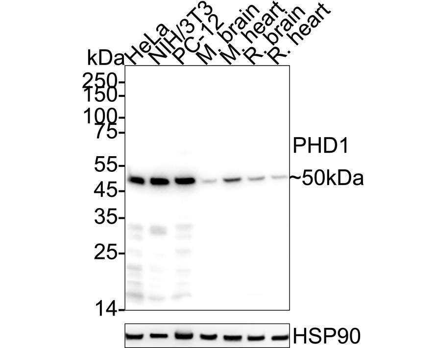
Western blot analysis of PHD1 / prolyl hydroxylase on different lysates with Rabbit anti-PHD1 / prolyl hydroxylase antibody at 1/1,000 dilution. Lane 1: HeLa cell lysate (20 µg/Lane), Lane 2: NIH/3T3 cell lysate (20 µg/Lane), Lane 3: PC-12 cell lysate (20 µg/Lane), Lane 4: Mouse brain tissue lysate (40 µg/Lane), Lane 5: Mouse heart tissue lysate (40 µg/Lane), Lane 6: Rat brain tissue lysate (40 µg/Lane), Lane 7: Rat heart tissue lysate (40 µg/Lane), Exposure time: 10 seconds; 4-20% SDS-PAGE gel. Proteins were transferred to a PVDF membrane and blocked with 5% NFDM/TBST for 1 hour at room temperature. The primary antibody at 1/1,000 dilution was used in 5% NFDM/TBST at 4℃ overnight. Goat Anti-Rabbit IgG - HRP Secondary Antibody at 1/50,000 dilution was used for 1 hour at room temperature.ICC/IF
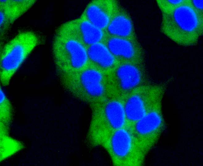
ICC staining PHD1 in Hela cells (green). The nuclear counter stain is DAPI (blue). Cells were fixed in paraformaldehyde, permeabilised with 0.25% Triton X100/PBS.ICC/IF
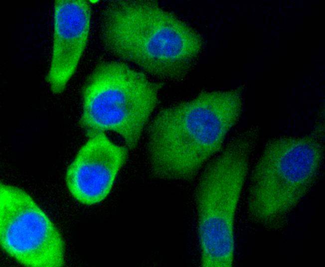
ICC staining PHD1 in A549 cells (green). The nuclear counter stain is DAPI (blue). Cells were fixed in paraformaldehyde, permeabilised with 0.25% Triton X100/PBS.ICC/IF
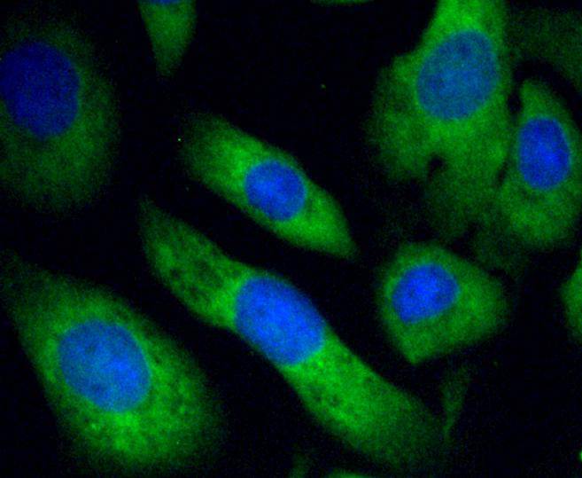
ICC staining PHD1 in SKOV-3 cells (green). The nuclear counter stain is DAPI (blue). Cells were fixed in paraformaldehyde, permeabilised with 0.25% Triton X100/PBS.IHC
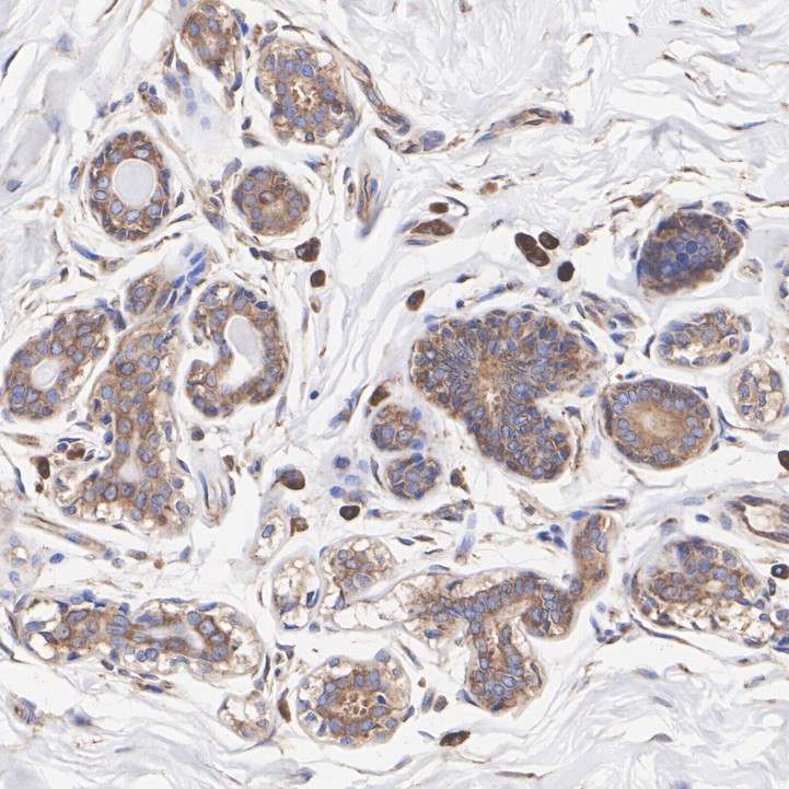
Immunohistochemical analysis of paraffin-embedded human breast cancer tissue with Rabbit anti-PHD1 / prolyl hydroxylase antibody at 1/1,000 dilution. The section was pre-treated using heat mediated antigen retrieval with Tris-EDTA buffer (pH 9.0) for 20 minutes. The tissues were blocked in 1% BSA for 20 minutes at room temperature, washed with ddH2O and PBS, and then probed with the primary antibody at 1/1,000 dilution for 1 hour at room temperature. The detection was performed using an HRP conjugated compact polymer system. DAB was used as the chromogen. Tissues were counterstained with hematoxylin and mounted with DPX.IHC
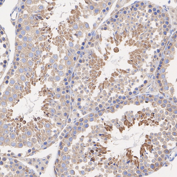
Immunohistochemical analysis of paraffin-embedded mouse testis tissue with Rabbit anti-PHD1 / prolyl hydroxylase antibody at 1/1,000 dilution. The section was pre-treated using heat mediated antigen retrieval with Tris-EDTA buffer (pH 9.0) for 20 minutes. The tissues were blocked in 1% BSA for 20 minutes at room temperature, washed with ddH2O and PBS, and then probed with the primary antibody at 1/1,000 dilution for 1 hour at room temperature. The detection was performed using an HRP conjugated compact polymer system. DAB was used as the chromogen. Tissues were counterstained with hematoxylin and mounted with DPX.FC
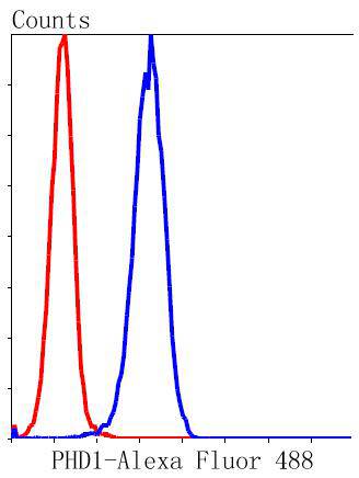
Flow cytometric analysis of Hela cells with PHD1 antibody at 1/50 dilution (blue) compared with an unlabelled control (cells without incubation with primary antibody; red). Alexa Fluor 488-conjugated goat anti rabbit IgG was used as the secondary antibody.| Product Name | PHD1 / prolyl hydroxylase Recombinant Rabbit Monoclonal Antibody |
|---|---|
| Antibody Type | Primary Antibodies |
| Product description | Prolyl hydroxylase domain proteins HIF PHD1, HIF PHD2 and HIF PHD3 (known as PHD1, PHD2 and PHD3 in rodents, respectively) can hydroxylate HIF-α subunits. Hypoxia-inducible factor (HIF) is a transcriptional regulator important in several aspects of oxygen homeostasis. The prolyl hydroxylases catalyze the posttranslational formation of 4-hydroxyproline in HIF-α proteins. HIF PHD1, which is widely expressed, with highest levels of expression in testis, functions as a cellular oxygen sensor and is important in cell growth regulation. HIF PHD1 can localize to the nucleus or the cytoplasm and is also detected in hormone responsive tissues, such as normal and cancerous mammary, ovarian and prostate epithelium. HIF PHD1 is encoded by EGLN2, which maps to chromosome 19q13.3. HIF PHD2 is regarded as the main cellular oxygen sensor, as RNA interference against HIF PHD2, but not HIF PHD1 or HIF PHD3, is enough to stabilize HIF-1α in normoxia. HIF PHD2, a direct HIF target gene, is expressed mainly in skeletal muscle, heart, kidney and brain. HIF PHD3 may play a role in the regulation of cell growth in muscle cells and in apoptosis in neuronal tissue. HIF PHD3 is widely expressed, although the highest levels can be detected in placenta and he |
| Immunogen | Recombinant protein within Human PHD1 aa 1-91 / 407. |
| Clonality | Monoclonal |
|---|---|
| Isotype | IgG |
| Host Species | Recombinant rabbit |
| Tested Applications | FCICC/IFIHCWB |
| WB:1:1000-1:5000 IHC:1:1000 ICC/IF:1:50-1:200 FC:1:50-1:100 |
|
| Species Reactivity | HumanMouseRat |
| Concentration | 1mg/ml |
| Purification | Protein A |
| Alternative Names | DKFZp434E026 antibody EGL nine (C.elegans) homolog 2 antibody Egl nine homolog 2 (C. elegans) antibody Egl nine homolog 2 antibody EGLN 2 antibody EGLN2 antibody EGLN2_HUMAN antibody EIT 6 antibody EIT6 antibody Estrogen-induced tag 6 antibody HIF P4H 1 antibody HIF PH1 antibody HIF prolyl hydroxylase 1 antibody HIF-PH1 antibody HIF-prolyl hydroxylase 1 antibody HIFPH 1 antibody HIFPH1 antibody HPH 3 antibody HPH-1 antibody HPH-3 antibody HPH3 antibody Hypoxia inducible factor prolyl hydroxylase 1 antibody Hypoxia-inducible factor prolyl hydroxylase 1 antibody P4H1 antibody PHD 1 antibody PhD1 antibody prolyl hydroxylase domain containing protein 1 antibody Prolyl hydroxylase domain-containing protein 1 antibody |
|---|---|
| Molecular Weight(MW) | 44 kDa(Observed band size: 50 kDa) |
| Cellular Localization | Nucleus, cytosol. |
WB
Western blot analysis of PHD1 / prolyl hydroxylase on different lysates with Rabbit anti-PHD1 / prolyl hydroxylase antibody at 1/1,000 dilution. Lane 1: HeLa cell lysate (20 µg/Lane), Lane 2: NIH/3T3 cell lysate (20 µg/Lane), Lane 3: PC-12 cell lysate (20 µg/Lane), Lane 4: Mouse brain tissue lysate (40 µg/Lane), Lane 5: Mouse heart tissue lysate (40 µg/Lane), Lane 6: Rat brain tissue lysate (40 µg/Lane), Lane 7: Rat heart tissue lysate (40 µg/Lane), Exposure time: 10 seconds; 4-20% SDS-PAGE gel. Proteins were transferred to a PVDF membrane and blocked with 5% NFDM/TBST for 1 hour at room temperature. The primary antibody at 1/1,000 dilution was used in 5% NFDM/TBST at 4℃ overnight. Goat Anti-Rabbit IgG - HRP Secondary Antibody at 1/50,000 dilution was used for 1 hour at room temperature.
ICC/IF
ICC staining PHD1 in Hela cells (green). The nuclear counter stain is DAPI (blue). Cells were fixed in paraformaldehyde, permeabilised with 0.25% Triton X100/PBS.
ICC/IF
ICC staining PHD1 in A549 cells (green). The nuclear counter stain is DAPI (blue). Cells were fixed in paraformaldehyde, permeabilised with 0.25% Triton X100/PBS.
ICC/IF
ICC staining PHD1 in SKOV-3 cells (green). The nuclear counter stain is DAPI (blue). Cells were fixed in paraformaldehyde, permeabilised with 0.25% Triton X100/PBS.
IHC
Immunohistochemical analysis of paraffin-embedded human breast cancer tissue with Rabbit anti-PHD1 / prolyl hydroxylase antibody at 1/1,000 dilution. The section was pre-treated using heat mediated antigen retrieval with Tris-EDTA buffer (pH 9.0) for 20 minutes. The tissues were blocked in 1% BSA for 20 minutes at room temperature, washed with ddH2O and PBS, and then probed with the primary antibody at 1/1,000 dilution for 1 hour at room temperature. The detection was performed using an HRP conjugated compact polymer system. DAB was used as the chromogen. Tissues were counterstained with hematoxylin and mounted with DPX.
IHC
Immunohistochemical analysis of paraffin-embedded mouse testis tissue with Rabbit anti-PHD1 / prolyl hydroxylase antibody at 1/1,000 dilution. The section was pre-treated using heat mediated antigen retrieval with Tris-EDTA buffer (pH 9.0) for 20 minutes. The tissues were blocked in 1% BSA for 20 minutes at room temperature, washed with ddH2O and PBS, and then probed with the primary antibody at 1/1,000 dilution for 1 hour at room temperature. The detection was performed using an HRP conjugated compact polymer system. DAB was used as the chromogen. Tissues were counterstained with hematoxylin and mounted with DPX.
FC
Flow cytometric analysis of Hela cells with PHD1 antibody at 1/50 dilution (blue) compared with an unlabelled control (cells without incubation with primary antibody; red). Alexa Fluor 488-conjugated goat anti rabbit IgG was used as the secondary antibody.| Application Notes | WB:1:1000-1:5000 IHC:1:1000 ICC/IF:1:50-1:200 FC:1:50-1:100 |
|---|
| Form | Liquid |
|---|---|
| Storage Instructions | Store at +4℃ after thawing. Aliquot store at -20℃ or -80℃. Avoid repeated freeze / thaw cycles. |
| Storage Buffer | 1*TBS (pH7.4), 1%BSA, 40%Glycerol. Preservative: 0.05% Sodium Azide. |


