Application
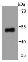
Fig1: Western blot analysis of p53 on 293 cells lysates using anti-p53 antibody at 1/1,000 dilution.Application
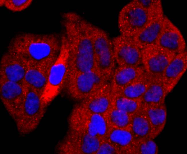
Fig2: ICC staining p53 in Hela cells (red). The nuclear counter stain is DAPI (blue). Cells were fixed in paraformaldehyde, permeabilised with 0.25% Triton X100/PBS.Application
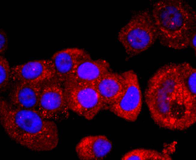
Fig3: ICC staining p53 in MCF-7 cells (red). The nuclear counter stain is DAPI (blue). Cells were fixed in paraformaldehyde, permeabilised with 0.25% Triton X100/PBS.Application
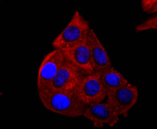
Fig4: ICC staining p53 in HepG2 cells (red). The nuclear counter stain is DAPI (blue). Cells were fixed in paraformaldehyde, permeabilised with 0.25% Triton X100/PBS.Application
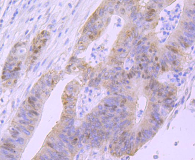
Fig5: Immunohistochemical analysis of paraffin-embedded human colon cancer tissue using anti-p53 antibody. Counter stained with hematoxylin.| Product Name | p53 [SY010-6] |
|---|---|
| Antibody Type | Primary Antibodies |
| Antigen Alias | Antigen NY-CO-13 antibody BCC7 antibody Cellular tumor antigen p53 antibody FLJ92943 antibody LFS1 antibody Mutant tumor protein 53 antibody p53 antibody p53 tumor suppressor antibody P53_HUMAN antibody Phosphoprotein p53 antibody Tp53 antibody Transformation related protein 53 antibody TRP53 antibody Tumor protein 53 antibody Tumor protein p53 antibody Tumor suppressor p53 antibody |
| Product description | p53, a DNA-binding, oligomerization domain- and transcription activation domain containing tumor suppressor, upregulates growth arrest and apoptosis-related genes in response to stress signals, thereby influencing programmed cell death, cell differentiation, and cell cycle control mechanisms. p53 localizes to the nucleus, yet can be chaperoned to the cytoplasm by the negative regulator, MDM2. MDM2 is an E3 ubiquitin ligase that is upregulated in the presence of active p53, where it poly-ubiquitinates p53 for proteasome targeting. p53 fluctuates between latent and active DNA-binding conformations and is differentially activated through posttranslational modifications, including phosphorylation and acetylation. Mutations in the DNA-binding domain (DBD) of p53, amino acids 110-286, can compromise energetically-favorable association with cis elements and are implicated in several human cancers. |
| Immunogen | recombinant protein |
| Clonality | Monoclonal |
|---|---|
| Isotype | IgG |
| Host Species | Recombinant rabbit |
| Tested Applications | WBICC/IFIHCIP |
| WB:1:1,000-1:2,000 ICC:1:50-1:200 IHC:1:50-1:200 |
|
| Species Reactivity | Human |
| Concentration | 1mg/ml |
| Alternative Names | Antigen NY-CO-13 antibody BCC7 antibody Cellular tumor antigen p53 antibody FLJ92943 antibody LFS1 antibody Mutant tumor protein 53 antibody p53 antibody p53 tumor suppressor antibody P53_HUMAN antibody Phosphoprotein p53 antibody Tp53 antibody Transformation related protein 53 antibody TRP53 antibody Tumor protein 53 antibody Tumor protein p53 antibody Tumor suppressor p53 antibody |
|---|---|
| Molecular Weight(MW) | 53 kDa |
| Cellular Localization | Cytoplasm, Nucleus, Endoplasmic reticulum, Mitochondrion matrix. |
| SwissProt ID | P04637 |
|---|
Application
Fig1: Western blot analysis of p53 on 293 cells lysates using anti-p53 antibody at 1/1,000 dilution.
Application
Fig2: ICC staining p53 in Hela cells (red). The nuclear counter stain is DAPI (blue). Cells were fixed in paraformaldehyde, permeabilised with 0.25% Triton X100/PBS.
Application
Fig3: ICC staining p53 in MCF-7 cells (red). The nuclear counter stain is DAPI (blue). Cells were fixed in paraformaldehyde, permeabilised with 0.25% Triton X100/PBS.
Application
Fig4: ICC staining p53 in HepG2 cells (red). The nuclear counter stain is DAPI (blue). Cells were fixed in paraformaldehyde, permeabilised with 0.25% Triton X100/PBS.
Application
Fig5: Immunohistochemical analysis of paraffin-embedded human colon cancer tissue using anti-p53 antibody. Counter stained with hematoxylin.| Positive Control | 293, MCF-7, Hela, HepG2, human colon cancer tissue. |
|---|---|
| Application Notes | WB:1:1,000-1:2,000 ICC:1:50-1:200 IHC:1:50-1:200 |
| Form | Liquid |
|---|---|
| Storage Instructions | Store at +4℃ after thawing. Aliquot store at -20℃ or -80℃. Avoid repeated freeze / thaw cycles. |
| Storage Buffer | 1*TBS (pH7.4), 1%BSA, 40%Glycerol. Preservative: 0.05% Sodium Azide. |


