WB
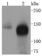
Fig1: Western blot analysis of TACC3 on different lysates using anti-TACC3 antibody at 1/1,000 dilution. Positive control: Lane 1: HT29 Lane 2: HelaICC/IF
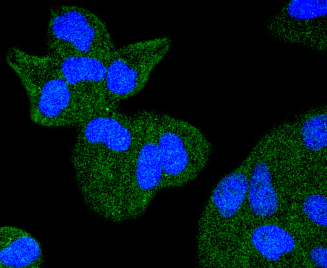
ICC staining TACC3 in Hela cells (green). The nuclear counter stain is DAPI (blue). Cells were fixed in paraformaldehyde, permeabilised with 0.25% Triton X100/PBS.ICC/IF
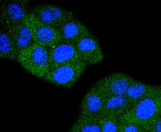
ICC staining TACC3 in HepG2 cells (green). The nuclear counter stain is DAPI (blue). Cells were fixed in paraformaldehyde, permeabilised with 0.25% Triton X100/PBS.IHC
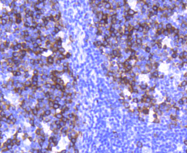
Immunohistochemical analysis of paraffin-embedded human tonsil tissue using anti-TACC3 antibody. Counter stained with hematoxylin.IHC
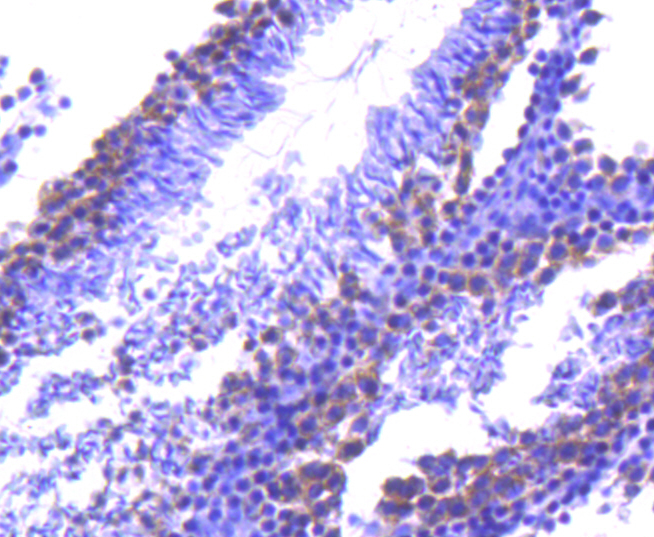
Immunohistochemical analysis of paraffin-embedded mouse testis tissue using anti-TACC3 antibody. Counter stained with hematoxylin.IHC
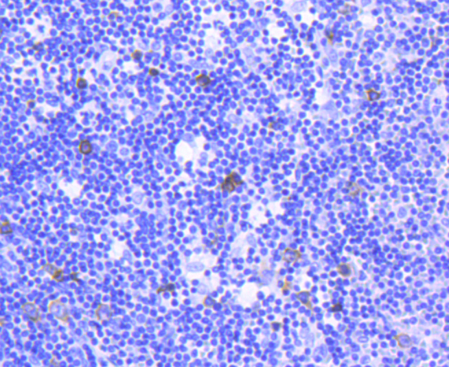
Immunohistochemical analysis of paraffin-embedded human spleen tissue using anti-TACC3 antibody. Counter stained with hematoxylin.| Product Name | TACC3 Recombinant Rabbit Monoclonal Antibody |
|---|---|
| Antibody Type | Primary Antibodies |
| Product description | TACC1 (transforming acidic coiled coil gene 1) is one of three TACC family members, which are thought to be involved in breast tumorigenesis. TACC1 is located on 8p11 chromosomal region that is amplified in approximately 15% of all breast tumor samples. The short arm of chromosome 8 also contains FGFR1 whose expression is enhanced in most breast cancer tumors. TACC family members, TACC1, TACC2, and TACC3, map very closely to the corresponding FGFR1, FGFR2, FGFR3 genes on chromosomes 8, 10, and 4. Subsequently, since they are phylogenetically related, it is proposed that TACC and FGFR have similar roles in cell growth and differentiation. Also, TACC1 contains a conserved C-terminal region as in the Drosophila homolog, D-TACC. It has been shown that D-TACC is necessary for normal spindle function, and the mammalian TACC proteins appears to interact with centrosomes and microtubules in a similar manner. |
| Immunogen | Synthetic peptide within Human TACC3 aa 40-89 / 838. |
| Clonality | Monoclonal |
|---|---|
| Isotype | IgG |
| Host Species | Recombinant rabbit |
| Tested Applications | ICC/IFIHCWB |
| WB:1:1000-1:5000 IHC:1:50-1:200 ICC/IF:1:50-1:200 |
|
| Species Reactivity | HumanMouseRat |
| Concentration | 1mg/ml |
| Purification | Protein A |
| Alternative Names | ERIC 1 antibody ERIC-1 antibody ERIC1 antibody MGC117382 antibody MGC133242 antibody OTTHUMP00000113796 antibody TACC3 antibody TACC3_HUMAN antibody Transforming acidic coiled coil containing protein 3 antibody Transforming acidic coiled-coil-containing protein 3 antibody |
|---|---|
| Molecular Weight(MW) | 90 kDa(Observed band size: 140 kDa) |
| Cellular Localization | Cytoplasm. |
WB
Fig1: Western blot analysis of TACC3 on different lysates using anti-TACC3 antibody at 1/1,000 dilution. Positive control: Lane 1: HT29 Lane 2: Hela
ICC/IF
ICC staining TACC3 in Hela cells (green). The nuclear counter stain is DAPI (blue). Cells were fixed in paraformaldehyde, permeabilised with 0.25% Triton X100/PBS.
ICC/IF
ICC staining TACC3 in HepG2 cells (green). The nuclear counter stain is DAPI (blue). Cells were fixed in paraformaldehyde, permeabilised with 0.25% Triton X100/PBS.
IHC
Immunohistochemical analysis of paraffin-embedded human tonsil tissue using anti-TACC3 antibody. Counter stained with hematoxylin.
IHC
Immunohistochemical analysis of paraffin-embedded mouse testis tissue using anti-TACC3 antibody. Counter stained with hematoxylin.
IHC
Immunohistochemical analysis of paraffin-embedded human spleen tissue using anti-TACC3 antibody. Counter stained with hematoxylin.| Application Notes | WB:1:1000-1:5000 IHC:1:50-1:200 ICC/IF:1:50-1:200 |
|---|
| Form | Liquid |
|---|---|
| Storage Instructions | Store at +4℃ after thawing. Aliquot store at -20℃ or -80℃. Avoid repeated freeze / thaw cycles. |
| Storage Buffer | 1*TBS (pH7.4), 1%BSA, 40%Glycerol. Preservative: 0.05% Sodium Azide. |


