Application
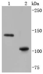
Fig1: Western blot analysis of SHIP on different lysates using anti-SHIP antibody at 1/1,000 dilution. Positive control: Lane 1: Daudi Lane 2: THP-1Application
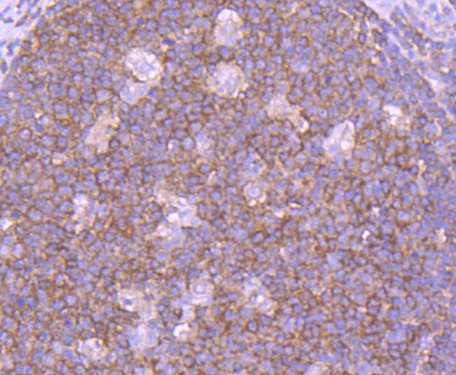
Fig2: Immunohistochemical analysis of paraffin-embedded human tonsil tissue using anti-SHIP antibody. Counter stained with hematoxylin.Application
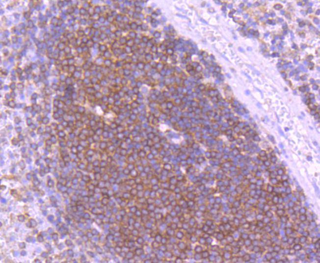
Fig3: Immunohistochemical analysis of paraffin-embedded human spleen tissue using anti-SHIP antibody. Counter stained with hematoxylin.Application
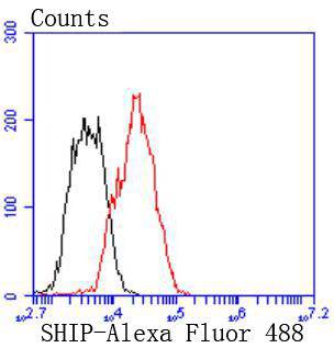
Fig4: Flow cytometric analysis of Raji cells with SHIP antibody at 1/50 dilution (red) compared with an unlabelled control (cells without incubation with primary antibody; black). Alexa Fluor 488-conjugated goat anti rabbit IgG was used as the secondary antibody| Product Name | SHIP [SY11-08] |
|---|---|
| Antibody Type | Primary Antibodies |
| Antigen Alias | Inositol polyphosphate 5 phosphatase of 145kDa antibody 4 antibody 5-trisphosphate 5-phosphatase 1 antibody hp51CN antibody Inositol polyphosphate 5 phosphatase 145kDa antibody Inositol polyphosphate 5 phosphatase antibody Inositol polyphosphate-5-phosphatase of 145 kDa antibody INPP5D antibody MGC104855 antibody MGC142140 antibody MGC142142 antibody p150Ship antibody Phosphatidylinositol 3,4,5 trisphosphate 5 phosphatase 1 antibody Phosphatidylinositol-3 antibody SH2 containing inositol phosphatase isoform b antibody SH2 domain containing inositol 5' phosphatase 1 antibody SH2 domain containing inositol phosphatase 1 antibody SH2 domain-containing inositol phosphatase 1 antibody SH2 domain-containing inositol-5''-phosphatase 1 antibody SHIP-1 antibody SHIP1 antibody SHIP1_HUMAN antibody Signaling inositol polyphosphate 5 phosphatase SIP 145 antibody SIP-145 antibody SIP145 antibody |
| Product description | The major translational product of the v-Fms oncogene, originally isolated from the McDonough strain of feline sarcoma virus, has been identified as a glycoprotein with intrinsic tyrosine kinase activity. The v-Fms human cellular homolog, c-Fms, has been molecularly cloned and mapped to band q34 on chromosome 5, and identified as the receptor for hematopoietic ligand, CSF-1. Ligand-induced activation of the intrinsic CSF-1R protein tyrosine kinase triggers its interaction with cytoplasmic effector molecules. One such effector molecule, SHIP-1 p145 (SH2-containing-inositol phosphatase), associates with activated Fms. SHIP-1 contains two phosphotyrosine-binding domains (PTB), a unique amino terminal SH2 domain, a proline-rich region, and two highly conserved motifs found among inositol phosphate 5-phosphatases. SHIP-1 displays both phosphatidylinositol 3,4,5-triphosphate and inositol 1,3,4,5-tetrakisphosphate polyphosphate 5-phosphatase activity. Evidence suggests that SHIP-1 may modulate Ras signaling in addition to inositol signaling pathways. |
| Immunogen | recombinant protein |
| Clonality | Monoclonal |
|---|---|
| Isotype | IgG |
| Host Species | Recombinant rabbit |
| Tested Applications | WBIHCIPFC |
| WB:1:1,000 FC:1:50-1:100 IHC:1:50-1:200 |
|
| Species Reactivity | Human |
| Concentration | 1mg/ml |
| Alternative Names | Inositol polyphosphate 5 phosphatase of 145kDa antibody
4 antibody
5-trisphosphate 5-phosphatase 1 antibody
hp51CN antibody
Inositol polyphosphate 5 phosphatase 145kDa antibody
Inositol polyphosphate 5 phosphatase antibody
Inositol polyphosphate-5-phosphatase of 145 kDa antibody
INPP5D antibody
MGC104855 antibody
MGC142140 antibody
MGC142142 antibody
p150Ship antibody
Phosphatidylinositol 3 4 5 trisphosphate 5 phosphatase 1 antibody Phosphatidylinositol-3 antibody SH2 containing inositol phosphatase isoform b antibody SH2 domain containing inositol 5' phosphatase 1 antibody SH2 domain containing inositol phosphatase 1 antibody SH2 domain-containing inositol phosphatase 1 antibody SH2 domain-containing inositol-5''-phosphatase 1 antibody SHIP-1 antibody SHIP1 antibody SHIP1_HUMAN antibody Signaling inositol polyphosphate 5 phosphatase SIP 145 antibody SIP-145 antibody SIP145 antibody |
|---|---|
| Molecular Weight(MW) | 133/109 kDa |
| Cellular Localization | Cytoplasm, Cell membrane, Membrane raft, Membrane. |
| SwissProt ID | Q92835 |
|---|
Application
Fig1: Western blot analysis of SHIP on different lysates using anti-SHIP antibody at 1/1,000 dilution. Positive control: Lane 1: Daudi Lane 2: THP-1
Application
Fig2: Immunohistochemical analysis of paraffin-embedded human tonsil tissue using anti-SHIP antibody. Counter stained with hematoxylin.
Application
Fig3: Immunohistochemical analysis of paraffin-embedded human spleen tissue using anti-SHIP antibody. Counter stained with hematoxylin.
Application
Fig4: Flow cytometric analysis of Raji cells with SHIP antibody at 1/50 dilution (red) compared with an unlabelled control (cells without incubation with primary antibody; black). Alexa Fluor 488-conjugated goat anti rabbit IgG was used as the secondary antibody| Positive Control | Daudi, THP-1, human tonsil tissue, human spleen tissue. |
|---|---|
| Application Notes | WB:1:1,000 FC:1:50-1:100 IHC:1:50-1:200 |
| Form | Liquid |
|---|---|
| Storage Instructions | Store at +4℃ after thawing. Aliquot store at -20℃ or -80℃. Avoid repeated freeze / thaw cycles. |
| Storage Buffer | 1*TBS (pH7.4), 1%BSA, 40%Glycerol. Preservative: 0.05% Sodium Azide. |


