WB
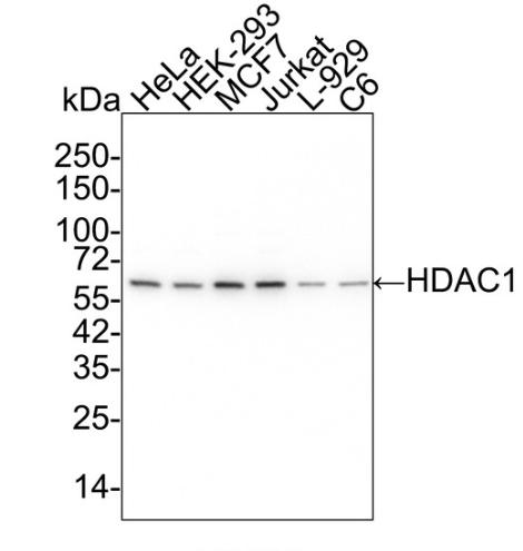
Western blot analysis of HDAC1 on different lysates with Rabbit anti-HDAC1 antibody at 1/5,000 dilution. Lane 1: HeLa cell lysate, Lane 2: HEK-293 cell lysate, Lane 3: MCF7 cell lysate, Lane 4: Jurkat cell lysate, Lane 5: L-929 cell lysate, Lane 6: C6 cell lysate, Lysates/proteins at 15 µg/Lane. Exposure time: 30 seconds; 4-20% SDS-PAGE gel. Proteins were transferred to a PVDF membrane and blocked with 5% NFDM/TBST for 1 hour at room temperature. The primary antibody at 1/5,000 dilution was used in 5% BSA at 4℃ overnight. Goat Anti-Rabbit IgG - HRP Secondary Antibody at 1/50,000 dilution was used for 1 hour at room temperature.IHC
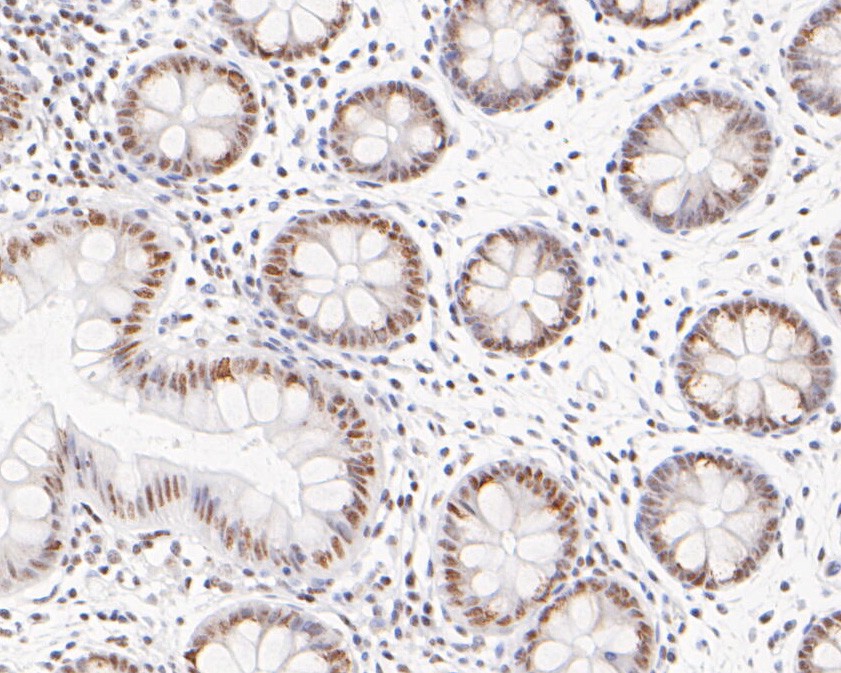
Immunohistochemical analysis of paraffin-embedded human colon tissue using anti-HDAC1 antibody. The section was pre-treated using heat mediated antigen retrieval with sodium citrate buffer (pH 6.0) (high pressure) for 2 minutes. The tissues were blocked in 5% BSA for 30 minutes at room temperature, washed with ddH2O and PBS, and then probed with the primary antibody (1/200) for 30 minutes at room temperature. The detection was performed using an HRP conjugated compact polymer system. DAB was used as the chromogen. Tissues were counterstained with hematoxylin and mounted with DPX.IHC
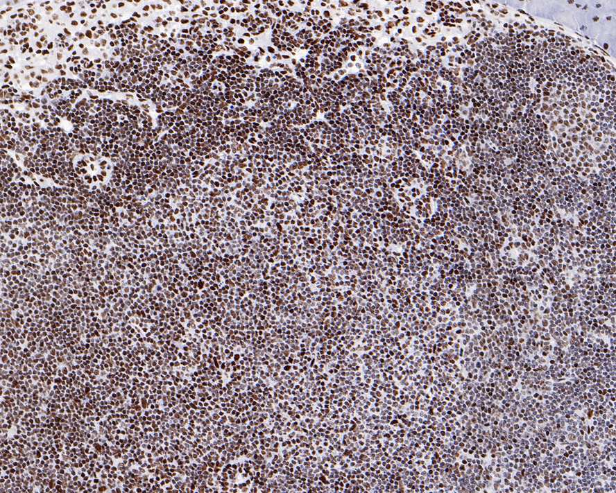
Immunohistochemical analysis of paraffin-embedded mouse lymph nodes tissue with Rabbit anti-HDAC1 antibody at 1/800 dilution. The section was pre-treated using heat mediated antigen retrieval with sodium citrate buffer (pH 6.0) (high pressure) for 2 minutes. The tissues were blocked in 1% BSA for 20 minutes at room temperature, washed with ddH2O and PBS, and then probed with the primary antibody at 1/800 dilution for 1 hour at room temperature. The detection was performed using an HRP conjugated compact polymer system. DAB was used as the chromogen. Tissues were counterstained with hematoxylin and mounted with DPX.IHC
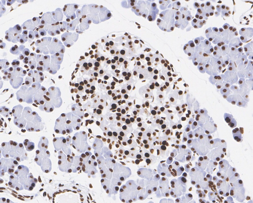
Immunohistochemical analysis of paraffin-embedded rat pancreas tissue with Rabbit anti-HDAC1 antibody at 1/1,000 dilution. The section was pre-treated using heat mediated antigen retrieval with sodium citrate buffer (pH 6.0) (high pressure) for 2 minutes. The tissues were blocked in 1% BSA for 20 minutes at room temperature, washed with ddH2O and PBS, and then probed with the primary antibody at 1/1,000 dilution for 1 hour at room temperature. The detection was performed using an HRP conjugated compact polymer system. DAB was used as the chromogen. Tissues were counterstained with hematoxylin and mounted with DPX.ICC/IF
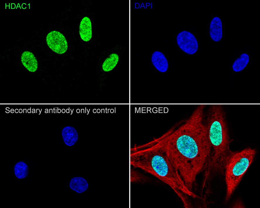
Immunocytochemistry analysis of C6 cells labeling HDAC1 with Rabbit anti-HDAC1 antibody at 1/100 dilution. Cells were fixed in 4% paraformaldehyde for 20 minutes at room temperature, permeabilized with 0.1% Triton X-100 in PBS for 5 minutes at room temperature, then blocked with 1% BSA in 10% negative goat serum for 1 hour at room temperature. Cells were then incubated with Rabbit anti-HDAC1 antibody at 1/100 dilution in 1% BSA in PBST overnight at 4 ℃. Goat Anti-Rabbit IgG H&L (488) was used as the secondary antibody at 1/1,000 dilution. PBS instead of the primary antibody was used as the secondary antibody only control. Nuclear DNA was labelled in blue with DAPI. Beta tubulin (red) was stained at 1/100 dilution overnight at +4℃. Goat Anti-Mouse IgG H&L (594) was used as the secondary antibody at 1/1,000 dilution.IP
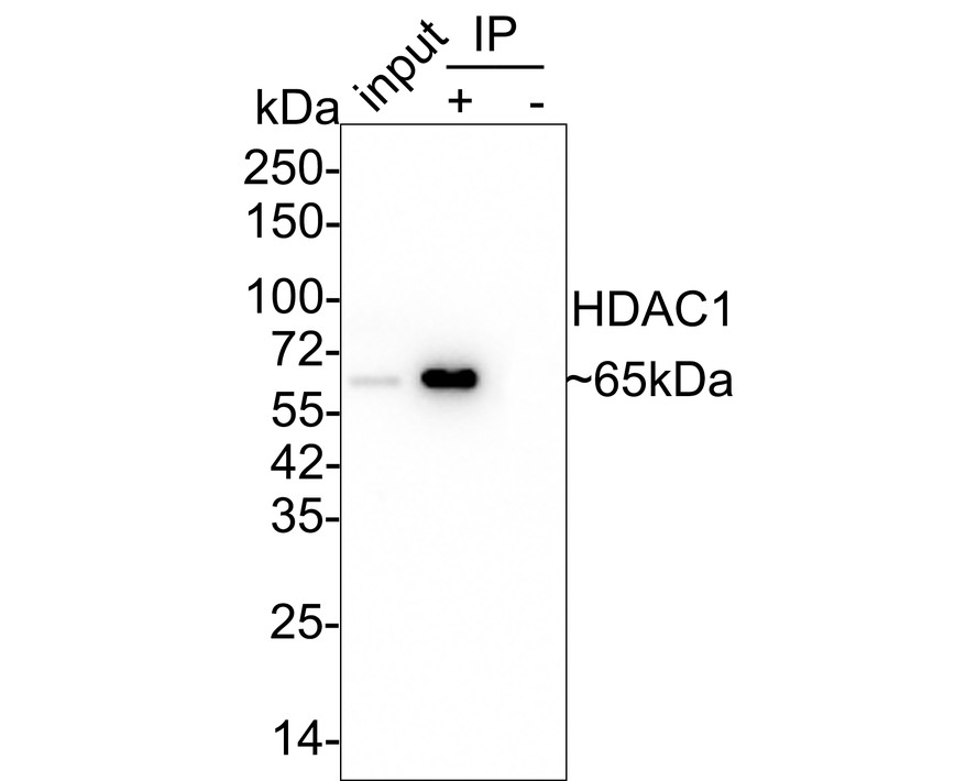
HDAC1 was immunoprecipitated in 0.2mg HeLa cell lysate with Rabbit anti-HDAC1 antibody at 2 µg/10 µl beads. Western blot was performed from the immunoprecipitate using Rabbit anti-HDAC1 antibody at 1/5,000 dilution. Anti-Rabbit IgG for IP Nano-secondary antibody at 1/5,000 dilution was used for 1 hour at room temperature. Lane 1: HeLa cell lysate (input), Lane 2: Rabbit anti-HDAC1 antibody IP in HeLa cell lysate, Lane 3: Rabbit IgG instead of Rabbit anti-HDAC1 antibody in HeLa cell lysate, Blocking/Dilution buffer: 5% NFDM/TBST, Exposure time: 43 seconds.| Product Name | HDAC1 Recombinant Rabbit Monoclonal Antibody |
|---|---|
| Antibody Type | Primary Antibodies |
| Product description | Acetylation of the histone tail causes chromatin to adopt an "open" conformation, allowing increased accessibility of transcription factors to DNA. The identification of histone acetyltransferases (HATs) and their large multiprotein complexes has yielded important insights into how these enzymes regulate transcription. HAT complexes interact with sequence-specific activator proteins to target specific genes. In addition to histones, HATs can acetylate nonhistone proteins, suggesting multiple roles for these enzymes. In contrast, histone deacetylation promotes a "closed" chromatin conformation and typically leads to repression of gene activity. Mammalian histone deacetylases can be divided into three classes on the basis of their similarity to various yeast deacetylases. Class I proteins (HDACs 1, 2, 3, and 8) are related to the yeast Rpd3-like proteins, those in class II (HDACs 4, 5, 6, 7, 9, and 10) are related to yeast Hda1-like proteins, and class III proteins are related to the yeast protein Sir2. Inhibitors of HDAC activity are now being explored as potential therapeutic cancer agents. |
| Immunogen | Synthetic peptide within human HDAC1 aa 420-460. |
| Clonality | Monoclonal |
|---|---|
| Isotype | IgG |
| Host Species | Recombinant rabbit |
| Tested Applications | ICC/IFIHCIPWB |
| WB:1:5000 IHC:1:200-1:1000 ICC/IF:1:100-1:500 IP:1-2μg/sample |
|
| Species Reactivity | HumanMouseRat |
| Concentration | 1mg/ml |
| Purification | Protein A |
| Alternative Names | DKFZp686H12203 antibody GON 10 antibody HD1 antibody HDAC 1 antibody HDAC1 antibody HDAC1_HUMAN antibody Histone deacetylase 1 antibody Reduced potassium dependency yeast homolog like 1 antibody RPD3 antibody RPD3L1 antibody |
|---|---|
| Molecular Weight(MW) | 55 kDa(Observed band size: 65 kDa) |
| Cellular Localization | Nucleus. |
WB
Western blot analysis of HDAC1 on different lysates with Rabbit anti-HDAC1 antibody at 1/5,000 dilution. Lane 1: HeLa cell lysate, Lane 2: HEK-293 cell lysate, Lane 3: MCF7 cell lysate, Lane 4: Jurkat cell lysate, Lane 5: L-929 cell lysate, Lane 6: C6 cell lysate, Lysates/proteins at 15 µg/Lane. Exposure time: 30 seconds; 4-20% SDS-PAGE gel. Proteins were transferred to a PVDF membrane and blocked with 5% NFDM/TBST for 1 hour at room temperature. The primary antibody at 1/5,000 dilution was used in 5% BSA at 4℃ overnight. Goat Anti-Rabbit IgG - HRP Secondary Antibody at 1/50,000 dilution was used for 1 hour at room temperature.
IHC
Immunohistochemical analysis of paraffin-embedded human colon tissue using anti-HDAC1 antibody. The section was pre-treated using heat mediated antigen retrieval with sodium citrate buffer (pH 6.0) (high pressure) for 2 minutes. The tissues were blocked in 5% BSA for 30 minutes at room temperature, washed with ddH2O and PBS, and then probed with the primary antibody (1/200) for 30 minutes at room temperature. The detection was performed using an HRP conjugated compact polymer system. DAB was used as the chromogen. Tissues were counterstained with hematoxylin and mounted with DPX.
IHC
Immunohistochemical analysis of paraffin-embedded mouse lymph nodes tissue with Rabbit anti-HDAC1 antibody at 1/800 dilution. The section was pre-treated using heat mediated antigen retrieval with sodium citrate buffer (pH 6.0) (high pressure) for 2 minutes. The tissues were blocked in 1% BSA for 20 minutes at room temperature, washed with ddH2O and PBS, and then probed with the primary antibody at 1/800 dilution for 1 hour at room temperature. The detection was performed using an HRP conjugated compact polymer system. DAB was used as the chromogen. Tissues were counterstained with hematoxylin and mounted with DPX.
IHC
Immunohistochemical analysis of paraffin-embedded rat pancreas tissue with Rabbit anti-HDAC1 antibody at 1/1,000 dilution. The section was pre-treated using heat mediated antigen retrieval with sodium citrate buffer (pH 6.0) (high pressure) for 2 minutes. The tissues were blocked in 1% BSA for 20 minutes at room temperature, washed with ddH2O and PBS, and then probed with the primary antibody at 1/1,000 dilution for 1 hour at room temperature. The detection was performed using an HRP conjugated compact polymer system. DAB was used as the chromogen. Tissues were counterstained with hematoxylin and mounted with DPX.
ICC/IF
Immunocytochemistry analysis of C6 cells labeling HDAC1 with Rabbit anti-HDAC1 antibody at 1/100 dilution. Cells were fixed in 4% paraformaldehyde for 20 minutes at room temperature, permeabilized with 0.1% Triton X-100 in PBS for 5 minutes at room temperature, then blocked with 1% BSA in 10% negative goat serum for 1 hour at room temperature. Cells were then incubated with Rabbit anti-HDAC1 antibody at 1/100 dilution in 1% BSA in PBST overnight at 4 ℃. Goat Anti-Rabbit IgG H&L (488) was used as the secondary antibody at 1/1,000 dilution. PBS instead of the primary antibody was used as the secondary antibody only control. Nuclear DNA was labelled in blue with DAPI. Beta tubulin (red) was stained at 1/100 dilution overnight at +4℃. Goat Anti-Mouse IgG H&L (594) was used as the secondary antibody at 1/1,000 dilution.
IP
HDAC1 was immunoprecipitated in 0.2mg HeLa cell lysate with Rabbit anti-HDAC1 antibody at 2 µg/10 µl beads. Western blot was performed from the immunoprecipitate using Rabbit anti-HDAC1 antibody at 1/5,000 dilution. Anti-Rabbit IgG for IP Nano-secondary antibody at 1/5,000 dilution was used for 1 hour at room temperature. Lane 1: HeLa cell lysate (input), Lane 2: Rabbit anti-HDAC1 antibody IP in HeLa cell lysate, Lane 3: Rabbit IgG instead of Rabbit anti-HDAC1 antibody in HeLa cell lysate, Blocking/Dilution buffer: 5% NFDM/TBST, Exposure time: 43 seconds.| Positive Control | K562, Hela, Jurkat, human tonsil tissue, mouse colon tissue, mouse pancreas tissue. |
|---|---|
| Application Notes | WB:1:5000 IHC:1:200-1:1000 ICC/IF:1:100-1:500 IP:1-2μg/sample |
| Form | Liquid |
|---|---|
| Storage Instructions | Store at +4℃ after thawing. Aliquot store at -20℃ or -80℃. Avoid repeated freeze / thaw cycles. |
| Storage Buffer | 1*TBS (pH7.4), 1%BSA, 40%Glycerol. Preservative: 0.05% Sodium Azide. |


