Application
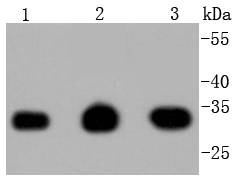
Fig1: Western blot analysis of PCNA on different lysates using anti-PCNA antibody at 1/1,000 dilution. Positive control: Lane 1: Hela Lane 2: 293 Lane 3: A431Application
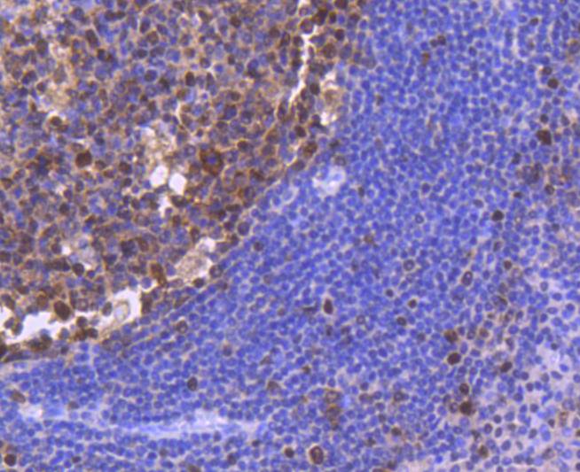
Fig2: Immunohistochemical analysis of paraffin-embedded human tonsil tissue using anti-PCNA antibody. Counter stained with hematoxylin.Application
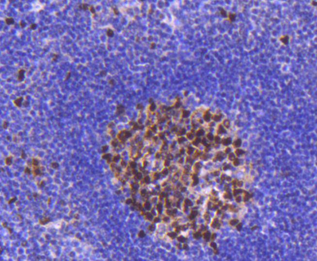
Fig3: Immunohistochemical analysis of paraffin-embedded human spleen tissue using anti-PCNA antibody. Counter stained with hematoxylin.Application
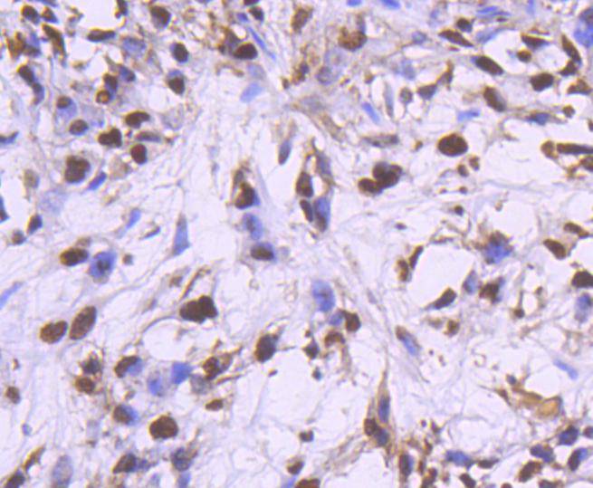
Fig4: Immunohistochemical analysis of paraffin-embedded human breast carcinama tissue using anti-PCNA antibody. Counter stained with hematoxylin.Application
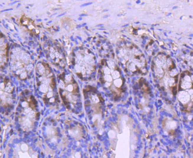
Fig5: Immunohistochemical analysis of paraffin-embedded mouse colon tissue using anti-PCNA antibody. Counter stained with hematoxylin.Application
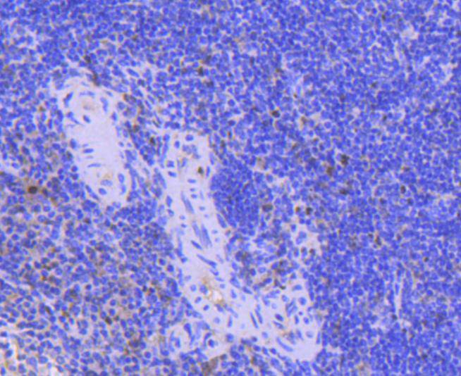
Fig6: Immunohistochemical analysis of paraffin-embedded mouse spleen tissue using anti-PCNA antibody. Counter stained with hematoxylin.Application
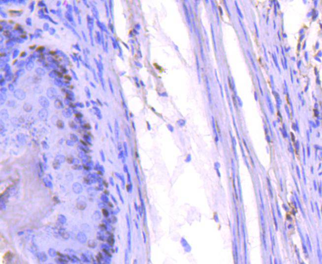
Fig7: Immunohistochemical analysis of paraffin-embedded mouse stomach tissue using anti-PCNA antibody. Counter stained with hematoxylin.Application
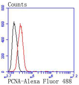
Fig8: Flow cytometric analysis of Hela cells with PCNA antibody at 1/50 dilution (red) compared with an unlabelled control (cells without incubation with primary antibody; black). Alexa Fluor 488-conjugated goat anti rabbit IgG was used as the secondary antibody.| Product Name | PCNA [SY12-07] |
|---|---|
| Antibody Type | Primary Antibodies |
| Antigen Alias | ATLD2 antibody cb16 antibody Cyclin antibody DNA polymerase delta auxiliary protein antibody etID36690.10 antibody fa28e03 antibody fb36g03 antibody HGCN8729 antibody MGC8367 antibody Mutagen-sensitive 209 protein antibody OTTHUMP00000030189 antibody OTTHUMP00000030190 antibody PCNA antibody Pcna/cyclin antibody PCNA_HUMAN antibody PCNAR antibody Polymerase delta accessory protein antibody Proliferating cell nuclear antigen antibody wu:fa28e03 antibody wu:fb36g03 antibody |
| Product description | The proliferating cell nuclear antigen (PCNA), a protein synthesized in early G1 and S phases of the cell cycle, functions in cell cycle progression, DNA replication and DNA repair. In early S phase, PCNA exhibits granular distribution and is absent from the nucleoli; however, in late S phase, it relocates to the nucleoli. PCNA exists in two basic forms: one involved in ongoing DNA replication, which localizes specifically to the nucleus, and a second, soluble form, not implicated in constant synthesis. Interestingly, the latter form degrades in the presence of organic solvents, rendering it undetectable by histological methods in tissues using organic fixatives, and thus also providing a method of visualizing only the synthesizing form. |
| Immunogen | recombinant protein |
| Clonality | Monoclonal |
|---|---|
| Isotype | IgG |
| Host Species | Recombinant rabbit |
| Tested Applications | WBICC/IFIHCIPFC |
| WB:1:1,000-1:5,000 ICC:1:50-1:200 IHC:1:100-1:500 FC:1:50-1:100 |
|
| Species Reactivity | HumanMouseRat |
| Concentration | 1mg/ml |
| Alternative Names | ATLD2 antibody cb16 antibody Cyclin antibody DNA polymerase delta auxiliary protein antibody etID36690.10 antibody fa28e03 antibody fb36g03 antibody HGCN8729 antibody MGC8367 antibody Mutagen-sensitive 209 protein antibody OTTHUMP00000030189 antibody OTTHUMP00000030190 antibody PCNA antibody Pcna/cyclin antibody PCNA_HUMAN antibody PCNAR antibody Polymerase delta accessory protein antibody Proliferating cell nuclear antigen antibody wu:fa28e03 antibody wu:fb36g03 antibody |
|---|---|
| Molecular Weight(MW) | 29 kDa |
| Cellular Localization | Nucleus. |
Application
Fig1: Western blot analysis of PCNA on different lysates using anti-PCNA antibody at 1/1,000 dilution. Positive control: Lane 1: Hela Lane 2: 293 Lane 3: A431
Application
Fig2: Immunohistochemical analysis of paraffin-embedded human tonsil tissue using anti-PCNA antibody. Counter stained with hematoxylin.
Application
Fig3: Immunohistochemical analysis of paraffin-embedded human spleen tissue using anti-PCNA antibody. Counter stained with hematoxylin.
Application
Fig4: Immunohistochemical analysis of paraffin-embedded human breast carcinama tissue using anti-PCNA antibody. Counter stained with hematoxylin.
Application
Fig5: Immunohistochemical analysis of paraffin-embedded mouse colon tissue using anti-PCNA antibody. Counter stained with hematoxylin.
Application
Fig6: Immunohistochemical analysis of paraffin-embedded mouse spleen tissue using anti-PCNA antibody. Counter stained with hematoxylin.
Application
Fig7: Immunohistochemical analysis of paraffin-embedded mouse stomach tissue using anti-PCNA antibody. Counter stained with hematoxylin.
Application
Fig8: Flow cytometric analysis of Hela cells with PCNA antibody at 1/50 dilution (red) compared with an unlabelled control (cells without incubation with primary antibody; black). Alexa Fluor 488-conjugated goat anti rabbit IgG was used as the secondary antibody.| Positive Control | A431, 293, Hela, human tonsil tissue, human spleen tissue, human breast carcinama tissue, mouse stomach tissue, mouse colon tissue, mouse spleen tissue. |
|---|---|
| Application Notes | WB:1:1,000-1:5,000 ICC:1:50-1:200 IHC:1:100-1:500 FC:1:50-1:100 |
| Form | Liquid |
|---|---|
| Storage Instructions | Store at +4℃ after thawing. Aliquot store at -20℃ or -80℃. Avoid repeated freeze / thaw cycles. |
| Storage Buffer | 1*TBS (pH7.4), 1%BSA, 40%Glycerol. Preservative: 0.05% Sodium Azide. |


