Application
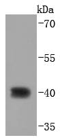
Fig1: Western blot analysis of CDX2 on AGS cells lysates using anti-CDX2 antibody at 1/1,000 dilution.Application
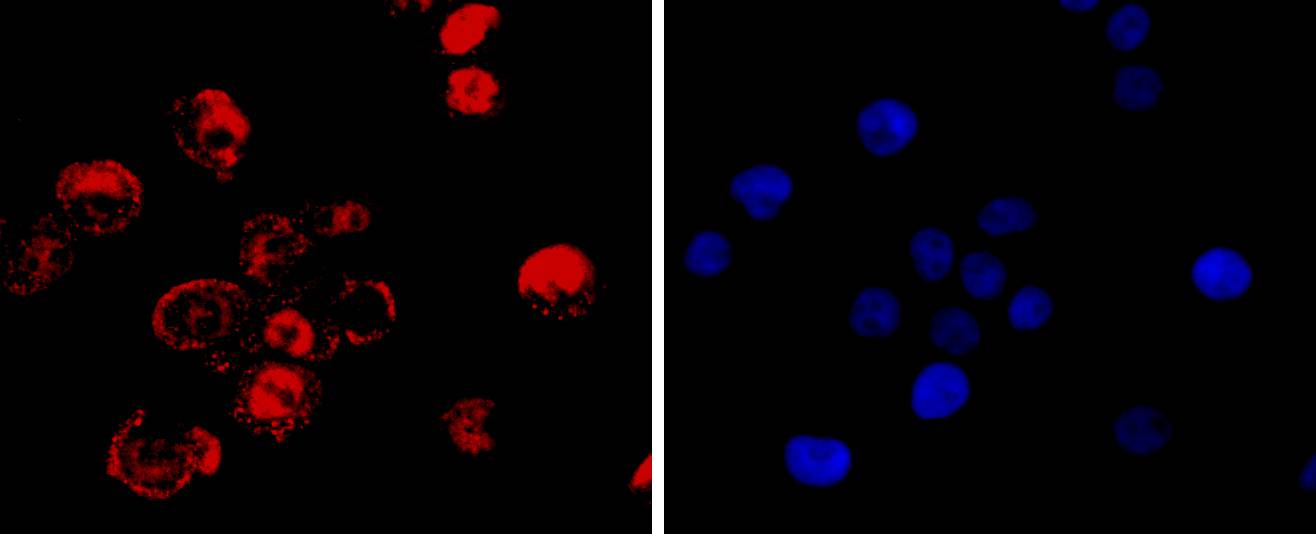
Fig2: ICC staining CDX2 in LOVO cells (red). The nuclear counter stain is DAPI (blue). Cells were fixed in paraformaldehyde, permeabilised with 0.25% Triton X100/PBS.Application
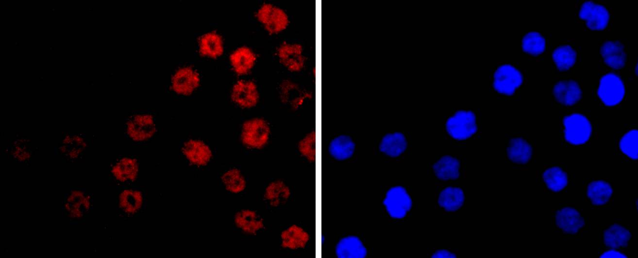
Fig3: ICC staining CDX2 in AGS cells (red). The nuclear counter stain is DAPI (blue). Cells were fixed in paraformaldehyde, permeabilised with 0.25% Triton X100/PBS.Application
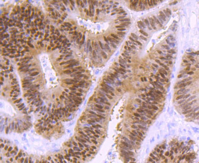
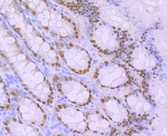
Fig4: Immunohistochemical analysis of paraffin-embedded human colon cancer tissue using anti-CDX2 antibody. Counter stained with hematoxylin.Application
Fig5: Immunohistochemical analysis of paraffin-embedded rat large intestine tissue using anti-CDX2 antibody. Counter stained with hematoxylin.Application
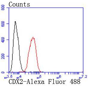
Fig6: Flow cytometric analysis of Hela cells with CDX2 antibody at 1/50 dilution (red) compared with an unlabelled control (cells without incubation with primary antibody; black). Alexa Fluor 488-conjugated goat anti rabbit IgG was used as the secondary antibody| Product Name | CDX2 [SY09-02] |
|---|---|
| Antibody Type | Primary Antibodies |
| Antigen Alias | Caudal type homeo box 2 antibody Caudal type homeo box transcription factor 2 antibody Caudal type homeobox 2 antibody Caudal type homeobox protein 2 antibody Caudal type homeobox transcription factor 2 antibody Caudal-type homeobox protein 2 antibody CDX 2 antibody CDX 3 antibody CDX-3 antibody Cdx2 antibody CDX2_HUMAN antibody CDX3 antibody Homeobox protein CDX 2 antibody Homeobox protein CDX-2 antibody |
| Product description | The members of the murine Cdx family (Cdx1, Cdx2, and Cdx4) are members of the caudal-type homeobox family of genes, which are homologues of the Drosophila 'caudal' gene required for anterior-posterior regional identity. The intestine-specific transcription factors Cdx1 and Cdx2 are candidate genes for directing intestinal development, differentiation, proliferation and maintenance of the intestinal phenotype. The relative expression of Cdx1 to Cdx2 protein may be important in the anterior to posterior patterning of the intestinal epithelium and in defining patterns of proliferation and differentiation along the crypt-villus axis. Cdx1 and Cdx2 are expressed in the small intestine and colon of fetus and adult. A decrease in human Cdx1 and/or Cdx2 expression is associated with colorectal tumorigenesis. Both Cdx1 and Cdx2 genes must be expressed to reduce tumorigenic potential, to increase sensitivity to apoptosis, and to reduce cell migration, suggesting that the two genes control the normal phenotype by independent pathways. |
| Immunogen | recombinant protein |
| Clonality | Monoclonal |
|---|---|
| Isotype | IgG |
| Host Species | Recombinant rabbit |
| Tested Applications | WBICC/IFIHCFC |
| WB:1:1,000-1:2,000 ICC:1:50-1:200 IHC:1:50-1:200 FC:1:50-1:100 |
|
| Species Reactivity | HumanRatRabbit |
| Concentration | 1mg/ml |
| Alternative Names | Caudal type homeo box 2 antibody Caudal type homeo box transcription factor 2 antibody Caudal type homeobox 2 antibody Caudal type homeobox protein 2 antibody Caudal type homeobox transcription factor 2 antibody Caudal-type homeobox protein 2 antibody CDX 2 antibody CDX 3 antibody CDX-3 antibody Cdx2 antibody CDX2_HUMAN antibody CDX3 antibody Homeobox protein CDX 2 antibody Homeobox protein CDX-2 antibody |
|---|---|
| Molecular Weight(MW) | 34 kDa |
| Cellular Localization | Nucleus. |
Application
Fig1: Western blot analysis of CDX2 on AGS cells lysates using anti-CDX2 antibody at 1/1,000 dilution.
Application
Fig2: ICC staining CDX2 in LOVO cells (red). The nuclear counter stain is DAPI (blue). Cells were fixed in paraformaldehyde, permeabilised with 0.25% Triton X100/PBS.
Application
Fig3: ICC staining CDX2 in AGS cells (red). The nuclear counter stain is DAPI (blue). Cells were fixed in paraformaldehyde, permeabilised with 0.25% Triton X100/PBS.
Application
Fig4: Immunohistochemical analysis of paraffin-embedded human colon cancer tissue using anti-CDX2 antibody. Counter stained with hematoxylin.
Application
Fig5: Immunohistochemical analysis of paraffin-embedded rat large intestine tissue using anti-CDX2 antibody. Counter stained with hematoxylin.
Application
Fig6: Flow cytometric analysis of Hela cells with CDX2 antibody at 1/50 dilution (red) compared with an unlabelled control (cells without incubation with primary antibody; black). Alexa Fluor 488-conjugated goat anti rabbit IgG was used as the secondary antibody| Positive Control | AGS, LOVO, rat large intestine tissue, human colon cancer tissue. |
|---|---|
| Application Notes | WB:1:1,000-1:2,000 ICC:1:50-1:200 IHC:1:50-1:200 FC:1:50-1:100 |
| Form | Liquid |
|---|---|
| Storage Instructions | Store at +4℃ after thawing. Aliquot store at -20℃ or -80℃. Avoid repeated freeze / thaw cycles. |
| Storage Buffer | 1*TBS (pH7.4), 1%BSA, 40%Glycerol. Preservative: 0.05% Sodium Azide. |


