Application
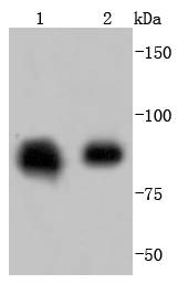
Fig1: Western blot analysis of Hsp90 beta on different lysates using anti-Hsp90 beta antibody at 1/1,000 dilution. Positive control: Lane 1: Hela Lane 2: K562Application
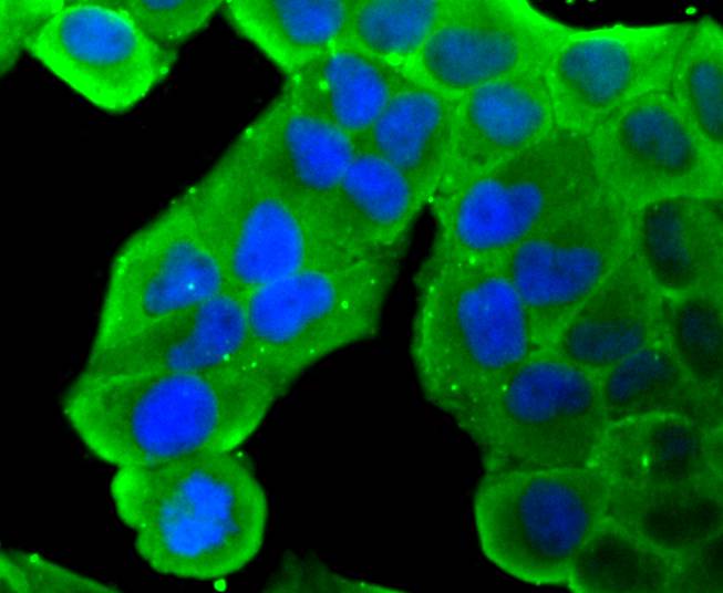
Fig2: ICC staining Hsp90 beta in Hela cells (green). The nuclear counter stain is DAPI (blue). Cells were fixed in paraformaldehyde, permeabilised with 0.25% Triton X100/PBS.Application
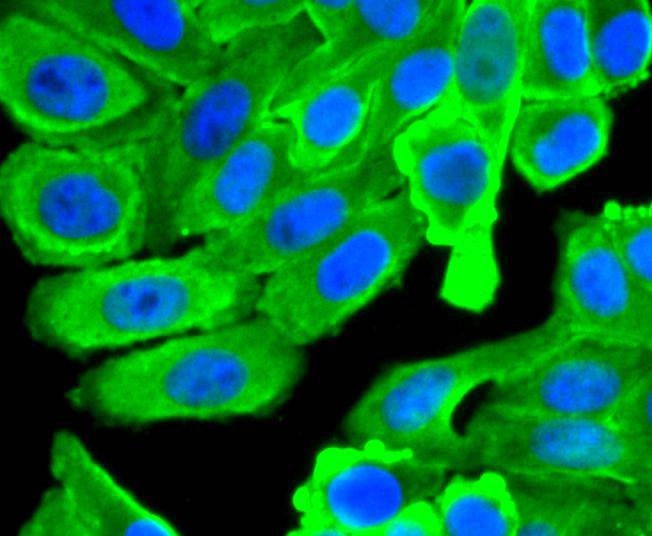
Fig3: ICC staining Hsp90 beta in HepG2 cells (green). The nuclear counter stain is DAPI (blue). Cells were fixed in paraformaldehyde, permeabilised with 0.25% Triton X100/PBS.Application
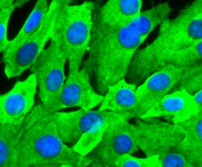
Fig4: ICC staining Hsp90 beta in NIH/3T3 cells (green). The nuclear counter stain is DAPI (blue). Cells were fixed in paraformaldehyde, permeabilised with 0.25% Triton X100/PBS.Application
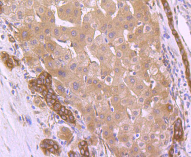
Fig5: Immunohistochemical analysis of paraffin-embedded human liver cancer tissue using anti-Hsp90 beta antibody. Counter stained with hematoxylin.Application
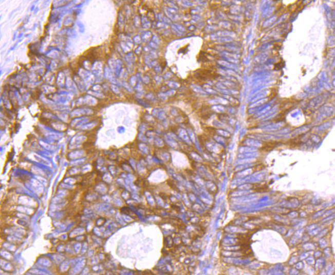
Fig6: Immunohistochemical analysis of paraffin-embedded human colon cancer tissue using anti-Hsp90 beta antibody. Counter stained with hematoxylin.Application
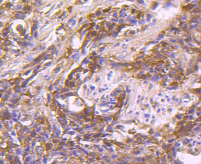
Fig7: Immunohistochemical analysis of paraffin-embedded human breast carcinoma tissue using anti-Hsp90 beta antibody. Counter stained with hematoxylin.Application
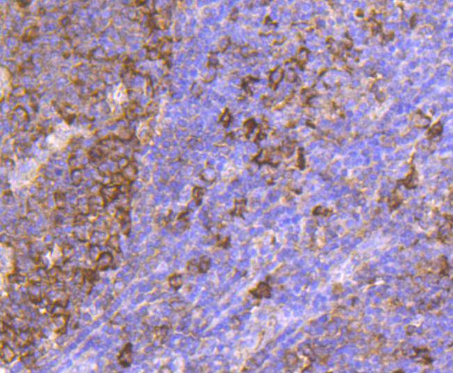
Fig8: Immunohistochemical analysis of paraffin-embedded human tonsil tissue using anti-Hsp90 beta antibody. Counter stained with hematoxylin.Application
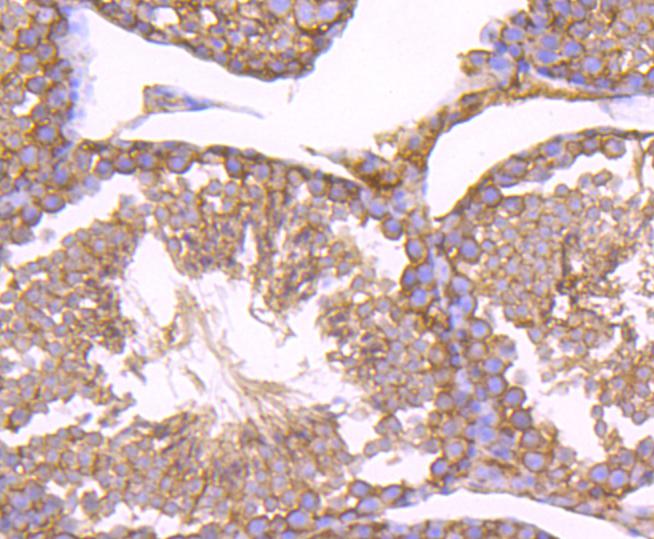
Fig9: Immunohistochemical analysis of paraffin-embedded mouse testis tissue using anti-Hsp90 beta antibody. Counter stained with hematoxylin.Application
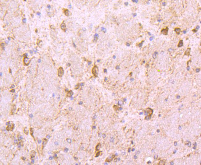
Fig10: Immunohistochemical analysis of paraffin-embedded mouse brain tissue using anti-Hsp90 beta antibody. Counter stained with hematoxylin.Application
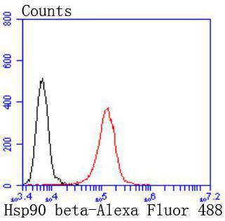
Fig11: Flow cytometric analysis of Jurkat cells with Hsp90 beta antibody at 1/50 dilution (red) compared with an unlabelled control (cells without incubation with primary antibody; black). Alexa Fluor 488-conjugated goat anti rabbit IgG was used as the secondary antibody| Product Name | Hsp90 beta [SY46-01] |
|---|---|
| Antibody Type | Primary Antibodies |
| Antigen Alias | 90 kda heat shock protein beta HSP90 beta antibody D6S182 antibody FLJ26984 antibody Heat shock 84 kDa antibody Heat shock 90kD protein 1, beta antibody Heat shock 90kDa protein 1 beta antibody Heat shock protein 90 alpha family class B member 1 antibody Heat shock protein 90 kDa antibody Heat shock protein 90kDa alpha (cytosolic) class B member 1 antibody Heat shock protein 90kDa alpha family class B member 1 antibody Heat shock protein beta antibody Heat shock protein HSP 90 beta antibody Heat shock protein HSP 90-beta antibody HS90B_HUMAN antibody HSP 84 antibody HSP 90 antibody HSP 90 b antibody HSP 90b antibody HSP84 antibody HSP90 BETA antibody hsp90ab1 antibody HSP90B antibody HSPC2 antibody HSPCB antibody |
| Product description | The heat shock response was first described for Drosophila salivary gland cells and morphologically consists of a change in their polytene chromosome puffing patterns that involves de novo synthesis of a few proteins. Similar heat shock proteins were later discovered in bacterial chicken and mammalian cells, and have been subsequently studied in other organisms. A series of proteins including HSP 90, HSP 70, HSP 20-30 and ubiquitin are induced by insults such as temperature shock, chemicals and other environmental stress. A major function of HSP 90 and other HSPs is to act as molecular chaperones. HSP 90 forms a complex with glucocorticoid receptor (GR), rendering the non ligand-bound receptor transcriptionally inactive. HSP 90 binds the GR as a heterocomplex composed of either HSP 56 or Cyclophilin D, forming an aporeceptor comiplex. HSP 90 also exists as a dimer with other proteins such as p60/sti1 and p23, forming an apo-receptor complex with estrogen and androgen receptors. |
| Immunogen | recombinant protein |
| Clonality | Monoclonal |
|---|---|
| Isotype | IgG |
| Host Species | Recombinant rabbit |
| Tested Applications | WBICC/IFIHCIPFC |
| WB:1:1,000-1:2,000 ICC:1:100-1:500 IHC:1:50-1:200 FC:1:50-1:100 |
|
| Species Reactivity | HumanMouseRat |
| Concentration | 1mg/ml |
| Alternative Names | 90 kda heat shock protein beta HSP90 beta antibody
D6S182 antibody
FLJ26984 antibody
Heat shock 84 kDa antibody
Heat shock 90kD protein 1 beta antibody Heat shock 90kDa protein 1 beta antibody Heat shock protein 90 alpha family class B member 1 antibody Heat shock protein 90 kDa antibody Heat shock protein 90kDa alpha (cytosolic) class B member 1 antibody Heat shock protein 90kDa alpha family class B member 1 antibody Heat shock protein beta antibody Heat shock protein HSP 90 beta antibody Heat shock protein HSP 90-beta antibody HS90B_HUMAN antibody HSP 84 antibody HSP 90 antibody HSP 90 b antibody HSP 90b antibody HSP84 antibody HSP90 BETA antibody hsp90ab1 antibody HSP90B antibody HSPC2 antibody HSPCB antibody |
|---|---|
| Molecular Weight(MW) | 83 kDa |
| Cellular Localization | Cytoplasm, Melanosome. |
Application
Fig1: Western blot analysis of Hsp90 beta on different lysates using anti-Hsp90 beta antibody at 1/1,000 dilution. Positive control: Lane 1: Hela Lane 2: K562
Application
Fig2: ICC staining Hsp90 beta in Hela cells (green). The nuclear counter stain is DAPI (blue). Cells were fixed in paraformaldehyde, permeabilised with 0.25% Triton X100/PBS.
Application
Fig3: ICC staining Hsp90 beta in HepG2 cells (green). The nuclear counter stain is DAPI (blue). Cells were fixed in paraformaldehyde, permeabilised with 0.25% Triton X100/PBS.
Application
Fig4: ICC staining Hsp90 beta in NIH/3T3 cells (green). The nuclear counter stain is DAPI (blue). Cells were fixed in paraformaldehyde, permeabilised with 0.25% Triton X100/PBS.
Application
Fig5: Immunohistochemical analysis of paraffin-embedded human liver cancer tissue using anti-Hsp90 beta antibody. Counter stained with hematoxylin.
Application
Fig6: Immunohistochemical analysis of paraffin-embedded human colon cancer tissue using anti-Hsp90 beta antibody. Counter stained with hematoxylin.
Application
Fig7: Immunohistochemical analysis of paraffin-embedded human breast carcinoma tissue using anti-Hsp90 beta antibody. Counter stained with hematoxylin.
Application
Fig8: Immunohistochemical analysis of paraffin-embedded human tonsil tissue using anti-Hsp90 beta antibody. Counter stained with hematoxylin.
Application
Fig9: Immunohistochemical analysis of paraffin-embedded mouse testis tissue using anti-Hsp90 beta antibody. Counter stained with hematoxylin.
Application
Fig10: Immunohistochemical analysis of paraffin-embedded mouse brain tissue using anti-Hsp90 beta antibody. Counter stained with hematoxylin.
Application
Fig11: Flow cytometric analysis of Jurkat cells with Hsp90 beta antibody at 1/50 dilution (red) compared with an unlabelled control (cells without incubation with primary antibody; black). Alexa Fluor 488-conjugated goat anti rabbit IgG was used as the secondary antibody| Positive Control | K562, NIH/3T3, Hela, HepG2, human tonsil tissue, human liver cancer tissue, human colon cancer tissue, human breast carcinoma tissue, mouse testis tissue, mouse brain tissue. |
|---|---|
| Application Notes | WB:1:1,000-1:2,000 ICC:1:100-1:500 IHC:1:50-1:200 FC:1:50-1:100 |
| Form | Liquid |
|---|---|
| Storage Instructions | Store at +4℃ after thawing. Aliquot store at -20℃ or -80℃. Avoid repeated freeze / thaw cycles. |
| Storage Buffer | 1*TBS (pH7.4), 1%BSA, 40%Glycerol. Preservative: 0.05% Sodium Azide. |


