Application
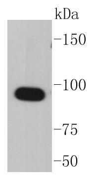
Fig1: Western blot analysis of Hsp90 alpha on COS-1 cell lysates using anti-Hsp90 alpha antibody at 1/1,000 dilution.Application
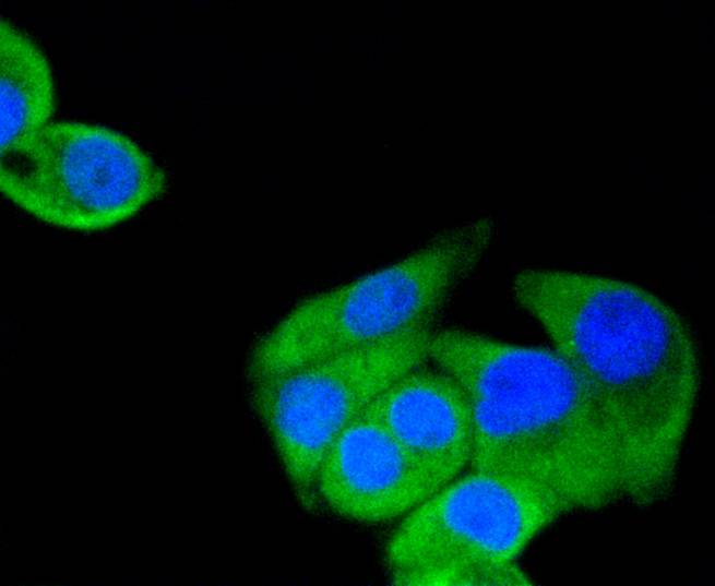
Fig2: ICC staining Hsp90 alpha in Hela cells (green). The nuclear counter stain is DAPI (blue). Cells were fixed in paraformaldehyde, permeabilised with 0.25% Triton X100/PBS.Application
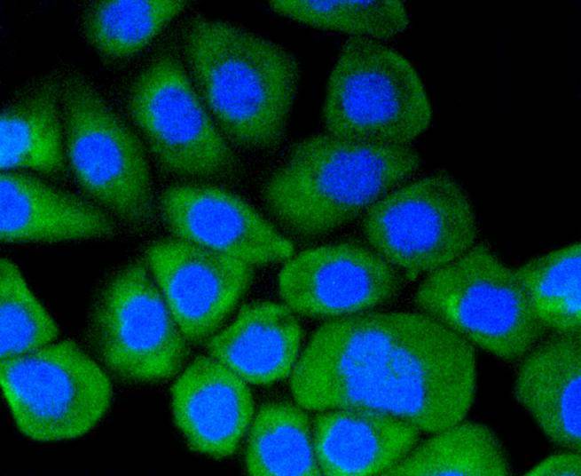
Fig3: ICC staining Hsp90 alpha in Ags cells (green). The nuclear counter stain is DAPI (blue). Cells were fixed in paraformaldehyde, permeabilised with 0.25% Triton X100/PBS.Application
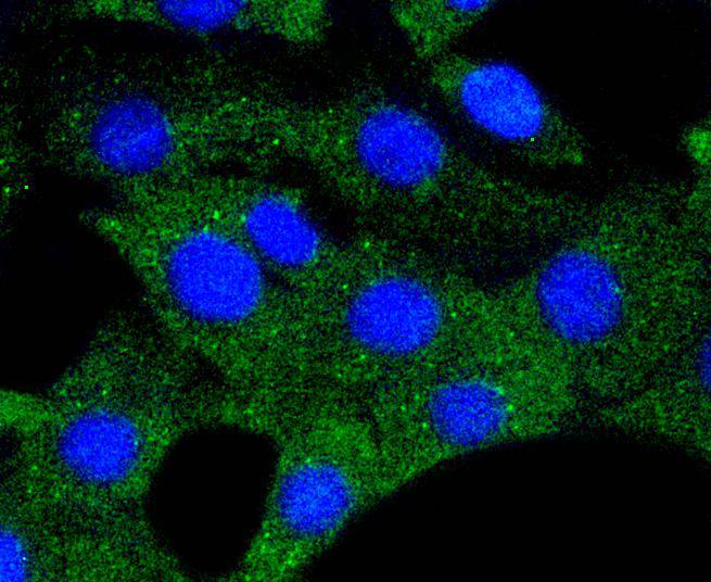
Fig4: ICC staining Hsp90 alpha in NIH/3T3 cells (green). The nuclear counter stain is DAPI (blue). Cells were fixed in paraformaldehyde, permeabilised with 0.25% Triton X100/PBS.Application
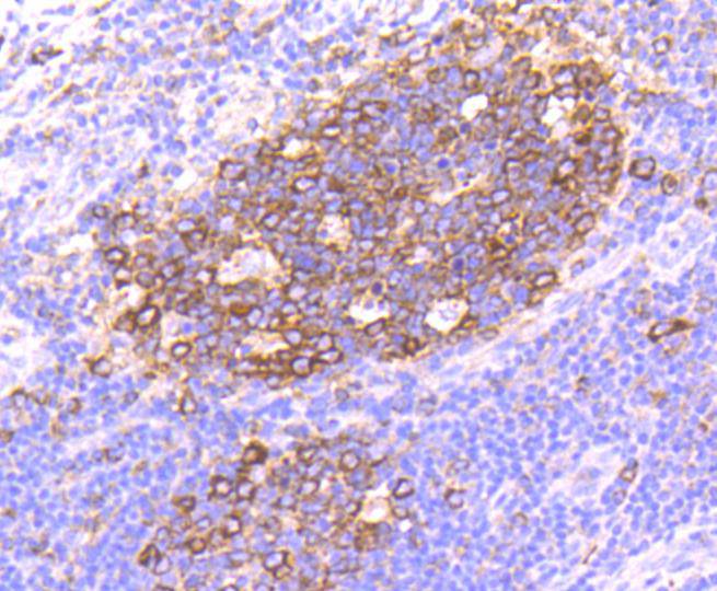
Fig5: Immunohistochemical analysis of paraffin-embedded human tonsil tissue using anti-Hsp90 alpha antibody. Counter stained with hematoxylin.Application
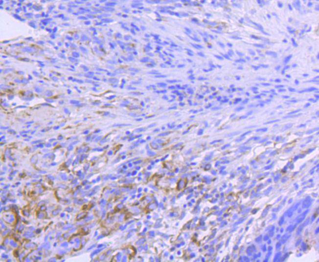
Fig6: Immunohistochemical analysis of paraffin-embedded human colon cancer tissue using anti-Hsp90 alpha antibody. Counter stained with hematoxylin.Application
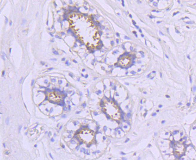
Fig7: Immunohistochemical analysis of paraffin-embedded human breast tissue using anti-Hsp90 alpha antibody. Counter stained with hematoxylin.Application
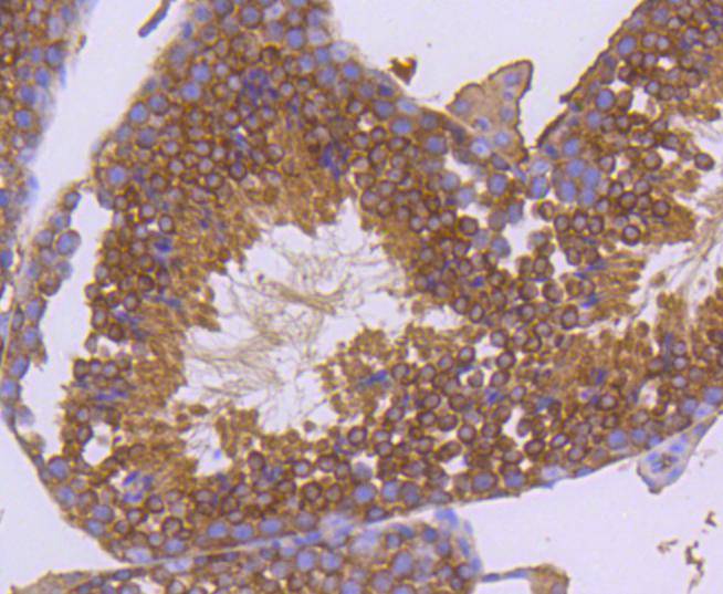
Fig8: Immunohistochemical analysis of paraffin-embedded mouse testis tissue using anti-Hsp90 alpha antibody. Counter stained with hematoxylin.Application
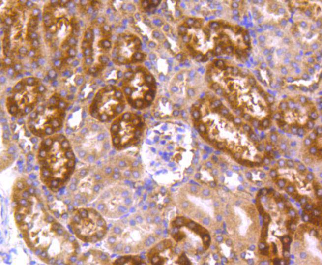
Fig9: Immunohistochemical analysis of paraffin-embedded human kidney tissue using anti-Hsp90 alpha antibody. Counter stained with hematoxylin.| Product Name | Hsp90 alpha [SY14-06] |
|---|---|
| Antibody Type | Primary Antibodies |
| Antigen Alias | EL52 antibody epididymis luminal secretory protein 52 antibody Heat shock 86 kDa antibody heat shock 90kD protein 1, alpha antibody Heat shock 90kD protein 1, alpha like 4 antibody heat shock 90kD protein, alpha-like 4 antibody Heat shock 90kDa protein 1 alpha antibody Heat shock protein 90kDa alpha (cytosolic) class A member 1 antibody Heat shock protein HSP 90-alpha antibody HS90A_HUMAN antibody HSP 86 antibody HSP86 antibody Hsp89 antibody HSP89A antibody Hsp90 antibody HSP90A antibody HSP90AA1 antibody HSP90ALPHA antibody HSP90N antibody HSPC1 antibody HSPCA antibody HSPCAL1 antibody HSPCAL4 antibody HSPN antibody LAP 2 antibody LAP2 antibody lipopolysaccharide-associated protein 2 antibody LPS-associated protein 2 antibody Renal carcinoma antigen NY-REN-38 antibody |
| Product description | The heat shock response was first described for Drosophila salivary gland cells and morphologically consists of a change in their polytene chromosome puffing patterns that involves de novo synthesis of a few proteins. Similar heat shock proteins were later discovered in bacterial chicken and mammalian cells, and have been subsequently studied in other organisms. A series of proteins including HSP 90, HSP 70, HSP 20-30 and ubiquitin are induced by insults such as temperature shock, chemicals and other environmental stress. A major function of HSP 90 and other HSPs is to act as molecular chaperones. HSP 90 forms a complex with glucocorticoid receptor (GR), rendering the non ligand-bound receptor transcriptionally inactive. HSP 90 binds the GR as a heterocomplex composed of either HSP 56 or Cyclophilin D, forming an aporeceptor comiplex. HSP 90 also exists as a dimer with other proteins such as p60/sti1 and p23, forming an apo-receptor complex with estrogen and androgen receptors. |
| Immunogen | recombinant protein |
| Clonality | Monoclonal |
|---|---|
| Isotype | IgG |
| Host Species | Recombinant rabbit |
| Tested Applications | WBICC/IFIHC |
| WB:1:1,000-1:2,000 ICC:1:50-1:200 IHC:1:50-1:200 |
|
| Species Reactivity | HumanMouseRat |
| Concentration | 1mg/ml |
| Alternative Names | EL52 antibody
epididymis luminal secretory protein 52 antibody
Heat shock 86 kDa antibody
heat shock 90kD protein 1 alpha antibody Heat shock 90kD protein 1 alpha like 4 antibody heat shock 90kD protein alpha-like 4 antibody Heat shock 90kDa protein 1 alpha antibody Heat shock protein 90kDa alpha (cytosolic) class A member 1 antibody Heat shock protein HSP 90-alpha antibody HS90A_HUMAN antibody HSP 86 antibody HSP86 antibody Hsp89 antibody HSP89A antibody Hsp90 antibody HSP90A antibody HSP90AA1 antibody HSP90ALPHA antibody HSP90N antibody HSPC1 antibody HSPCA antibody HSPCAL1 antibody HSPCAL4 antibody HSPN antibody LAP 2 antibody LAP2 antibody lipopolysaccharide-associated protein 2 antibody LPS-associated protein 2 antibody Renal carcinoma antigen NY-REN-38 antibody |
|---|---|
| Molecular Weight(MW) | 90 kDa |
| Cellular Localization | Cytoplasm, Melanosome, Cell membrane. |
Application
Fig1: Western blot analysis of Hsp90 alpha on COS-1 cell lysates using anti-Hsp90 alpha antibody at 1/1,000 dilution.
Application
Fig2: ICC staining Hsp90 alpha in Hela cells (green). The nuclear counter stain is DAPI (blue). Cells were fixed in paraformaldehyde, permeabilised with 0.25% Triton X100/PBS.
Application
Fig3: ICC staining Hsp90 alpha in Ags cells (green). The nuclear counter stain is DAPI (blue). Cells were fixed in paraformaldehyde, permeabilised with 0.25% Triton X100/PBS.
Application
Fig4: ICC staining Hsp90 alpha in NIH/3T3 cells (green). The nuclear counter stain is DAPI (blue). Cells were fixed in paraformaldehyde, permeabilised with 0.25% Triton X100/PBS.
Application
Fig5: Immunohistochemical analysis of paraffin-embedded human tonsil tissue using anti-Hsp90 alpha antibody. Counter stained with hematoxylin.
Application
Fig6: Immunohistochemical analysis of paraffin-embedded human colon cancer tissue using anti-Hsp90 alpha antibody. Counter stained with hematoxylin.
Application
Fig7: Immunohistochemical analysis of paraffin-embedded human breast tissue using anti-Hsp90 alpha antibody. Counter stained with hematoxylin.
Application
Fig8: Immunohistochemical analysis of paraffin-embedded mouse testis tissue using anti-Hsp90 alpha antibody. Counter stained with hematoxylin.
Application
Fig9: Immunohistochemical analysis of paraffin-embedded human kidney tissue using anti-Hsp90 alpha antibody. Counter stained with hematoxylin.| Positive Control | Ags, NIH/3T3, Hela, mouse testis tissue, human tonsil tissue, human kidney tissue, human colon cancer tissue, human breast tissue. |
|---|---|
| Application Notes | WB:1:1,000-1:2,000 ICC:1:50-1:200 IHC:1:50-1:200 |
| Form | Liquid |
|---|---|
| Storage Instructions | Store at +4℃ after thawing. Aliquot store at -20℃ or -80℃. Avoid repeated freeze / thaw cycles. |
| Storage Buffer | 1*TBS (pH7.4), 1%BSA, 40%Glycerol. Preservative: 0.05% Sodium Azide. |


