Application
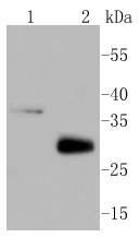
Fig1: Western blot analysis of MCL1 on different lysates using anti-MCL1 antibody at 1/1,000 dilution. Positive control: Lane 1: Raji Lane 2: Mouse heartApplication
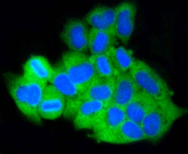
Fig2: ICC staining MCL1 in Hela cells (green). The nuclear counter stain is DAPI (blue). Cells were fixed in paraformaldehyde, permeabilised with 0.25% Triton X100/PBS.Application
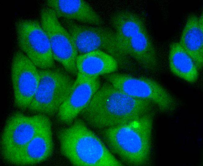
Fig3: ICC staining MCL1 in HepG2 cells (green). The nuclear counter stain is DAPI (blue). Cells were fixed in paraformaldehyde, permeabilised with 0.25% Triton X100/PBS.Application
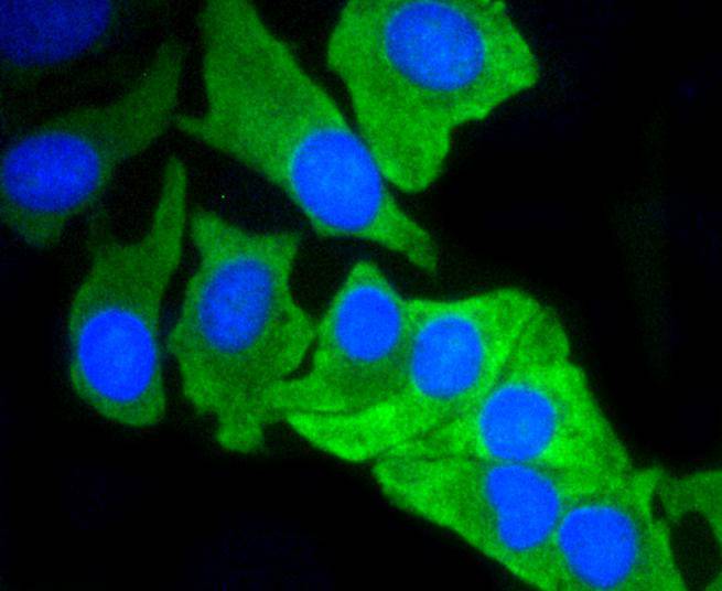
Fig4: ICC staining MCL1 in BT-20 cells (green). The nuclear counter stain is DAPI (blue). Cells were fixed in paraformaldehyde, permeabilised with 0.25% Triton X100/PBS.Application
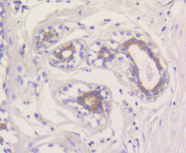
Fig5: Immunohistochemical analysis of paraffin-embedded human breast tissue using anti-MCL1 antibody. Counter stained with hematoxylin.Application
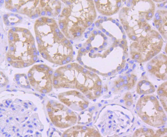
Fig6: Immunohistochemical analysis of paraffin-embedded human kidney tissue using anti-MCL1 antibody. Counter stained with hematoxylin.Application
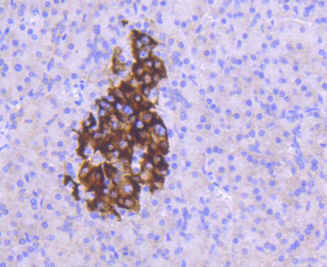
Fig7: Immunohistochemical analysis of paraffin-embedded human pancreas tissue using anti-MCL1 antibody. Counter stained with hematoxylin.Application
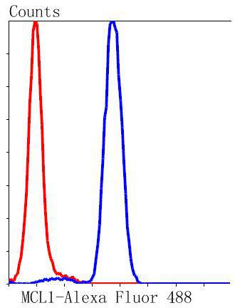
Fig8: Flow cytometric analysis of Jurkat cells with MCL1 antibody at 1/50 dilution (blue) compared with an unlabelled control (cells without incubation with primary antibody; red). Alexa Fluor 488-conjugated goat anti rabbit IgG was used as the secondary antibody.| Product Name | MCL1 [SI16-04] |
|---|---|
| Antibody Type | Primary Antibodies |
| Antigen Alias | Bcl 2 related protein EAT/mcl1 antibody Bcl-2-like protein 3 antibody Bcl-2-related protein EAT/mcl1 antibody BCL2 related antibody Bcl2-L-3 antibody BCL2L3 antibody EAT antibody Induced myeloid leukemia cell differentiation protein Mcl 1 antibody Induced myeloid leukemia cell differentiation protein Mcl-1 antibody MCL 1 antibody MCL1 antibody MCL1-ES antibody mcl1/EAT antibody MCL1_HUMAN antibody MCL1L antibody MCL1S antibody MGC104264 antibody MGC1839 antibody Myeloid Cell Leukemia 1 antibody Myeloid cell leukemia ES antibody Myeloid cell leukemia sequence 1 antibody Myeloid cell leukemia sequence 1 BCL2 related antibody Myeloid cell leukemia sequence 1 isoform 1 antibody OTTHUMP00000032794 antibody OTTHUMP00000032795 antibody TM antibody |
| Product description | B-cell CLL/lymphoma 2 (Bcl-2) blocks cell death following a variety of stimuli and confers a death-sparing effect to certain hematopoietic cell lines following growth factor withdrawal. Myeloid cell leukemia 1 (Mcl-1) shares sequence homology with Bcl-2 and further resembles Bcl-2 in that its expression promotes cell viability. p53 and Mcl-1 demonstrate opposing effects on mitochondrial apoptosis by mediating Bcl-2 antagonist killer (Bak) activity. Mcl-1 is an important and specific regulator that is necessary for the homeostasis of early hematopoietic progenitors. Glycogen synthase kinase 3 (GSK3) controls Mcl-1 stability, which has an effect on the regulation of apoptosis by growth factors, PI 3-kinase and AKT. Mice with a deficiency of the Mcl-1 protein show a significant reduction in B and T lymphocytes similar to the effects observed in IL-7- or IL-7R-deficient mice. |
| Immunogen | recombinant protein |
| Clonality | Monoclonal |
|---|---|
| Isotype | IgG |
| Host Species | Recombinant rabbit |
| Tested Applications | WBICC/IFIHCIPFC |
| WB:1:1,000 ICC:1:50-1:200 IHC:1:50-1:200 FC:1:50-1:100 |
|
| Species Reactivity | HumanMouseRat |
| Concentration | 1mg/ml |
| Alternative Names | Bcl 2 related protein EAT/mcl1 antibody Bcl-2-like protein 3 antibody Bcl-2-related protein EAT/mcl1 antibody BCL2 related antibody Bcl2-L-3 antibody BCL2L3 antibody EAT antibody Induced myeloid leukemia cell differentiation protein Mcl 1 antibody Induced myeloid leukemia cell differentiation protein Mcl-1 antibody MCL 1 antibody MCL1 antibody MCL1-ES antibody mcl1/EAT antibody MCL1_HUMAN antibody MCL1L antibody MCL1S antibody MGC104264 antibody MGC1839 antibody Myeloid Cell Leukemia 1 antibody Myeloid cell leukemia ES antibody Myeloid cell leukemia sequence 1 antibody Myeloid cell leukemia sequence 1 BCL2 related antibody Myeloid cell leukemia sequence 1 isoform 1 antibody OTTHUMP00000032794 antibody OTTHUMP00000032795 antibody TM antibody |
|---|---|
| Molecular Weight(MW) | 37/29 kDa |
| Cellular Localization | Membrane, Cytoplasm, Mitochondrion, Nucleus. |
Application
Fig1: Western blot analysis of MCL1 on different lysates using anti-MCL1 antibody at 1/1,000 dilution. Positive control: Lane 1: Raji Lane 2: Mouse heart
Application
Fig2: ICC staining MCL1 in Hela cells (green). The nuclear counter stain is DAPI (blue). Cells were fixed in paraformaldehyde, permeabilised with 0.25% Triton X100/PBS.
Application
Fig3: ICC staining MCL1 in HepG2 cells (green). The nuclear counter stain is DAPI (blue). Cells were fixed in paraformaldehyde, permeabilised with 0.25% Triton X100/PBS.
Application
Fig4: ICC staining MCL1 in BT-20 cells (green). The nuclear counter stain is DAPI (blue). Cells were fixed in paraformaldehyde, permeabilised with 0.25% Triton X100/PBS.
Application
Fig5: Immunohistochemical analysis of paraffin-embedded human breast tissue using anti-MCL1 antibody. Counter stained with hematoxylin.
Application
Fig6: Immunohistochemical analysis of paraffin-embedded human kidney tissue using anti-MCL1 antibody. Counter stained with hematoxylin.
Application
Fig7: Immunohistochemical analysis of paraffin-embedded human pancreas tissue using anti-MCL1 antibody. Counter stained with hematoxylin.
Application
Fig8: Flow cytometric analysis of Jurkat cells with MCL1 antibody at 1/50 dilution (blue) compared with an unlabelled control (cells without incubation with primary antibody; red). Alexa Fluor 488-conjugated goat anti rabbit IgG was used as the secondary antibody.| Positive Control | Raji, BT-20, Hela, HepG2, human pancreas tissue, human breast tissue, human kidney tissue. |
|---|---|
| Application Notes | WB:1:1,000 ICC:1:50-1:200 IHC:1:50-1:200 FC:1:50-1:100 |
| Form | Liquid |
|---|---|
| Storage Instructions | Store at +4℃ after thawing. Aliquot store at -20℃ or -80℃. Avoid repeated freeze / thaw cycles. |
| Storage Buffer | 1*TBS (pH7.4), 1%BSA, 40%Glycerol. Preservative: 0.05% Sodium Azide. |


