Application
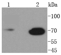
Fig1: Western blot analysis of Lamin B1 on different lysates using anti-Lamin B1 antibody at 1/1,000 dilution. Positive control: Lane 1: Mouse liver Lane 2: Mouse brainApplication
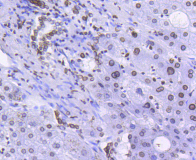
Fig2: Immunohistochemical analysis of paraffin-embedded human liver tissue using anti-Lamin B1 antibody. Counter stained with hematoxylin.Application
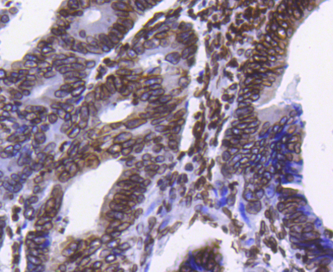
Fig3: Immunohistochemical analysis of paraffin-embedded human colon cancer tissue using anti-Lamin B1 antibody. Counter stained with hematoxylin.Application
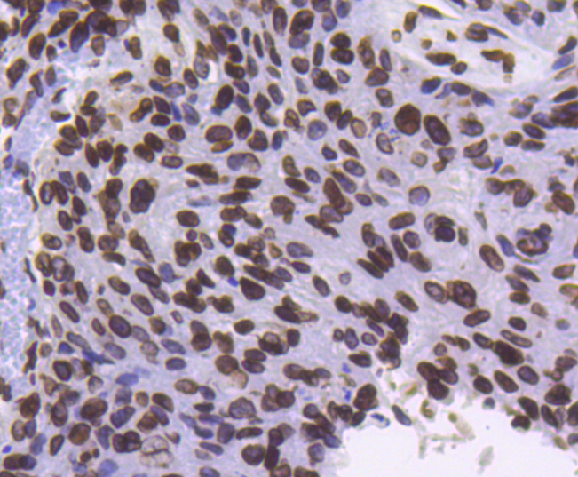
Fig4: Immunohistochemical analysis of paraffin-embedded human breast carcinoma tissue using anti-Lamin B1 antibody. Counter stained with hematoxylin.Application
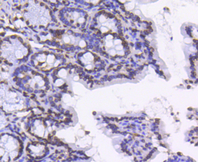
Fig5: Immunohistochemical analysis of paraffin-embedded mouse colon tissue using anti-Lamin B1 antibody. Counter stained with hematoxylin.Application
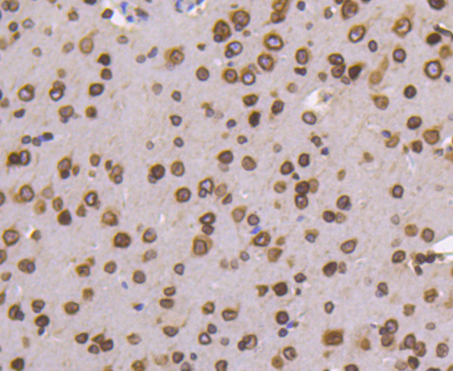
Fig6: Immunohistochemical analysis of paraffin-embedded mouse brain tissue using anti-Lamin B1 antibody. Counter stained with hematoxylin.| Product Name | Lamin B1 [SI17-06] |
|---|---|
| Antibody Type | Primary Antibodies |
| Antigen Alias | ADLD antibody lamin B1 antibody Lamin-B1 antibody LMN antibody LMN2 antibody LMNB antibody Lmnb1 antibody LMNB1_HUMAN antibody MGC111419 antibody OTTHUMP00000159218 antibody |
| Product description | A unique family of cysteine proteases has been described that differs in sequence, structure and substrate specificity from any previously described protease family. This family, termed Ced-3/ICE, functions as key components of the apoptotic machinery and act to destroy specific target proteins which are critical to cellular longevity. Nuclear lamins are critical to maintaining the integrity of the nuclear envelope and cellular morphology as components of the nuclear lamina, a fibrous layer on the nucleoplasmic side of the inner nuclear membrane, which is thought to provide a framework for the nuclear envelope and may also interact with chromatin. B-type lamins undergo a series of modifications, such as farnesylation and phosphorylation. Increased phosphorylation of the lamins occurs before envelope disintegration and probably plays a role in regulating lamin associations. Nuclear Lamin B is fragmented as a consequence of apoptosis by an unidentified member of the ICE family. |
| Immunogen | recombinant protein |
| Clonality | Monoclonal |
|---|---|
| Isotype | IgG |
| Host Species | Recombinant rabbit |
| Tested Applications | WBICC/IFIHCIP |
| WB:1:1,000 ICC:1:50-1:200 IHC:1:100-1:500 |
|
| Species Reactivity | HumanMouseRat |
| Concentration | 1mg/ml |
| Alternative Names | ADLD antibody lamin B1 antibody Lamin-B1 antibody LMN antibody LMN2 antibody LMNB antibody Lmnb1 antibody LMNB1_HUMAN antibody MGC111419 antibody OTTHUMP00000159218 antibody |
|---|---|
| Molecular Weight(MW) | 70 kDa |
| Cellular Localization | Nucleus inner membrane. |
Application
Fig1: Western blot analysis of Lamin B1 on different lysates using anti-Lamin B1 antibody at 1/1,000 dilution. Positive control: Lane 1: Mouse liver Lane 2: Mouse brain
Application
Fig2: Immunohistochemical analysis of paraffin-embedded human liver tissue using anti-Lamin B1 antibody. Counter stained with hematoxylin.
Application
Fig3: Immunohistochemical analysis of paraffin-embedded human colon cancer tissue using anti-Lamin B1 antibody. Counter stained with hematoxylin.
Application
Fig4: Immunohistochemical analysis of paraffin-embedded human breast carcinoma tissue using anti-Lamin B1 antibody. Counter stained with hematoxylin.
Application
Fig5: Immunohistochemical analysis of paraffin-embedded mouse colon tissue using anti-Lamin B1 antibody. Counter stained with hematoxylin.
Application
Fig6: Immunohistochemical analysis of paraffin-embedded mouse brain tissue using anti-Lamin B1 antibody. Counter stained with hematoxylin.| Positive Control | Human liver tissue, human colon cancer tissue, human breast carcinoma tissue, mouse brain tissue, mouse colon tissue, mouse liver tissue. |
|---|---|
| Application Notes | WB:1:1,000 ICC:1:50-1:200 IHC:1:100-1:500 |
| Form | Liquid |
|---|---|
| Storage Instructions | Store at +4℃ after thawing. Aliquot store at -20℃ or -80℃. Avoid repeated freeze / thaw cycles. |
| Storage Buffer | 1*TBS (pH7.4), 1%BSA, 40%Glycerol. Preservative: 0.05% Sodium Azide. |


