Application
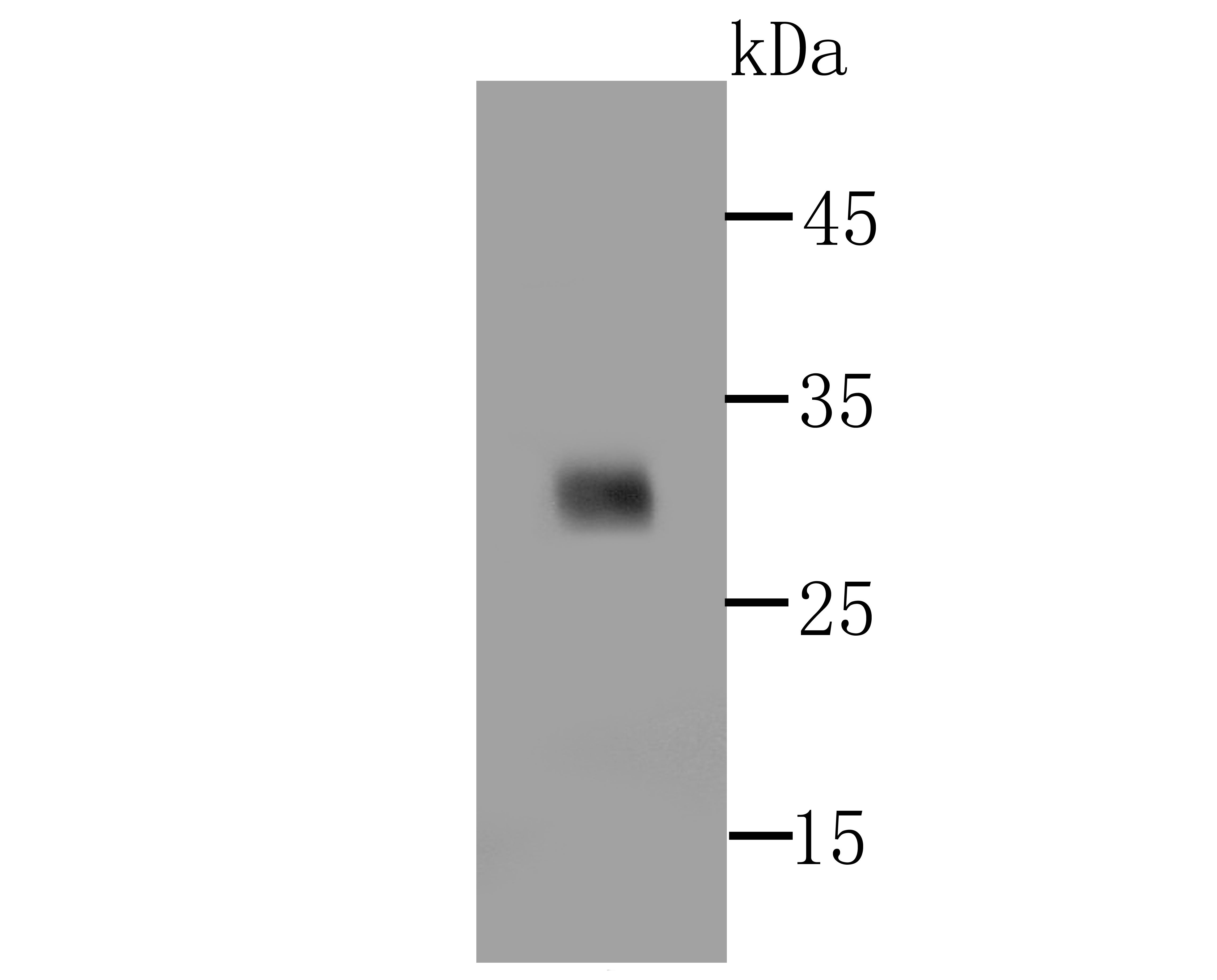
Fig1: Western blot analysis of over-expressed PD1(whole extracellular domain) on 293T cell lysate using anti-PD1 antibody at 1/500 dilution.Application
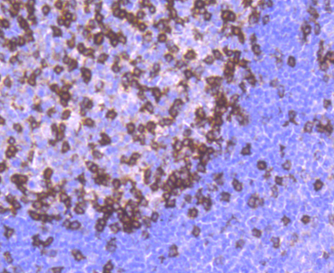
Fig2: Immunohistochemical analysis of paraffin-embedded human tonsil tissue using anti-PD1 antibody. Counter stained with hematoxylin.| Product Name | PD1 [SJ01-91] |
|---|---|
| Antibody Type | Primary Antibodies |
| Antigen Alias | CD279 antibody CD279 antigen antibody hPD 1 antibody hPD l antibody hPD-1 antibody hSLE1 antibody PD 1 antibody PD-1 antibody PD1 antibody PDCD 1 antibody PDCD1 antibody PDCD1_HUMAN antibody Programmed cell death 1 antibody Programmed cell death 1 protein antibody Programmed cell death protein 1 antibody Protein PD 1 antibody Protein PD-1 antibody SLEB2 antibody Systemic lupus erythematosus susceptibility 2 antibody |
| Product description | Pdcd-1 (Programmed Cell Death-1 protein), also designated CD279, is a type I transmembrane receptor and a member of the immunoglobin gene superfamily. Pdcd-1 contains an immunoreceptor tyrosine-based inhibitory motif (ITIM) within the cytoplasmic domain, which is conserved between the mouse and human homologs. Expression of Pdcd-1 is detected in mouse thymus, and it is induced in stimulated B and T cell lines, where it may play a role in the negative regulation of various immune responses. Receptors such as Pdcd-1 function by recruiting tyrosine phosphatases, including SHP-1 and SHIP, which are responsible for altering various B cell responses. Additionally, in activated lymphocytes, Pdcd-1 mediates the activation of the classical type of programmed cell death. |
| Immunogen | recombinant protein |
| Clonality | Monoclonal |
|---|---|
| Isotype | IgG |
| Host Species | Recombinant rabbit |
| Tested Applications | WBIHC |
| WB:1:500-1:1,000 IHC:1:50-1:200 |
|
| Species Reactivity | Human |
| Concentration | 1mg/ml |
| Alternative Names | CD279 antibody CD279 antigen antibody hPD 1 antibody hPD l antibody hPD-1 antibody hSLE1 antibody PD 1 antibody PD-1 antibody PD1 antibody PDCD 1 antibody PDCD1 antibody PDCD1_HUMAN antibody Programmed cell death 1 antibody Programmed cell death 1 protein antibody Programmed cell death protein 1 antibody Protein PD 1 antibody Protein PD-1 antibody SLEB2 antibody Systemic lupus erythematosus susceptibility 2 antibody |
|---|---|
| Molecular Weight(MW) | 32 kDa |
| Cellular Localization | Membrane. |
| SwissProt ID | Q15116 |
|---|
Application
Fig1: Western blot analysis of over-expressed PD1(whole extracellular domain) on 293T cell lysate using anti-PD1 antibody at 1/500 dilution.
Application
Fig2: Immunohistochemical analysis of paraffin-embedded human tonsil tissue using anti-PD1 antibody. Counter stained with hematoxylin.| Positive Control | Human tonsil tissue. |
|---|---|
| Application Notes | WB:1:500-1:1,000 IHC:1:50-1:200 |
| Form | Liquid |
|---|---|
| Storage Instructions | Store at +4℃ after thawing. Aliquot store at -20℃ or -80℃. Avoid repeated freeze / thaw cycles. |
| Storage Buffer | 1*TBS (pH7.4), 1%BSA, 40%Glycerol. Preservative: 0.05% Sodium Azide. |


