

WB
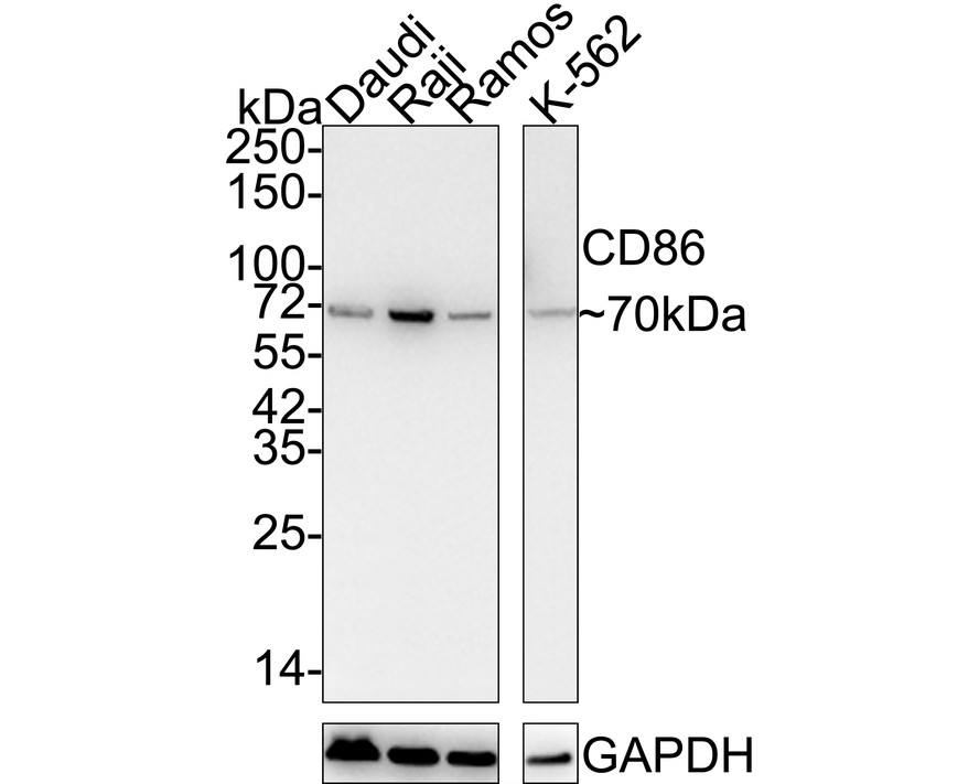
Western blot analysis of CD86 on different lysates with Rabbit anti-CD86 antibody at 1/5,000 dilution. Lane 1: Daudi cell lysate, Lane 2: Raji cell lysate, Lane 3: Ramos cell lysate, Lane 4: K-562 cell lysate Lysates/proteins at 15 µg/Lane. Exposure time: 2 minutes 37 seconds; 4-20% SDS-PAGE gel. Proteins were transferred to a PVDF membrane and blocked with 5% NFDM/TBST for 1 hour at room temperature. The primary antibody at 1/5,000 dilution was used in 5% NFDM/TBST at room temperature for 2 hours. Goat Anti-Rabbit IgG - HRP Secondary Antibody at 1/100,000 dilution was used for 1 hour at room temperature.WB
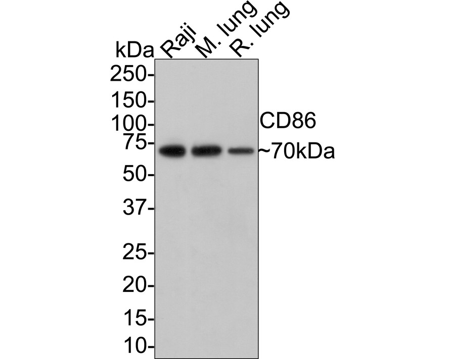
Western blot analysis of CD86 on different lysates with Rabbit anti-CD86 antibody at 1/5,000 dilution. Lane 1: Raji cell lysate (10 µg/Lane), Lane 2: Mouse lung tissue lysate (20 µg/Lane), Lane 3: Rat lung tissue lysate (20 µg/Lane), Exposure time: 1 minute; 4-20% SDS-PAGE gel. Proteins were transferred to a PVDF membrane and blocked with 5% NFDM/TBST for 1 hour at room temperature. The primary antibody at 1/5,000 dilution was used in 5% NFDM/TBST at room temperature for 2 hours. Goat Anti-Rabbit IgG - HRP Secondary Antibody at 1/100,000 dilution was used for 1 hour at room temperature.ICC/IF
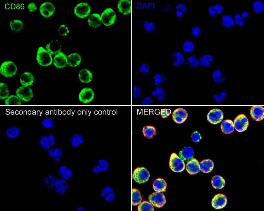
Immunocytochemistry analysis of Raji cells labeling CD86 with Rabbit anti-CD86 antibody at 1/500 dilution. Cells were fixed in 80% precooled methanol for 5 minutes at room temperature, then blocked with 1% BSA in 10% negative goat serum for 1 hour at room temperature. Cells were then incubated with Rabbit anti-CD86 antibody at 1/500 dilution in 1% BSA in PBST overnight at 4 ℃. Goat Anti-Rabbit IgG H&L (488) was used as the secondary antibody at 1/1,000 dilution. PBS instead of the primary antibody was used as the secondary antibody only control. Nuclear DNA was labelled in blue with DAPI. Beta tubulin (red) was stained at 1/100 dilution overnight at +4℃. Goat Anti-Mouse IgG H&L (594) was used as the secondary antibody at 1/1,000 dilution.ICC/IF
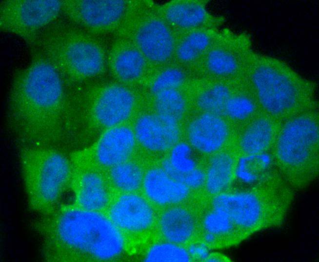
ICC staining CD86 in JAR cells (green). The nuclear counter stain is DAPI (blue). Cells were fixed in paraformaldehyde, permeabilised with 0.25% Triton X100/PBS.IHC
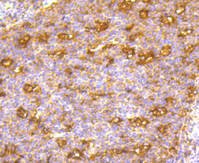
Immunohistochemical analysis of paraffin-embedded human tonsil tissue using anti-CD86 antibody. Counter stained with hematoxylin.FC
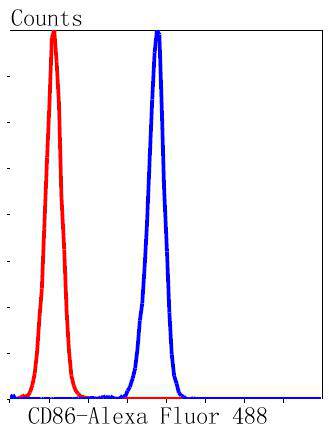
Flow cytometric analysis of K562 cells with CD86 antibody at 1/50 dilution (blue) compared with an unlabelled control (cells without incubation with primary antibody; red). Alexa Fluor 488-conjugated goat anti rabbit IgG was used as the secondary antibody.WB
Illustrative pictures and western-blot quantitative data of (E) IBA1, (F) CD86, (G) CD206.WB
Schematic photographs and statistical analysis of (F) IBA1, (G) CD86 and (H) CD206.WB
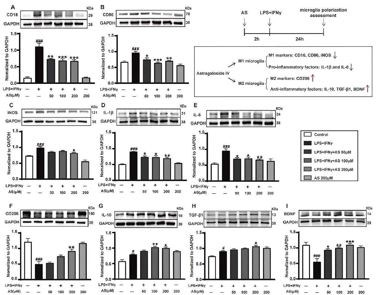
Illustrative photographs and quantitative analysis of (A–E) M1 markers (CD16, CD86, iNOS, IL-1β and IL-6), (F–I) M2 markers (CD206, IL-10, TGF-β1 and BDNF).WB
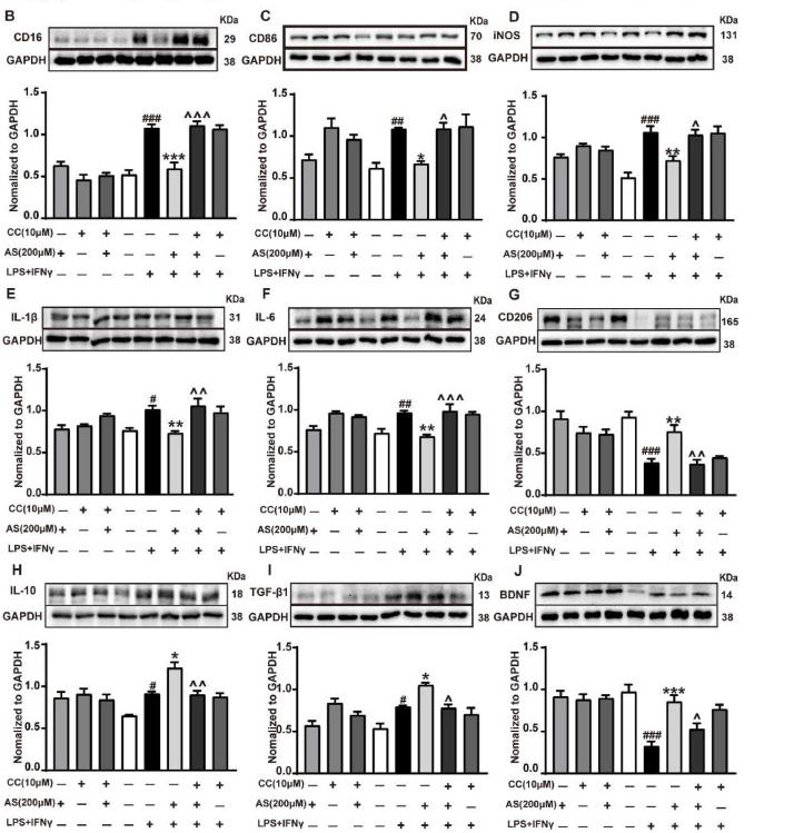
Typical immunoblots and statistical analysis of M1 markers (CD16, CD86, iNOS, IL-1β and IL-6) (B–F) and M2 markers (CD206, IL-10, TGF-β1 and BDNF) (G–J).| Product Name | CD86 Recombinant Rabbit Monoclonal Antibody |
|---|---|
| Antibody Type | Primary Antibodies |
| Product description | T cell proliferation and lymphokine production are triggered by occupation of the TCR by antigen, followed by a costimulatory signal that is delivered by a ligand expressed on antigen presenting cells. The B7-related cell surface proteins B7-1 (CD80) and B7-2 (CD86) expressed on antigen presenting cells bind the homologous T cell receptors CD28 and CTLA-4 (cytotoxic T lymphocyte-associated protein-4) and trigger costimulatory signals for optimal T cell activation. CTLA-4 shares 31% overall amino acid identity with CD28, and it has been proposed that CD28 and CTLA-4 are functionally redundant. SLAM is a novel receptor on T cells that, when engaged, potentiates T cell expansion in a CD28-independent manner. B7, also designated BB1, is another ligand or counterreceptor for CD28 and CTLA-4 that is expressed on the antigen-presenting cell. |
| Immunogen | Synthetic peptide within Human CD86 aa 1-50 / 329. |
| Clonality | Monoclonal |
|---|---|
| Isotype | IgG |
| Host Species | Recombinant rabbit |
| Tested Applications | FCICC/IFIHCWB |
| WB:1:1,000-1:2,000 ICC/IF:1:50-1:200 IHC:1:50-1:200 FC:1:50-1:100 |
|
| Species Reactivity | HumanMouseRat |
| Concentration | 1mg/ml |
| Purification | Protein A |
| Alternative Names | Activation B7-2 antigen 3 antibody
Activation B7-2 antigen antibody
B-lymphocyte activation antigen B7-2 2 antibody
B-lymphocyte activation antigen B7-2 antibody
B7 2 antibody
B7 antibody
B7-2 antibody
B7.2 antibody
B70 antibody
B72 antigen antibody
BU63 antibody
CD28 antigen ligand 2 2 antibody
CD28 antigen ligand 2 antibody
Cd28l2 antibody
CD28LG2 antibody
CD86 antibody
CD86 antigen (CD28 antigen ligand 2 B7-2 antigen) 1 2 antibody CD86 antigen (CD28 antigen ligand 2 B7-2 antigen) antibody CD86 antigen antibody CD86 molecule antibody CD86_HUMAN antibody CLS1 antibody CTLA-4 counter-receptor B7.2 2 3 antibody CTLA-4 counter-receptor B7.2 antibody Early T-cell costimulatory molecule 1 antibody ETC-1 antibody FUN 1 antibody FUN-1 antibody LAB72 antibody Ly-58 antibody Ly58 antibody MB7 antibody MB7-2 antibody Membrane glycoprotein antibody MGC34413 antibody T-lymphocyte activation antigen CD86 antibody TS/A-2 antibody |
|---|---|
| Molecular Weight(MW) | 70 kDa |
| Cellular Localization | Cell membrane. |
WB
Western blot analysis of CD86 on different lysates with Rabbit anti-CD86 antibody at 1/5,000 dilution. Lane 1: Daudi cell lysate, Lane 2: Raji cell lysate, Lane 3: Ramos cell lysate, Lane 4: K-562 cell lysate Lysates/proteins at 15 µg/Lane. Exposure time: 2 minutes 37 seconds; 4-20% SDS-PAGE gel. Proteins were transferred to a PVDF membrane and blocked with 5% NFDM/TBST for 1 hour at room temperature. The primary antibody at 1/5,000 dilution was used in 5% NFDM/TBST at room temperature for 2 hours. Goat Anti-Rabbit IgG - HRP Secondary Antibody at 1/100,000 dilution was used for 1 hour at room temperature.
WB
Western blot analysis of CD86 on different lysates with Rabbit anti-CD86 antibody at 1/5,000 dilution. Lane 1: Raji cell lysate (10 µg/Lane), Lane 2: Mouse lung tissue lysate (20 µg/Lane), Lane 3: Rat lung tissue lysate (20 µg/Lane), Exposure time: 1 minute; 4-20% SDS-PAGE gel. Proteins were transferred to a PVDF membrane and blocked with 5% NFDM/TBST for 1 hour at room temperature. The primary antibody at 1/5,000 dilution was used in 5% NFDM/TBST at room temperature for 2 hours. Goat Anti-Rabbit IgG - HRP Secondary Antibody at 1/100,000 dilution was used for 1 hour at room temperature.
ICC/IF
Immunocytochemistry analysis of Raji cells labeling CD86 with Rabbit anti-CD86 antibody at 1/500 dilution. Cells were fixed in 80% precooled methanol for 5 minutes at room temperature, then blocked with 1% BSA in 10% negative goat serum for 1 hour at room temperature. Cells were then incubated with Rabbit anti-CD86 antibody at 1/500 dilution in 1% BSA in PBST overnight at 4 ℃. Goat Anti-Rabbit IgG H&L (488) was used as the secondary antibody at 1/1,000 dilution. PBS instead of the primary antibody was used as the secondary antibody only control. Nuclear DNA was labelled in blue with DAPI. Beta tubulin (red) was stained at 1/100 dilution overnight at +4℃. Goat Anti-Mouse IgG H&L (594) was used as the secondary antibody at 1/1,000 dilution.
ICC/IF
ICC staining CD86 in JAR cells (green). The nuclear counter stain is DAPI (blue). Cells were fixed in paraformaldehyde, permeabilised with 0.25% Triton X100/PBS.
IHC
Immunohistochemical analysis of paraffin-embedded human tonsil tissue using anti-CD86 antibody. Counter stained with hematoxylin.
FC
Flow cytometric analysis of K562 cells with CD86 antibody at 1/50 dilution (blue) compared with an unlabelled control (cells without incubation with primary antibody; red). Alexa Fluor 488-conjugated goat anti rabbit IgG was used as the secondary antibody.
WB
Illustrative pictures and western-blot quantitative data of (E) IBA1, (F) CD86, (G) CD206.
WB
Schematic photographs and statistical analysis of (F) IBA1, (G) CD86 and (H) CD206.
WB
Illustrative photographs and quantitative analysis of (A–E) M1 markers (CD16, CD86, iNOS, IL-1β and IL-6), (F–I) M2 markers (CD206, IL-10, TGF-β1 and BDNF).
WB
Typical immunoblots and statistical analysis of M1 markers (CD16, CD86, iNOS, IL-1β and IL-6) (B–F) and M2 markers (CD206, IL-10, TGF-β1 and BDNF) (G–J).| Positive Control | HUVEC, JAR, Hela, Raji, Jurkat, human tonsil tissue. |
|---|---|
| Application Notes | WB:1:1,000-1:2,000 ICC/IF:1:50-1:200 IHC:1:50-1:200 FC:1:50-1:100 |
| Form | Liquid |
|---|---|
| Storage Instructions | Store at +4℃ after thawing. Aliquot store at -20℃ or -80℃. Avoid repeated freeze / thaw cycles. |
| Storage Buffer | 1*TBS (pH7.4), 1%BSA, 40%Glycerol. Preservative: 0.05% Sodium Azide. |


