Application
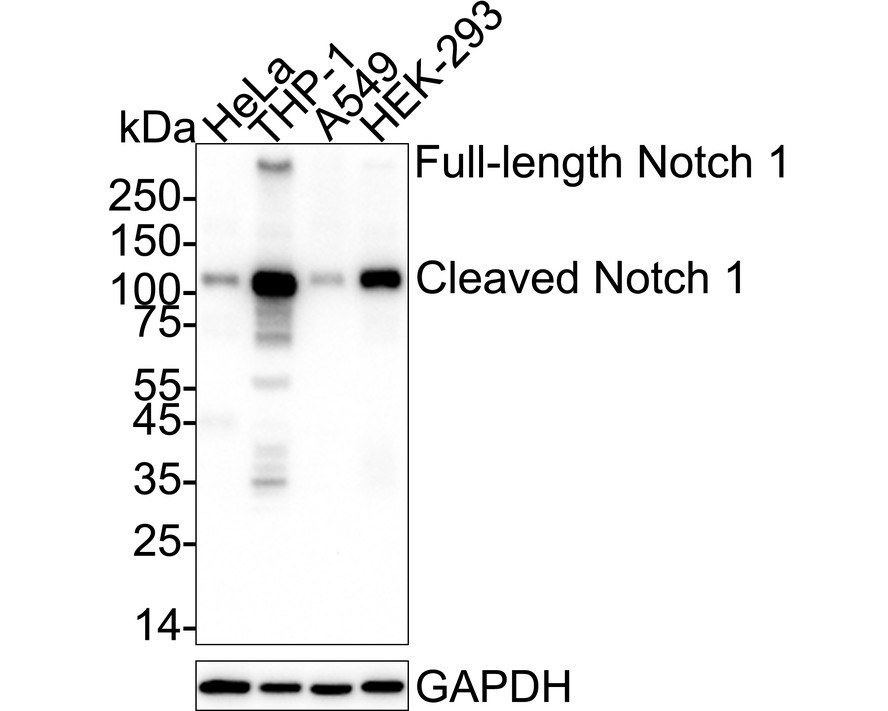
Fig1: Western blot analysis of Notch 1 on different lysates with Rabbit anti-Notch 1 antibody at 1/1,000 dilution. Lane 1: HeLa cell lysate, Lane 2: THP-1 cell lysate, Lane 3: A549 cell lysate, Lane 4: HEK-293 cell lysate, Lysates/proteins at 30 µg/Lane. Exposure time: 59 seconds; 4-20% SDS-PAGE gel. Proteins were transferred to a PVDF membrane and blocked with 5% NFDM/TBST for 1 hour at room temperature. The primary antibody at 1/1,000 dilution was used in 5% NFDM/TBST at 4℃ overnight. Goat Anti-Rabbit IgG - HRP Secondary Antibody at 1/50,000 dilution was used for 1 hour at room temperature.Application
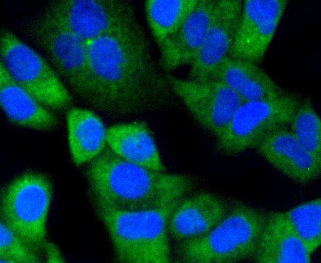
Fig2: ICC staining Notch 1 in Hela cells (green). The nuclear counter stain is DAPI (blue). Cells were fixed in paraformaldehyde, permeabilised with 0.25% Triton X100/PBS.Application
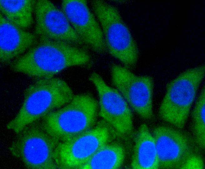
Fig3: ICC staining Notch 1 in HepG2 cells (green). The nuclear counter stain is DAPI (blue). Cells were fixed in paraformaldehyde, permeabilised with 0.25% Triton X100/PBS.Application
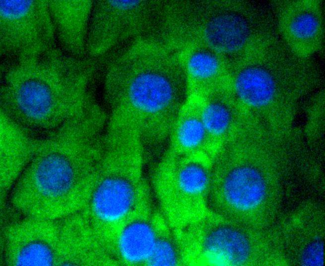
Fig4: ICC staining Notch 1 in NIH/3T3 cells (green). The nuclear counter stain is DAPI (blue). Cells were fixed in paraformaldehyde, permeabilised with 0.25% Triton X100/PBS.Application
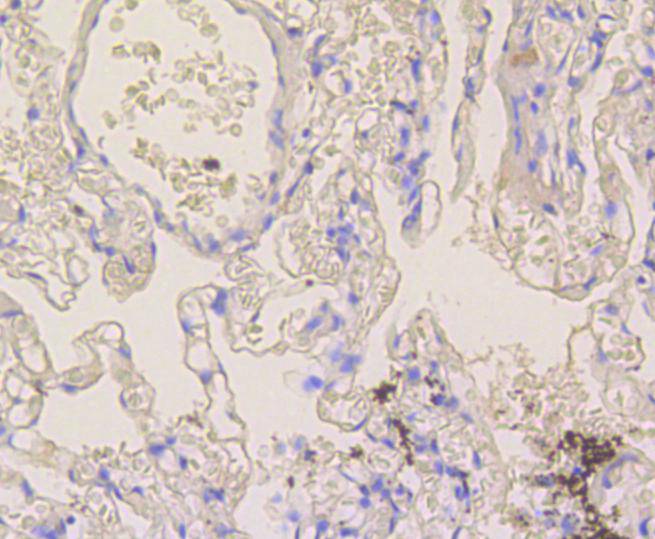
Fig5: Immunohistochemical analysis of paraffin-embedded human lung tissue using anti-Notch 1 antibody. Counter stained with hematoxylin.Application
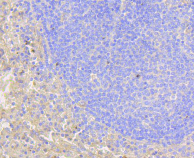
Fig6: Immunohistochemical analysis of paraffin-embedded human spleen tissue using anti-Notch 1 antibody. Counter stained with hematoxylin.Application
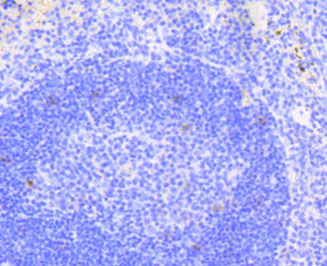
Fig7: Immunohistochemical analysis of paraffin-embedded mouse spleen tissue using anti-Notch 1 antibody. Counter stained with hematoxylin.Application
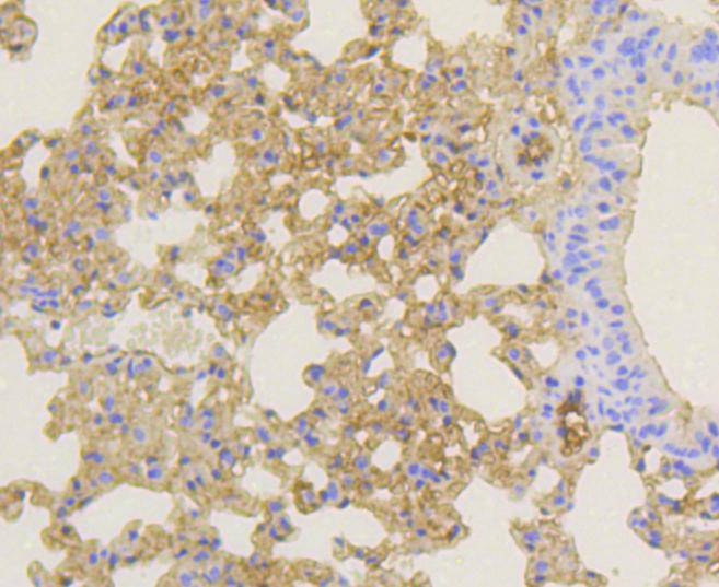
Fig8: Immunohistochemical analysis of paraffin-embedded mouse lung tissue using anti-Notch 1 antibody. Counter stained with hematoxylin.Application
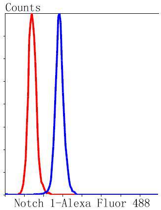
Fig9: Flow cytometric analysis of Hela cells with Notch 1 antibody at 1/50 dilution (blue) compared with an unlabelled control (cells without incubation with primary antibody; red). Alexa Fluor 488-conjugated goat anti rabbit IgG was used as the secondary antibody.Application
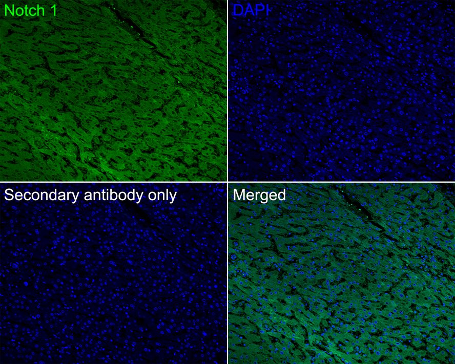
Fig10:Immunofluorescence analysis of paraffin-embedded human liver cancer tissue labeling Notch 1 with Rabbit anti-Notch 1 antibody at 1/50 dilution. The section was pre-treated using heat mediated antigen retrieval with sodium citrate buffer (pH 6.0) (high pressure) for 2 minutes. The tissues were blocked in 10% negative goat serum for 1 hour at room temperature, washed with PBS, and then probed with the primary antibody (green) at 1/50 dilution overnight at 4 ℃, washed with PBS. Goat Anti-Rabbit IgG H&L (488) was used as the secondary antibody at 1/1,000 dilution. Nuclei were counterstained with DAPI (blue).| Product Name | Notch 1 Recombinant Rabbit Monoclonal Antibody |
|---|---|
| Antibody Type | Primary Antibodies |
| Product description | The LIN-12/notch family of transmembrane receptors is believed to play a central role in development by regulating cell fate decisions. To date, four notch homologs have been identified in mammals and have been designated Notch 1, Notch 2, Notch 3 and Notch 4. The notch genes are expressed in a variety of tissues in both the embryonic and adult organism, suggesting that the genes are involved in multiple signaling pathways. The notch proteins have been found to be overexpressed or rearranged in human tumors. Ligands for notch include Jagged1, Jagged2 and Delta. Jagged can activate notch and prevent myoblast differentiation by inhibiting the expression of muscle regulatory and structural genes. Jagged2 is thought to be involved in the development of various tissues whose development is dependent upon epithelial-mesenchymal interactions. Normal Delta expression is restricted to the adrenal gland and placenta. Delta expression has also been found in neuroendocrine tumors such as neuroblastomas and pheochromocytomas. |
| Immunogen | recombinant protein |
| Clonality | Monoclonal |
|---|---|
| Isotype | IgG |
| Host Species | Recombinant rabbit |
| Tested Applications | FCICC/IFIF-PIHCIPWB |
| WB:1:1,000-1:2,000 ICC:1:50-1:200 IHC:1:50-1:200 FC:1:50-1:100 IF-P:1:50-1:200 |
|
| Species Reactivity | HumanMouseRat |
| Concentration | 1mg/ml |
| Purification | Protein A |
| Alternative Names | 9930111A19Rik antibody
AOS5 antibody
AOVD1 antibody
hN1 antibody
Lin-12 antibody
LIN12 antibody
Mis6 antibody
Motch A antibody
mT14 antibody
Neurogenic locus notch homolog protein 1 antibody
Neurogenic locus notch protein homolog antibody
NICD antibody
NOTC1_HUMAN antibody
Notch 1 antibody
Notch 1 intracellular domain antibody
NOTCH antibody
Notch gene homolog 1 (Drosophila) antibody
Notch homolog 1 translocation-associated (Drosophila) antibody NOTCH Drosophila homolog of 1 antibody notch1 antibody TAN1 antibody Translocation associated notch homolog antibody Translocation associated notch protein TAN 1 antibody Translocation-associated notch protein TAN-1 antibody xotch antibody |
|---|---|
| Molecular Weight(MW) | 273 kDa(Observed band size: 125/273 kDa) |
| Cellular Localization | Cell membrane, Nucleus(intracellular domain). |
Application
Fig1: Western blot analysis of Notch 1 on different lysates with Rabbit anti-Notch 1 antibody at 1/1,000 dilution. Lane 1: HeLa cell lysate, Lane 2: THP-1 cell lysate, Lane 3: A549 cell lysate, Lane 4: HEK-293 cell lysate, Lysates/proteins at 30 µg/Lane. Exposure time: 59 seconds; 4-20% SDS-PAGE gel. Proteins were transferred to a PVDF membrane and blocked with 5% NFDM/TBST for 1 hour at room temperature. The primary antibody at 1/1,000 dilution was used in 5% NFDM/TBST at 4℃ overnight. Goat Anti-Rabbit IgG - HRP Secondary Antibody at 1/50,000 dilution was used for 1 hour at room temperature.
Application
Fig2: ICC staining Notch 1 in Hela cells (green). The nuclear counter stain is DAPI (blue). Cells were fixed in paraformaldehyde, permeabilised with 0.25% Triton X100/PBS.
Application
Fig3: ICC staining Notch 1 in HepG2 cells (green). The nuclear counter stain is DAPI (blue). Cells were fixed in paraformaldehyde, permeabilised with 0.25% Triton X100/PBS.
Application
Fig4: ICC staining Notch 1 in NIH/3T3 cells (green). The nuclear counter stain is DAPI (blue). Cells were fixed in paraformaldehyde, permeabilised with 0.25% Triton X100/PBS.
Application
Fig5: Immunohistochemical analysis of paraffin-embedded human lung tissue using anti-Notch 1 antibody. Counter stained with hematoxylin.
Application
Fig6: Immunohistochemical analysis of paraffin-embedded human spleen tissue using anti-Notch 1 antibody. Counter stained with hematoxylin.
Application
Fig7: Immunohistochemical analysis of paraffin-embedded mouse spleen tissue using anti-Notch 1 antibody. Counter stained with hematoxylin.
Application
Fig8: Immunohistochemical analysis of paraffin-embedded mouse lung tissue using anti-Notch 1 antibody. Counter stained with hematoxylin.
Application
Fig9: Flow cytometric analysis of Hela cells with Notch 1 antibody at 1/50 dilution (blue) compared with an unlabelled control (cells without incubation with primary antibody; red). Alexa Fluor 488-conjugated goat anti rabbit IgG was used as the secondary antibody.
Application
Fig10:Immunofluorescence analysis of paraffin-embedded human liver cancer tissue labeling Notch 1 with Rabbit anti-Notch 1 antibody at 1/50 dilution. The section was pre-treated using heat mediated antigen retrieval with sodium citrate buffer (pH 6.0) (high pressure) for 2 minutes. The tissues were blocked in 10% negative goat serum for 1 hour at room temperature, washed with PBS, and then probed with the primary antibody (green) at 1/50 dilution overnight at 4 ℃, washed with PBS. Goat Anti-Rabbit IgG H&L (488) was used as the secondary antibody at 1/1,000 dilution. Nuclei were counterstained with DAPI (blue).| Positive Control | NIH/3T3, Hela, HepG2, mouse lung tissue, human lung tissue, mouse spleen tissue, human spleen tissue. |
|---|---|
| Application Notes | WB:1:1,000-1:2,000 ICC:1:50-1:200 IHC:1:50-1:200 FC:1:50-1:100 IF-P:1:50-1:200 |
| Form | Liquid |
|---|---|
| Storage Instructions | Store at +4℃ after thawing. Aliquot store at -20℃ or -80℃. Avoid repeated freeze / thaw cycles. |
| Storage Buffer | 1*TBS (pH7.4), 1%BSA, 40%Glycerol. Preservative: 0.05% Sodium Azide. |


