WB
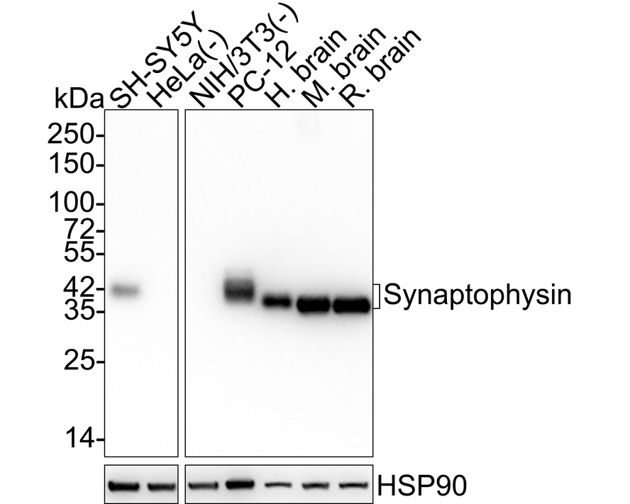
Western blot analysis of Synaptophysin on different lysates with Rabbit anti-Synaptophysin antibody at 1/5,000 dilution. Lane 1: SH-SY5Y cell lysate, Lane 2: HeLa cell lysate (negative), Lane 3: NIH/3T3 cell lysate (negative), Lane 4: PC-12 cell lysate, Lane 5: Human brain tissue lysate, Lane 6: Mouse brain tissue lysate, Lane 7: Rat brain tissue lysate, Lysates/proteins at 20 µg/Lane. Exposure time: 43 seconds; 4-20% SDS-PAGE gel. Proteins were transferred to a PVDF membrane and blocked with 5% NFDM/TBST for 1 hour at room temperature. The primary antibody at 1/5,000 dilution was used in 5% NFDM/TBST at 4℃ overnight. Goat Anti-Rabbit IgG - HRP Secondary Antibody at 1:50,000 dilution was used for 1 hour at room temperature.IHC
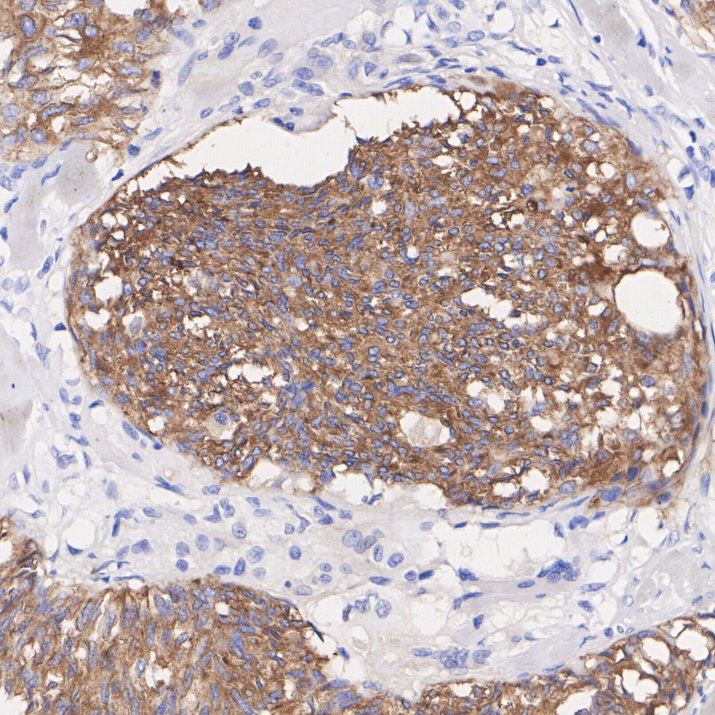
Immunohistochemical analysis of paraffin-embedded human medullary thyroid carcinoma tissue with Rabbit anti-Synaptophysin antibody at 1/2,000 dilution. The section was pre-treated using heat mediated antigen retrieval with Tris-EDTA buffer (pH 9.0) for 20 minutes. The tissues were blocked in 1% BSA for 20 minutes at room temperature, washed with ddH2O and PBS, and then probed with the primary antibody at 1/2,000 dilution for 1 hour at room temperature. The detection was performed using an HRP conjugated compact polymer system. DAB was used as the chromogen. Tissues were counterstained with hematoxylin and mounted with DPX.IHC
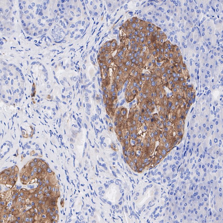
Immunohistochemical analysis of paraffin-embedded human pancreas tissue with Rabbit anti-Synaptophysin antibody at 1/2,000 dilution. The section was pre-treated using heat mediated antigen retrieval with Tris-EDTA buffer (pH 9.0) for 20 minutes. The tissues were blocked in 1% BSA for 20 minutes at room temperature, washed with ddH2O and PBS, and then probed with the primary antibody at 1/2,000 dilution for 1 hour at room temperature. The detection was performed using an HRP conjugated compact polymer system. DAB was used as the chromogen. Tissues were counterstained with hematoxylin and mounted with DPX.IHC
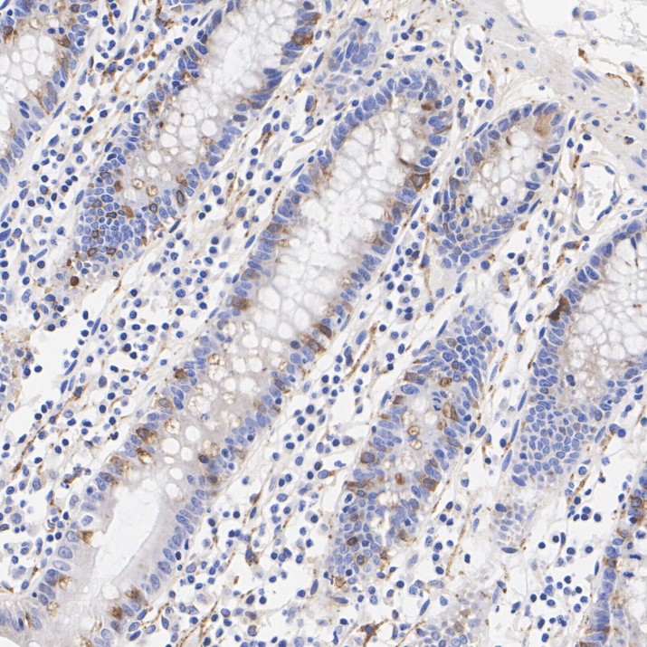
Immunohistochemical analysis of paraffin-embedded human colon tissue with Rabbit anti-Synaptophysin antibody at 1/2,000 dilution. The section was pre-treated using heat mediated antigen retrieval with Tris-EDTA buffer (pH 9.0) for 20 minutes. The tissues were blocked in 1% BSA for 20 minutes at room temperature, washed with ddH2O and PBS, and then probed with the primary antibody at 1/2,000 dilution for 1 hour at room temperature. The detection was performed using an HRP conjugated compact polymer system. DAB was used as the chromogen. Tissues were counterstained with hematoxylin and mounted with DPX.IHC
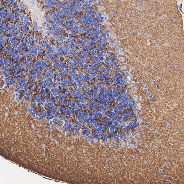
Immunohistochemical analysis of paraffin-embedded mouse cerebellum tissue with Rabbit anti-Synaptophysin antibody at 1/2,000 dilution. The section was pre-treated using heat mediated antigen retrieval with Tris-EDTA buffer (pH 9.0) for 20 minutes. The tissues were blocked in 1% BSA for 20 minutes at room temperature, washed with ddH2O and PBS, and then probed with the primary antibody at 1/2,000 dilution for 1 hour at room temperature. The detection was performed using an HRP conjugated compact polymer system. DAB was used as the chromogen. Tissues were counterstained with hematoxylin and mounted with DPX.IHC
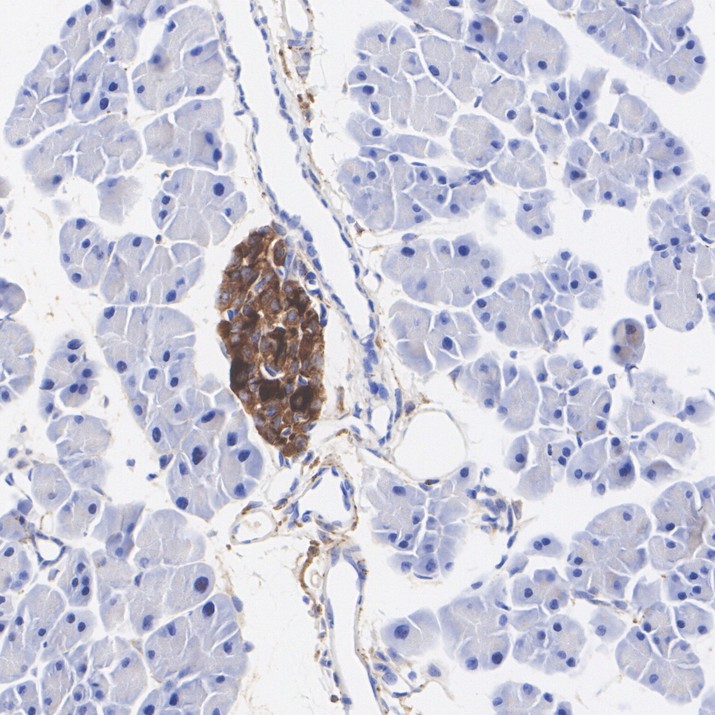
Immunohistochemical analysis of paraffin-embedded mouse pancreas tissue with Rabbit anti-Synaptophysin antibody at 1/2,000 dilution. The section was pre-treated using heat mediated antigen retrieval with Tris-EDTA buffer (pH 9.0) for 20 minutes. The tissues were blocked in 1% BSA for 20 minutes at room temperature, washed with ddH2O and PBS, and then probed with the primary antibody at 1/2,000 dilution for 1 hour at room temperature. The detection was performed using an HRP conjugated compact polymer system. DAB was used as the chromogen. Tissues were counterstained with hematoxylin and mounted with DPX.IHC
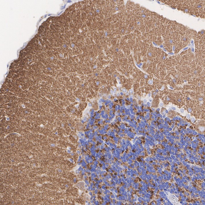
Immunohistochemical analysis of paraffin-embedded rat cerebellum tissue with Rabbit anti-Synaptophysin antibody at 1/2,000 dilution. The section was pre-treated using heat mediated antigen retrieval with Tris-EDTA buffer (pH 9.0) for 20 minutes. The tissues were blocked in 1% BSA for 20 minutes at room temperature, washed with ddH2O and PBS, and then probed with the primary antibody at 1/2,000 dilution for 1 hour at room temperature. The detection was performed using an HRP conjugated compact polymer system. DAB was used as the chromogen. Tissues were counterstained with hematoxylin and mounted with DPX.IF-P
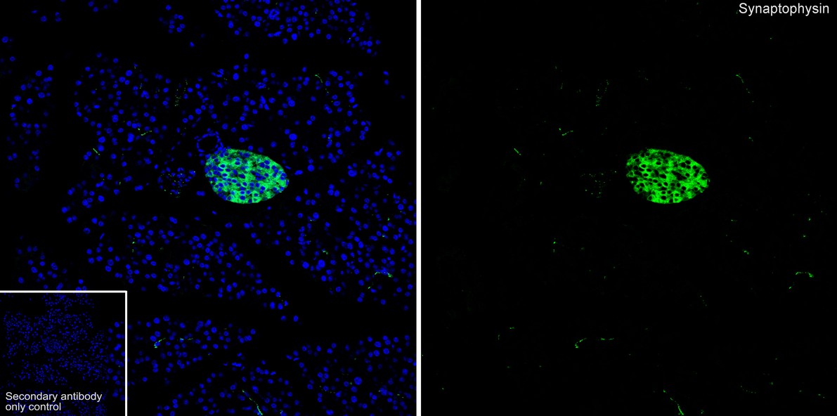
Species: Mouse, Site: Pancreas, Sample: Paraffin-embedded section, Antibody concentration: 1:500.IF-P
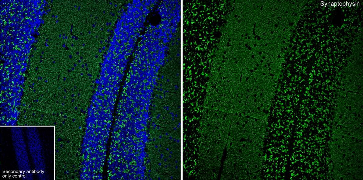
Species: Mouse, Site: Cerebellum, Sample: Paraffin-embedded section, Antibody concentration: 1:500.IF-F
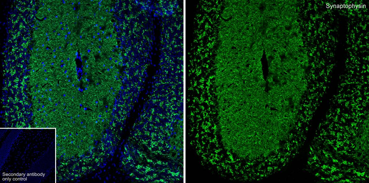
Species: Mouse, Site: Cerebellum, Sample: Frozen section, Antibody concentration: 1:1,000, Antigen retrieval: Not required.| Product Name | Synaptophysin Recombinant Rabbit Monoclonal Antibody |
|---|---|
| Antibody Type | Primary Antibodies |
| Product description | Synaptic vesicles participate in a cycle of fusion with the plasma membrane and reformation by endocytosis. Synaptic vesicle protein synaptophysin (SYP) is targeted to early endosomes in transfected fibroblasts and in neuroendocrine cells. SYP is an N-glycosylated intergral membrane protein found in neurons and endocrine cells that associates into hexamers to form a large conductance channel. SYP contains four transmembrane domains and may function as a gap juction-like channel. Membrane cholesterol specfically interacts with SYP to play a role in vesicle formation. Synaptobrevin (VAMP) also binds to SYP and the resultant complex is upregulated during neuronal development, but is absent in exocytosis fusion complex. Thus, the synaptophysin-synaptobrevin complex is not essential for exocytosis, but rather provides a pool of synaptobrevin for exocytosis. In addition, the tail domain of brain Myosin V also forms a stable complex with synaptobrevin II and SYP, and this complex is disassembled upon the depolarization-induced entry of Ca2+ into intact nerve endings. |
| Immunogen | Synthetic peptide within Human Synaptophysin aa 224 – 313 (Cytoplasmic). |
| Clonality | Monoclonal |
|---|---|
| Isotype | IgG |
| Host Species | Recombinant rabbit |
| Tested Applications | IF-FIF-PIHCWB |
| WB:1:5000-1:10000 IHC:1:2000 IF-P:1:500 IF-F:1:1000 |
|
| Species Reactivity | HumanMouseRat |
| Concentration | 1mg/ml |
| Purification | Protein A |
| Alternative Names | Major synaptic vesicle protein p38 antibody MRX96 antibody MRXSYP antibody Syn p38 antibody Synaptophysin antibody Syp antibody SYPH antibody SYPH_HUMAN antibody SypI antibody |
|---|---|
| Molecular Weight(MW) | 34kDa(Observed band size: 34/40kDa) |
| Cellular Localization | Cytoplasmic, Cell junction, synapse, synaptosome. |
WB
Western blot analysis of Synaptophysin on different lysates with Rabbit anti-Synaptophysin antibody at 1/5,000 dilution. Lane 1: SH-SY5Y cell lysate, Lane 2: HeLa cell lysate (negative), Lane 3: NIH/3T3 cell lysate (negative), Lane 4: PC-12 cell lysate, Lane 5: Human brain tissue lysate, Lane 6: Mouse brain tissue lysate, Lane 7: Rat brain tissue lysate, Lysates/proteins at 20 µg/Lane. Exposure time: 43 seconds; 4-20% SDS-PAGE gel. Proteins were transferred to a PVDF membrane and blocked with 5% NFDM/TBST for 1 hour at room temperature. The primary antibody at 1/5,000 dilution was used in 5% NFDM/TBST at 4℃ overnight. Goat Anti-Rabbit IgG - HRP Secondary Antibody at 1:50,000 dilution was used for 1 hour at room temperature.
IHC
Immunohistochemical analysis of paraffin-embedded human medullary thyroid carcinoma tissue with Rabbit anti-Synaptophysin antibody at 1/2,000 dilution. The section was pre-treated using heat mediated antigen retrieval with Tris-EDTA buffer (pH 9.0) for 20 minutes. The tissues were blocked in 1% BSA for 20 minutes at room temperature, washed with ddH2O and PBS, and then probed with the primary antibody at 1/2,000 dilution for 1 hour at room temperature. The detection was performed using an HRP conjugated compact polymer system. DAB was used as the chromogen. Tissues were counterstained with hematoxylin and mounted with DPX.
IHC
Immunohistochemical analysis of paraffin-embedded human pancreas tissue with Rabbit anti-Synaptophysin antibody at 1/2,000 dilution. The section was pre-treated using heat mediated antigen retrieval with Tris-EDTA buffer (pH 9.0) for 20 minutes. The tissues were blocked in 1% BSA for 20 minutes at room temperature, washed with ddH2O and PBS, and then probed with the primary antibody at 1/2,000 dilution for 1 hour at room temperature. The detection was performed using an HRP conjugated compact polymer system. DAB was used as the chromogen. Tissues were counterstained with hematoxylin and mounted with DPX.
IHC
Immunohistochemical analysis of paraffin-embedded human colon tissue with Rabbit anti-Synaptophysin antibody at 1/2,000 dilution. The section was pre-treated using heat mediated antigen retrieval with Tris-EDTA buffer (pH 9.0) for 20 minutes. The tissues were blocked in 1% BSA for 20 minutes at room temperature, washed with ddH2O and PBS, and then probed with the primary antibody at 1/2,000 dilution for 1 hour at room temperature. The detection was performed using an HRP conjugated compact polymer system. DAB was used as the chromogen. Tissues were counterstained with hematoxylin and mounted with DPX.
IHC
Immunohistochemical analysis of paraffin-embedded mouse cerebellum tissue with Rabbit anti-Synaptophysin antibody at 1/2,000 dilution. The section was pre-treated using heat mediated antigen retrieval with Tris-EDTA buffer (pH 9.0) for 20 minutes. The tissues were blocked in 1% BSA for 20 minutes at room temperature, washed with ddH2O and PBS, and then probed with the primary antibody at 1/2,000 dilution for 1 hour at room temperature. The detection was performed using an HRP conjugated compact polymer system. DAB was used as the chromogen. Tissues were counterstained with hematoxylin and mounted with DPX.
IHC
Immunohistochemical analysis of paraffin-embedded mouse pancreas tissue with Rabbit anti-Synaptophysin antibody at 1/2,000 dilution. The section was pre-treated using heat mediated antigen retrieval with Tris-EDTA buffer (pH 9.0) for 20 minutes. The tissues were blocked in 1% BSA for 20 minutes at room temperature, washed with ddH2O and PBS, and then probed with the primary antibody at 1/2,000 dilution for 1 hour at room temperature. The detection was performed using an HRP conjugated compact polymer system. DAB was used as the chromogen. Tissues were counterstained with hematoxylin and mounted with DPX.
IHC
Immunohistochemical analysis of paraffin-embedded rat cerebellum tissue with Rabbit anti-Synaptophysin antibody at 1/2,000 dilution. The section was pre-treated using heat mediated antigen retrieval with Tris-EDTA buffer (pH 9.0) for 20 minutes. The tissues were blocked in 1% BSA for 20 minutes at room temperature, washed with ddH2O and PBS, and then probed with the primary antibody at 1/2,000 dilution for 1 hour at room temperature. The detection was performed using an HRP conjugated compact polymer system. DAB was used as the chromogen. Tissues were counterstained with hematoxylin and mounted with DPX.
IF-P
Species: Mouse, Site: Pancreas, Sample: Paraffin-embedded section, Antibody concentration: 1:500.
IF-P
Species: Mouse, Site: Cerebellum, Sample: Paraffin-embedded section, Antibody concentration: 1:500.
IF-F
Species: Mouse, Site: Cerebellum, Sample: Frozen section, Antibody concentration: 1:1,000, Antigen retrieval: Not required.| Application Notes | WB:1:5000-1:10000 IHC:1:2000 IF-P:1:500 IF-F:1:1000 |
|---|
| Form | Liquid |
|---|---|
| Storage Instructions | Store at +4℃ after thawing. Aliquot store at -20℃ or -80℃. Avoid repeated freeze / thaw cycles. |
| Storage Buffer | 1*TBS (pH7.4), 1%BSA, 40%Glycerol. Preservative: 0.05% Sodium Azide. |


