WB
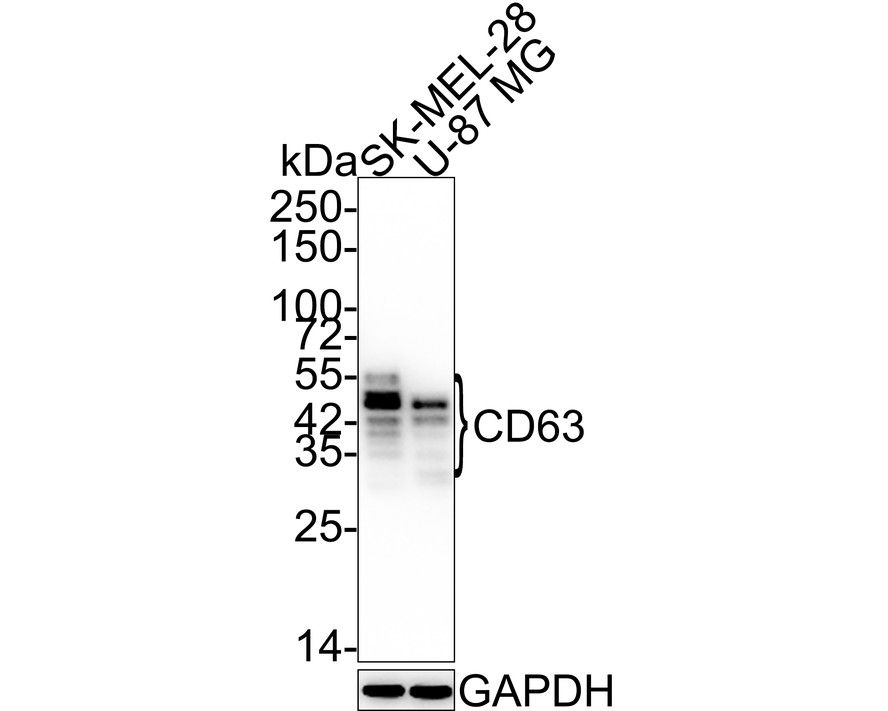
Western blot analysis of CD63 on different lysates with Rabbit anti-CD63 antibody at 1/2,000 dilution. Lane 1: SK-MEL-28 cell lysate, Lane 2: U-87 MG cell lysate, Lysates/proteins at 15 µg/Lane. Exposure time: 43 seconds; 4-20% SDS-PAGE gel. Proteins were transferred to a PVDF membrane and blocked with 5% NFDM/TBST for 1 hour at room temperature. The primary antibody at 1/2,000 dilution was used in 5% NFDM/TBST at 4℃ overnight. Goat Anti-Rabbit IgG - HRP Secondary Antibody at 1:50,000 dilution was used for 1 hour at room temperature.IHC
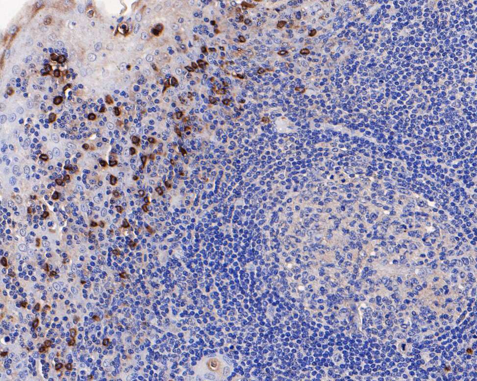
Immunohistochemical analysis of paraffin-embedded human tonsil tissue using anti-CD63 antibody. The section was pre-treated using heat mediated antigen retrieval with Tris-EDTA buffer (pH 8.0-8.4) for 20 minutes.The tissues were blocked in 5% BSA for 30 minutes at room temperature, washed with ddH2O and PBS, and then probed with the primary antibody (1/200) for 30 minutes at room temperature. The detection was performed using an HRP conjugated compact polymer system. DAB was used as the chromogen. Tissues were counterstained with hematoxylin and mounted with DPX.IHC
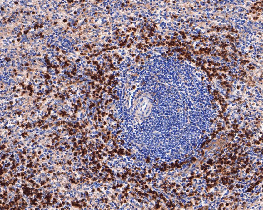
Immunohistochemical analysis of paraffin-embedded human spleen tissue using anti-CD63 antibody. The section was pre-treated using heat mediated antigen retrieval with Tris-EDTA buffer (pH 8.0-8.4) for 20 minutes.The tissues were blocked in 5% BSA for 30 minutes at room temperature, washed with ddH2O and PBS, and then probed with the primary antibody (1/400) for 30 minutes at room temperature. The detection was performed using an HRP conjugated compact polymer system. DAB was used as the chromogen. Tissues were counterstained with hematoxylin and mounted with DPX.WB
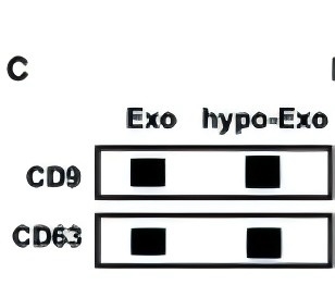
(C) Exosomal surface markers CD9 and CD63 detected by western blotting.| Product Name | CD63 Recombinant Rabbit Monoclonal Antibody |
|---|---|
| Antibody Type | Primary Antibodies |
| Product description | The tetraspanins are integral membrane proteins expressed on cell surface and granular membranes of hematopoietic cells and are components of multi-molecular complexes with specific integrins. The tetraspanin CD63 (also known as LAMP-3, Melanoma-associated antigen ME491, TSPAN30, MLA1 and OMA81H) is a lysosomal membrane glycoprotein that translocates to the plasma membrane after platelet activation. CD63 is expressed on activated platelets, monocytes and macrophages, and is weakly expressed on granulocytes, T cell and B cells. It is located on the basophilic granule membranes and on the plasma membranes of lymphocytes and granulocytes. CD63 is a member of the TM4 superfamily of leukocyte glycoproteins that includes CD9, CD37 and CD53, which contain four transmembrane regions. CD63 may play a role in phagocytic and intracellular lysosome-phagosome fusion events. CD63 deficiency is associated with Hermansky-Pudlak syndrome. |
| Immunogen | Recombinant protein within Human CD63 aa 88-216 / 238. |
| Clonality | Monoclonal |
|---|---|
| Isotype | IgG |
| Host Species | Recombinant rabbit |
| Tested Applications | IHCWB |
| WB:1:2000 IHC:1:100-1:500 |
|
| Species Reactivity | HumanMouse |
| Concentration | 1mg/ml |
| Purification | Protein A |
| Alternative Names | Lysosomal associated membrane protein 3 antibody CD 63 antibody CD63 antibody CD63 antigen (melanoma 1 antigen) antibody CD63 antigen antibody CD63 antigen melanoma 1 antigen antibody CD63 molecule antibody CD63_HUMAN antibody gp55 antibody Granulophysin antibody LAMP 3 antibody LAMP-3 antibody LAMP3 antibody LIMP antibody Lysosomal-associated membrane protein 3 antibody Lysosome associated membrane glycoprotein 3 antibody Mast cell antigen AD1 antibody ME491 antibody Melanoma 1 antigen antibody Melanoma associated antigen ME491 antibody Melanoma associated antigen MLA1 antibody Melanoma-associated antigen ME491 antibody MGC72893 antibody MLA 1 antibody MLA1 antibody NGA antibody Ocular melanoma associated antigen antibody Ocular melanoma-associated antigen antibody OMA81H antibody PTLGP40 antibody Tetraspanin 30 antibody Tetraspanin-30 antibody Tspan 30 antibody Tspan-30 antibody TSPAN30 antibody |
|---|---|
| Molecular Weight(MW) | 26kDa(Observed band size:30-60kDa) |
| Cellular Localization | Cell membrane, Lysosome membrane, Endosome, Melanosome, Secreted. |
| SwissProt ID | P08962 |
|---|
WB
Western blot analysis of CD63 on different lysates with Rabbit anti-CD63 antibody at 1/2,000 dilution. Lane 1: SK-MEL-28 cell lysate, Lane 2: U-87 MG cell lysate, Lysates/proteins at 15 µg/Lane. Exposure time: 43 seconds; 4-20% SDS-PAGE gel. Proteins were transferred to a PVDF membrane and blocked with 5% NFDM/TBST for 1 hour at room temperature. The primary antibody at 1/2,000 dilution was used in 5% NFDM/TBST at 4℃ overnight. Goat Anti-Rabbit IgG - HRP Secondary Antibody at 1:50,000 dilution was used for 1 hour at room temperature.
IHC
Immunohistochemical analysis of paraffin-embedded human tonsil tissue using anti-CD63 antibody. The section was pre-treated using heat mediated antigen retrieval with Tris-EDTA buffer (pH 8.0-8.4) for 20 minutes.The tissues were blocked in 5% BSA for 30 minutes at room temperature, washed with ddH2O and PBS, and then probed with the primary antibody (1/200) for 30 minutes at room temperature. The detection was performed using an HRP conjugated compact polymer system. DAB was used as the chromogen. Tissues were counterstained with hematoxylin and mounted with DPX.
IHC
Immunohistochemical analysis of paraffin-embedded human spleen tissue using anti-CD63 antibody. The section was pre-treated using heat mediated antigen retrieval with Tris-EDTA buffer (pH 8.0-8.4) for 20 minutes.The tissues were blocked in 5% BSA for 30 minutes at room temperature, washed with ddH2O and PBS, and then probed with the primary antibody (1/400) for 30 minutes at room temperature. The detection was performed using an HRP conjugated compact polymer system. DAB was used as the chromogen. Tissues were counterstained with hematoxylin and mounted with DPX.
WB
(C) Exosomal surface markers CD9 and CD63 detected by western blotting.| Application Notes | WB:1:2000 IHC:1:100-1:500 |
|---|
| Form | Liquid |
|---|---|
| Storage Instructions | Store at +4℃ after thawing. Aliquot store at -20℃ or -80℃. Avoid repeated freeze / thaw cycles. |
| Storage Buffer | 1*TBS (pH7.4), 1%BSA, 40%Glycerol. Preservative: 0.05% Sodium Azide. |


