WB
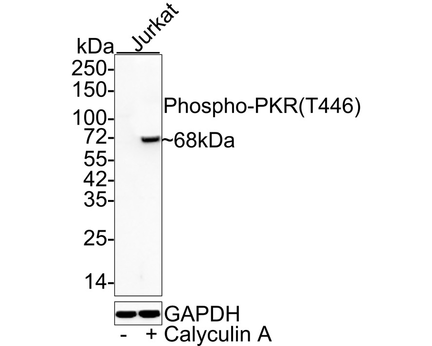
Western blot analysis of Phospho-PKR (T446) on different lysates with Rabbit anti-Phospho-PKR (T446) antibody at 1/5,000 dilution. Lane 1: Jurkat cell lysate, Lane 2: Jurkat treated with 100nM Calyculin A for 30 minutes cell lysate, Lysates/proteins at 20 µg/Lane. Exposure time: 30 seconds; 4-20% SDS-PAGE gel. Proteins were transferred to a PVDF membrane and blocked with 5% NFDM/TBST for 1 hour at room temperature. The primary antibody at 1/5,000 dilution was used in 5% NFDM/TBST at 4℃ overnight. Goat Anti-Rabbit IgG - HRP Secondary Antibody at 1/50,000 dilution was used for 1 hour at room temperature.IHC
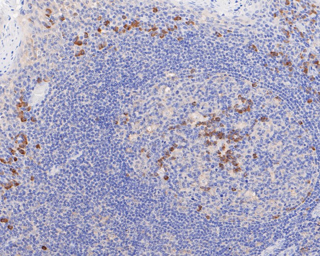
Immunohistochemical analysis of paraffin-embedded human tonsil tissue using anti-Phospho-PKR (T446) antibody. The section was pre-treated using heat mediated antigen retrieval with sodium citrate buffer (pH 6.0) (high pressure) for 2 minutes. The tissues were blocked in 5% BSA for 30 minutes at room temperature, washed with ddH2O and PBS, and then probed with the primary antibody (1/50) for 30 minutes at room temperature. The detection was performed using an HRP conjugated compact polymer system. DAB was used as the chromogen. Tissues were counterstained with hematoxylin and mounted with DPX.IHC
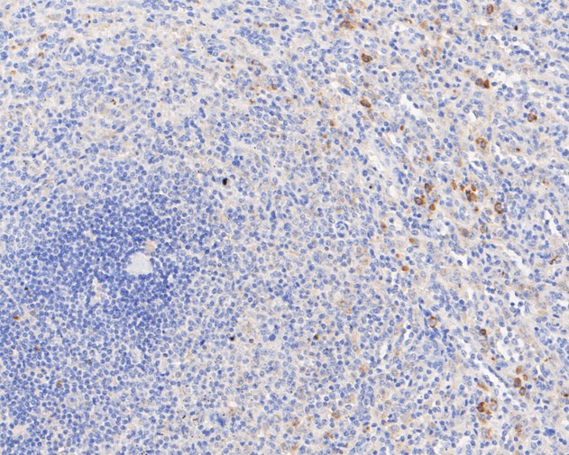
Immunohistochemical analysis of paraffin-embedded human spleen tissue using anti-Phospho-PKR (T446) antibody. The section was pre-treated using heat mediated antigen retrieval with sodium citrate buffer (pH 6.0) (high pressure) for 2 minutes. The tissues were blocked in 5% BSA for 30 minutes at room temperature, washed with ddH2O and PBS, and then probed with the primary antibody (1/50) for 30 minutes at room temperature. The detection was performed using an HRP conjugated compact polymer system. DAB was used as the chromogen. Tissues were counterstained with hematoxylin and mounted with DPX.IHC
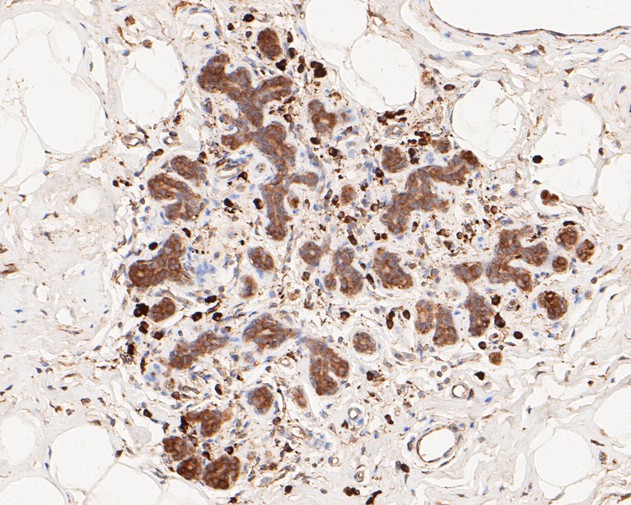
Immunohistochemical analysis of paraffin-embedded human breast tissue using anti-Phospho-PKR (T446) antibody. The section was pre-treated using heat mediated antigen retrieval with sodium citrate buffer (pH 6.0) (high pressure) for 2 minutes. The tissues were blocked in 5% BSA for 30 minutes at room temperature, washed with ddH2O and PBS, and then probed with the primary antibody (1/50) for 30 minutes at room temperature. The detection was performed using an HRP conjugated compact polymer system. DAB was used as the chromogen. Tissues were counterstained with hematoxylin and mounted with DPX.IHC
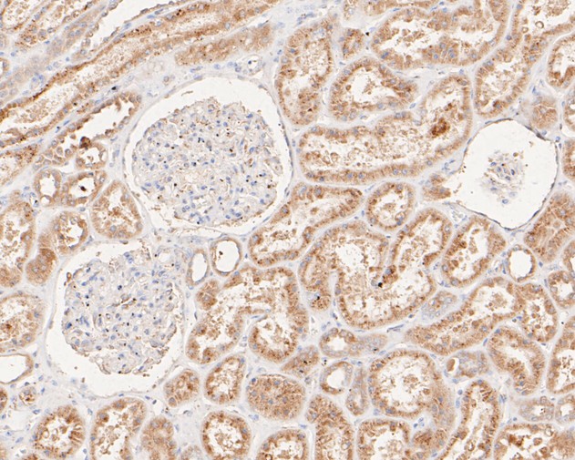
Immunohistochemical analysis of paraffin-embedded human kidney tissue using anti-Phospho-PKR (T446) antibody. The section was pre-treated using heat mediated antigen retrieval with sodium citrate buffer (pH 6.0) (high pressure) for 2 minutes. The tissues were blocked in 5% BSA for 30 minutes at room temperature, washed with ddH2O and PBS, and then probed with the primary antibody (1/50) for 30 minutes at room temperature. The detection was performed using an HRP conjugated compact polymer system. DAB was used as the chromogen. Tissues were counterstained with hematoxylin and mounted with DPX.IHC
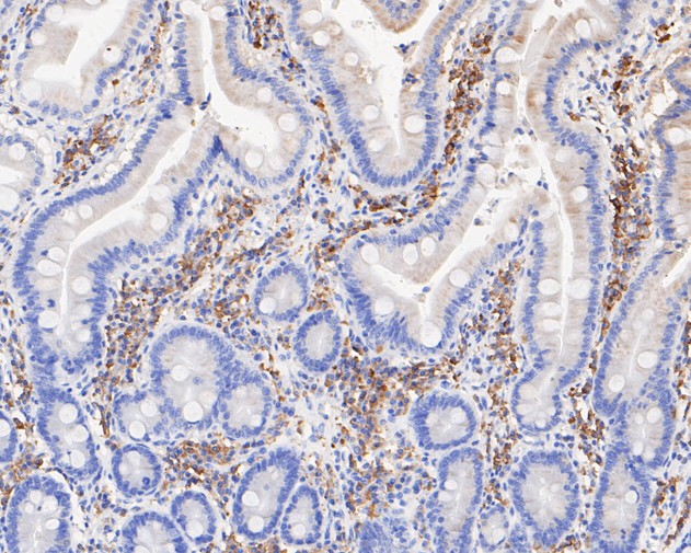
Immunohistochemical analysis of paraffin-embedded human small intestine tissue using anti-Phospho-PKR (T446) antibody. The section was pre-treated using heat mediated antigen retrieval with sodium citrate buffer (pH 6.0) (high pressure) for 2 minutes. The tissues were blocked in 5% BSA for 30 minutes at room temperature, washed with ddH2O and PBS, and then probed with the primary antibody (1/50) for 30 minutes at room temperature. The detection was performed using an HRP conjugated compact polymer system. DAB was used as the chromogen. Tissues were counterstained with hematoxylin and mounted with DPX.ICC/IF
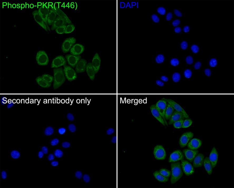
Immunocytochemistry analysis of SiHa cells labeling Phospho-PKR (T446) with Rabbit anti-Phospho-PKR (T446) antibody at 1/50 dilution. Cells were fixed in 4% paraformaldehyde for 10 minutes at 37 ℃, permeabilized with 0.05% Triton X-100 in PBS for 20 minutes, and then blocked with 2% negative goat serum for 30 minutes at room temperature. Cells were then incubated with Rabbit anti-Phospho-PKR (T446) antibody at 1/50 dilution in 2% negative goat serum overnight at 4 ℃. Goat Anti-Rabbit IgG H&L (488) was used as the secondary antibody at 1/1,000 dilution. PBS instead of the primary antibody was used as the secondary antibody only control. Nuclear DNA was labelled in blue with DAPI.| Product Name | Phospho-PKR (T446) Recombinant Rabbit Monoclonal Antibody |
|---|---|
| Antibody Type | Primary Antibodies |
| Product description | An interferon-inducible, RNA-dependent protein serine/threonine kinase, PKR has various designations. Mouse PKR is known as DAI, dsJ, PI kinase, p65, p67 or TIK, whereas human PKR is known as p68 or p69. PKR phosphorylates its substrate, a subunit of protein synthesis initiation factor eIF-2 on Ser 51 to inhibit translation. PKR contains two dsRNA binding motifs required for its activation by dsRNA. Three kinds of regulation of PKR enzymatic activity occur, and these include transcriptional regulation in response to interferon, an autoregulatory mechanism controlling PKR expression at the level of translation, and posttranslational regulation by RNA mediated autophosphorylation. Human PKR contains at least 15 autophosphorylation sites, but only Thr-446 and Thr-451 in the activation loop are critical for its kinase activity. Thr-446 is the in vivo autophosphorylation site of PKR. Mutation of threonine to alanine at position 446 substantially reduces PKR function, and mutant kinase containing Ala-451 is completely inactive. |
| Immunogen | Synthetic phospho-peptide corresponding to residues surrounding Thr446 of Human PKR aa 421-470 / 551. |
| Modification | p-T446 |
| Clonality | Monoclonal |
|---|---|
| Isotype | IgG |
| Host Species | Recombinant rabbit |
| Tested Applications | ICC/IFIHCWB |
| WB:1:500-1:5000 IHC:1:50-1:200 ICC/IF:1:50 |
|
| Species Reactivity | Human |
| Concentration | 1mg/ml |
| Purification | Protein A |
| Alternative Names | Double stranded RNA activated protein kinase antibody E2AK2_HUMAN antibody eIF-2A protein kinase 2 antibody EIF2AK1 antibody EIF2AK2 antibody Eukaryotic translation initiation factor 2 alpha kinase 2 antibody Eukaryotic translation initiation factor 2-alpha kinase 2 antibody HGNC:9437 antibody Interferon induced double stranded RNA activated protein kinase antibody Interferon inducible elF2 alpha kinase antibody Interferon inducible RNA dependent protein kinase antibody Interferon-induced double-stranded RNA-activated protein kinase antibody Interferon-inducible RNA-dependent protein kinase antibody MGC126524 antibody P1/eIF-2A protein kinase antibody P1/eIF2A protein kinase antibody p68 kinase antibody PKR antibody PPP1R83 antibody PRKR antibody Protein kinase interferon inducible double stranded RNA dependent antibody Protein kinase RNA activated antibody Protein kinase RNA-activated antibody Protein phosphatase 1 regulatory subunit 83 antibody Serine/threonine protein kinase TIK antibody Tyrosine protein kinase EIF2AK2 antibody |
|---|---|
| Molecular Weight(MW) | 62 kDa(Observed band size: 68 kDa) |
| Cellular Localization | Cytoplasm, Nucleus. |
| SwissProt ID | P19525 |
|---|
WB
Western blot analysis of Phospho-PKR (T446) on different lysates with Rabbit anti-Phospho-PKR (T446) antibody at 1/5,000 dilution. Lane 1: Jurkat cell lysate, Lane 2: Jurkat treated with 100nM Calyculin A for 30 minutes cell lysate, Lysates/proteins at 20 µg/Lane. Exposure time: 30 seconds; 4-20% SDS-PAGE gel. Proteins were transferred to a PVDF membrane and blocked with 5% NFDM/TBST for 1 hour at room temperature. The primary antibody at 1/5,000 dilution was used in 5% NFDM/TBST at 4℃ overnight. Goat Anti-Rabbit IgG - HRP Secondary Antibody at 1/50,000 dilution was used for 1 hour at room temperature.
IHC
Immunohistochemical analysis of paraffin-embedded human tonsil tissue using anti-Phospho-PKR (T446) antibody. The section was pre-treated using heat mediated antigen retrieval with sodium citrate buffer (pH 6.0) (high pressure) for 2 minutes. The tissues were blocked in 5% BSA for 30 minutes at room temperature, washed with ddH2O and PBS, and then probed with the primary antibody (1/50) for 30 minutes at room temperature. The detection was performed using an HRP conjugated compact polymer system. DAB was used as the chromogen. Tissues were counterstained with hematoxylin and mounted with DPX.
IHC
Immunohistochemical analysis of paraffin-embedded human spleen tissue using anti-Phospho-PKR (T446) antibody. The section was pre-treated using heat mediated antigen retrieval with sodium citrate buffer (pH 6.0) (high pressure) for 2 minutes. The tissues were blocked in 5% BSA for 30 minutes at room temperature, washed with ddH2O and PBS, and then probed with the primary antibody (1/50) for 30 minutes at room temperature. The detection was performed using an HRP conjugated compact polymer system. DAB was used as the chromogen. Tissues were counterstained with hematoxylin and mounted with DPX.
IHC
Immunohistochemical analysis of paraffin-embedded human breast tissue using anti-Phospho-PKR (T446) antibody. The section was pre-treated using heat mediated antigen retrieval with sodium citrate buffer (pH 6.0) (high pressure) for 2 minutes. The tissues were blocked in 5% BSA for 30 minutes at room temperature, washed with ddH2O and PBS, and then probed with the primary antibody (1/50) for 30 minutes at room temperature. The detection was performed using an HRP conjugated compact polymer system. DAB was used as the chromogen. Tissues were counterstained with hematoxylin and mounted with DPX.
IHC
Immunohistochemical analysis of paraffin-embedded human kidney tissue using anti-Phospho-PKR (T446) antibody. The section was pre-treated using heat mediated antigen retrieval with sodium citrate buffer (pH 6.0) (high pressure) for 2 minutes. The tissues were blocked in 5% BSA for 30 minutes at room temperature, washed with ddH2O and PBS, and then probed with the primary antibody (1/50) for 30 minutes at room temperature. The detection was performed using an HRP conjugated compact polymer system. DAB was used as the chromogen. Tissues were counterstained with hematoxylin and mounted with DPX.
IHC
Immunohistochemical analysis of paraffin-embedded human small intestine tissue using anti-Phospho-PKR (T446) antibody. The section was pre-treated using heat mediated antigen retrieval with sodium citrate buffer (pH 6.0) (high pressure) for 2 minutes. The tissues were blocked in 5% BSA for 30 minutes at room temperature, washed with ddH2O and PBS, and then probed with the primary antibody (1/50) for 30 minutes at room temperature. The detection was performed using an HRP conjugated compact polymer system. DAB was used as the chromogen. Tissues were counterstained with hematoxylin and mounted with DPX.
ICC/IF
Immunocytochemistry analysis of SiHa cells labeling Phospho-PKR (T446) with Rabbit anti-Phospho-PKR (T446) antibody at 1/50 dilution. Cells were fixed in 4% paraformaldehyde for 10 minutes at 37 ℃, permeabilized with 0.05% Triton X-100 in PBS for 20 minutes, and then blocked with 2% negative goat serum for 30 minutes at room temperature. Cells were then incubated with Rabbit anti-Phospho-PKR (T446) antibody at 1/50 dilution in 2% negative goat serum overnight at 4 ℃. Goat Anti-Rabbit IgG H&L (488) was used as the secondary antibody at 1/1,000 dilution. PBS instead of the primary antibody was used as the secondary antibody only control. Nuclear DNA was labelled in blue with DAPI.| Application Notes | WB:1:500-1:5000 IHC:1:50-1:200 ICC/IF:1:50 |
|---|
| Form | Liquid |
|---|---|
| Storage Instructions | Store at +4℃ after thawing. Aliquot store at -20℃ or -80℃. Avoid repeated freeze / thaw cycles. |
| Storage Buffer | 1*TBS (pH7.4), 1%BSA, 40%Glycerol. Preservative: 0.05% Sodium Azide. |


