Application
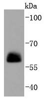
Fig1: Western blot analysis of Paxillin on NIH/3T3 cells lysates using anti-Paxillin antibody at 1/1,000 dilution.Application
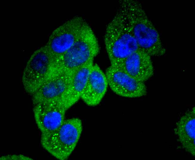
Fig2: ICC staining Paxillin in Hela cells (green). The nuclear counter stain is DAPI (blue). Cells were fixed in paraformaldehyde, permeabilised with 0.25% Triton X100/PBS.Application
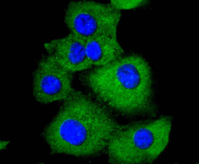
Fig3: ICC staining Paxillin in A549 cells (green). The nuclear counter stain is DAPI (blue). Cells were fixed in paraformaldehyde, permeabilised with 0.25% Triton X100/PBS.Application
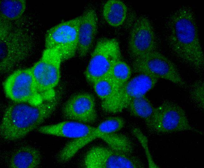
Fig4: ICC staining Paxillin in SKOV-3 cells (green). The nuclear counter stain is DAPI (blue). Cells were fixed in paraformaldehyde, permeabilised with 0.25% Triton X100/PBS.Application
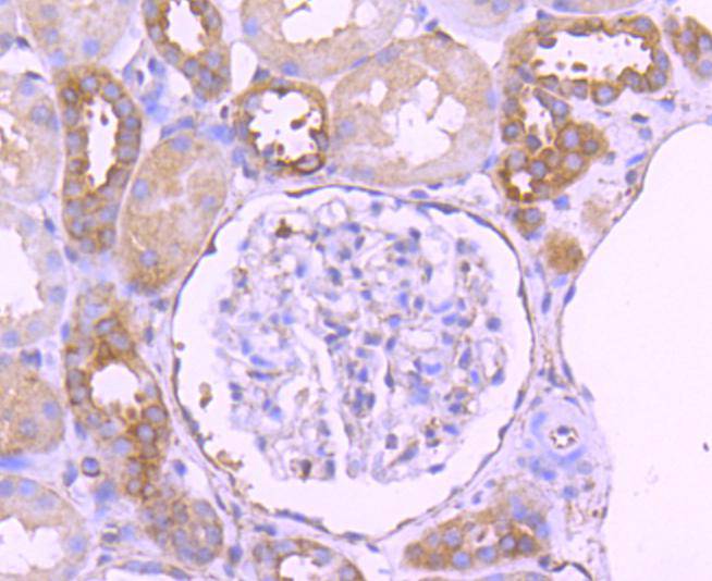
Fig5: Immunohistochemical analysis of paraffin-embedded human kideny tissue using anti-Paxillin antibody. Counter stained with hematoxylin.Application
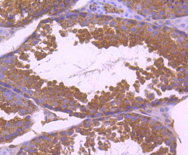
Fig6: Immunohistochemical analysis of paraffin-embedded mouse testis tissue using anti-Paxillin antibody. Counter stained with hematoxylin.Application
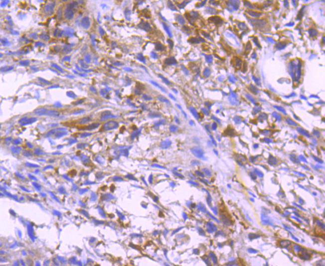
Fig7: Immunohistochemical analysis of paraffin-embedded human breast carcinoma tissue using anti-Paxillin antibody. Counter stained with hematoxylin.Application
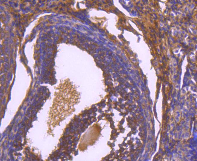
Fig8: Immunohistochemical analysis of paraffin-embedded mouse ovary tissue using anti-Paxillin antibody. Counter stained with hematoxylin.| Product Name | Paxillin [SY23-02] |
|---|---|
| Antibody Type | Primary Antibodies |
| Antigen Alias | FLJ16691 antibody FLJ23042 antibody Paired box protein Pax 1 antibody PAX 1 antibody PAX1 antibody PAXI_HUMAN antibody Paxillin alpha antibody Paxillin antibody PXN antibody PXN protein antibody |
| Product description | Paxillin is a focal adhesion phosphoprotein that is localized to the cytoskeleton. Phosphorylation of paxillin has been shown to occur in response to PDGF treatment, v-Src transformation or cross-linking of integrins. FAK (focal adhesion kinase) and PYK2 have been shown to phosphorylate paxillin. FAK phosphorylates paxillin specifically on Tyr 118 in vitro. However, FAK phosphorylation does not seem to be required for the recruitment of paxillin to cell adhesion sites. Paxillin may play a role in signal transduction, regulation of cell morphology and the recruitment of structural and signaling molecules to focal adhesions. It has been shown that the amount of paxillin is reduced in mitotic cells by proteolytic downregulation and that paxillin is alternatively phosphorylated on serine rather than on tyrosine and serine during mitosis. |
| Immunogen | recombinant protein |
| Clonality | Monoclonal |
|---|---|
| Isotype | IgG |
| Host Species | Recombinant rabbit |
| Tested Applications | WBICC/IFIHCIP |
| WB:1:1,000-1:2,000 ICC:1:50-1:200 IHC:1:50-1:200 |
|
| Species Reactivity | HumanMouseRat |
| Concentration | 1mg/ml |
| Alternative Names | FLJ16691 antibody FLJ23042 antibody Paired box protein Pax 1 antibody PAX 1 antibody PAX1 antibody PAXI_HUMAN antibody Paxillin alpha antibody Paxillin antibody PXN antibody PXN protein antibody |
|---|---|
| Molecular Weight(MW) | 65 kDa |
| Cellular Localization | Cytoplasm, Cell junction. |
Application
Fig1: Western blot analysis of Paxillin on NIH/3T3 cells lysates using anti-Paxillin antibody at 1/1,000 dilution.
Application
Fig2: ICC staining Paxillin in Hela cells (green). The nuclear counter stain is DAPI (blue). Cells were fixed in paraformaldehyde, permeabilised with 0.25% Triton X100/PBS.
Application
Fig3: ICC staining Paxillin in A549 cells (green). The nuclear counter stain is DAPI (blue). Cells were fixed in paraformaldehyde, permeabilised with 0.25% Triton X100/PBS.
Application
Fig4: ICC staining Paxillin in SKOV-3 cells (green). The nuclear counter stain is DAPI (blue). Cells were fixed in paraformaldehyde, permeabilised with 0.25% Triton X100/PBS.
Application
Fig5: Immunohistochemical analysis of paraffin-embedded human kideny tissue using anti-Paxillin antibody. Counter stained with hematoxylin.
Application
Fig6: Immunohistochemical analysis of paraffin-embedded mouse testis tissue using anti-Paxillin antibody. Counter stained with hematoxylin.
Application
Fig7: Immunohistochemical analysis of paraffin-embedded human breast carcinoma tissue using anti-Paxillin antibody. Counter stained with hematoxylin.
Application
Fig8: Immunohistochemical analysis of paraffin-embedded mouse ovary tissue using anti-Paxillin antibody. Counter stained with hematoxylin.| Positive Control | A549, SKOV-3, Hela, NIH/3T3, human breast carcinoma tissue, human kideny tissue, mouse ovary tissue, mouse testis tissue. |
|---|---|
| Application Notes | WB:1:1,000-1:2,000 ICC:1:50-1:200 IHC:1:50-1:200 |
| Form | Liquid |
|---|---|
| Storage Instructions | Store at +4℃ after thawing. Aliquot store at -20℃ or -80℃. Avoid repeated freeze / thaw cycles. |
| Storage Buffer | 1*TBS (pH7.4), 1%BSA, 40%Glycerol. Preservative: 0.05% Sodium Azide. |


