WB
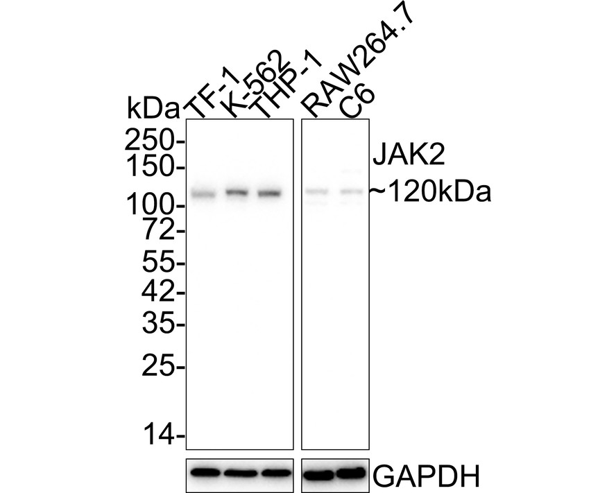
Western blot analysis of JAK2 on different lysates with Rabbit anti-JAK2 antibody at 1/2,000 dilution. Lane 1: TF-1 cell lysate, Lane 2: K-562 cell lysate, Lane 3: THP-1 cell lysate, Lane 4: RAW264.7 cell lysate, Lane 5: C6 cell lysate, Lysates/proteins at 15 µg/Lane. Exposure time: 5 minutes 10 seconds; 4-20% SDS-PAGE gel. Proteins were transferred to a PVDF membrane and blocked with 5% NFDM/TBST for 1 hour at room temperature. The primary antibody at 1/2,000 dilution was used in 5% NFDM/TBST at 4℃ overnight. Goat Anti-Rabbit IgG - HRP Secondary Antibody at 1/50,000 dilution was used for 1 hour at room temperature.ICC/IF
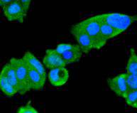
ICC staining JAK2 in MCF-7 cells (green). The nuclear counter stain is DAPI (blue). Cells were fixed in paraformaldehyde, permeabilised with 0.25% Triton X100/PBS.ICC/IF
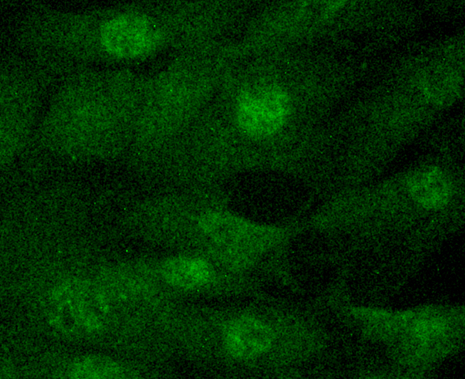
ICC staining JAK2 in NIH/3T3 cells (green). The nuclear counter stain is DAPI (blue). Cells were fixed in paraformaldehyde, permeabilised with 0.25% Triton X100/PBS.| Product Name | JAK2 Recombinant Rabbit Monoclonal Antibody |
|---|---|
| Antibody Type | Primary Antibodies |
| Product description | JAK2 (Janus kinase 2) belongs to the emerging family of non-receptor Janus tyrosine kinases, which regulate a spectrum of cellular functions downstream of activated cytokine receptors in the lympho-hematopoietic system. Immunological stimuli, such as interferons and cytokines, induce recruitment of Stat transcription factors to cytokine receptor-associated JAK2. JAK2 then phosphorylates proximal Stat factors, which subsequently dimerize, translocate to the nucleus and bind to cis elements upstream of target gene promoters to regulate transcription. The canonical JAK/Stat pathway is integral to maintaining a normal immune system by stimulating proliferation, differentiation, survival and host resistance to pathogens. Altering JAK/Stat signaling to reduce cytokine induced pro-inflammatory responses represents an attractive target for anti-inflammatory therapies. |
| Immunogen | Synthetic peptide within C-terminal human JAK2. |
| Clonality | monoclonal |
|---|---|
| Isotype | IgG |
| Host Species | Recombinant rabbit |
| Tested Applications | ICC/IFWB |
| WB:1:500-1:2000 ICC/IF:1:100-1:500 |
|
| Species Reactivity | HumanMouseRat |
| Concentration | 1mg/ml |
| Purification | Protein A |
| Alternative Names | JAK 2 antibody JAK-2 antibody JAK2 antibody JAK2_HUMAN antibody Janus Activating Kinase 2 antibody Janus kinase 2 (a protein tyrosine kinase) antibody Janus kinase 2 antibody JTK 10 antibody JTK10 antibody kinase Jak2 antibody OTTHUMP00000043260 antibody THCYT3 antibody Tyrosine protein kinase JAK2 antibody Tyrosine-protein kinase JAK2 antibody |
|---|---|
| Molecular Weight(MW) | 130kDa(Observed band size: 120kDa) |
| Cellular Localization | Endomembrane system, Cytoplasm, Nucleus. |
WB
Western blot analysis of JAK2 on different lysates with Rabbit anti-JAK2 antibody at 1/2,000 dilution. Lane 1: TF-1 cell lysate, Lane 2: K-562 cell lysate, Lane 3: THP-1 cell lysate, Lane 4: RAW264.7 cell lysate, Lane 5: C6 cell lysate, Lysates/proteins at 15 µg/Lane. Exposure time: 5 minutes 10 seconds; 4-20% SDS-PAGE gel. Proteins were transferred to a PVDF membrane and blocked with 5% NFDM/TBST for 1 hour at room temperature. The primary antibody at 1/2,000 dilution was used in 5% NFDM/TBST at 4℃ overnight. Goat Anti-Rabbit IgG - HRP Secondary Antibody at 1/50,000 dilution was used for 1 hour at room temperature.
ICC/IF
ICC staining JAK2 in MCF-7 cells (green). The nuclear counter stain is DAPI (blue). Cells were fixed in paraformaldehyde, permeabilised with 0.25% Triton X100/PBS.
ICC/IF
ICC staining JAK2 in NIH/3T3 cells (green). The nuclear counter stain is DAPI (blue). Cells were fixed in paraformaldehyde, permeabilised with 0.25% Triton X100/PBS.| Application Notes | WB:1:500-1:2000 ICC/IF:1:100-1:500 |
|---|
| Form | Liquid |
|---|---|
| Storage Instructions | Store at +4℃ after thawing. Aliquot store at -20℃ or -80℃. Avoid repeated freeze / thaw cycles. |
| Storage Buffer | 1*TBS (pH7.4), 1%BSA, 40%Glycerol. Preservative: 0.05% Sodium Azide. |


