WB
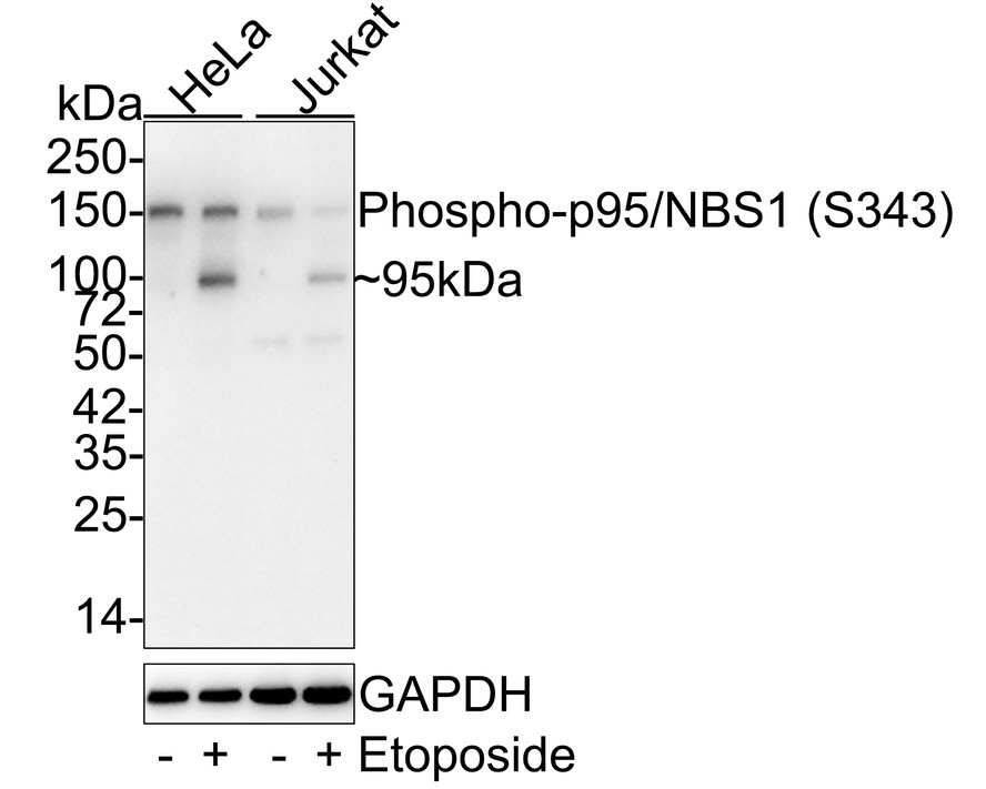
Western blot analysis of Phospho-p95/NBS1 (S343) on different lysates with Rabbit anti-Phospho-p95/NBS1 (S343) antibody at 1/1,000 dilution. Lane 1: HeLa whole cell lysate (20 µg/Lane), Lane 2: HeLa treated with 20μM Etoposide for 2 hours whole cell lysate (20 µg/Lane), Lane 3: Jurkat whole cell lysate (20 µg/Lane), Lane 4: Jurkattreated with 25μM Etoposide for 5 hours whole cell lysate (20 µg/Lane), Exposure time: 5 minutes; 4-20% SDS-PAGE gel. Proteins were transferred to a PVDF membrane and blocked with 5% NFDM/TBST for 1 hour at room temperature. The primary antibody at 1/1,000 dilution was used in 5% NFDM/TBST at room temperature for 2 hours. Goat Anti-Rabbit IgG - HRP Secondary Antibody at 1/50,000 dilution was used for 1 hour at room temperature.IHC
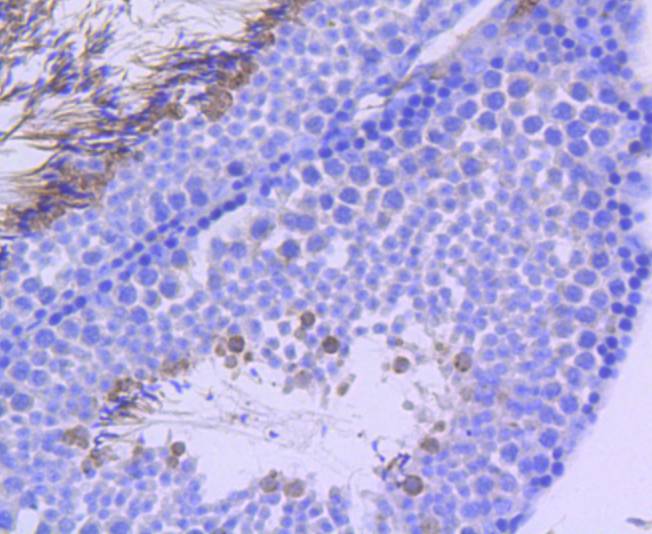
Immunohistochemical analysis of paraffin-embedded mouse testis tissue using anti-Phospho-p95/NBS1(S343) antibody. Counter stained with hematoxylin.| Product Name | Phospho-p95/NBS1 (S343) Recombinant Rabbit Monoclonal Antibody |
|---|---|
| Antibody Type | Primary Antibodies |
| Product description | Nijmegen breakage syndrome (NBS) is characterized by extreme radiation sensitivity, chromosomal instability and cancer. These phenotypes are similar to those of ataxia telangiectasia mutated (ATM) disease, where there is a deficiency in a protein kinase that is activated by DNA damage, indicating that the NBS1 (Nibrin) and ATM proteins may participate in common pathways. Nibrin is specifically phosphorylated in response to gamma-radiation, ultraviolet light and exposure to hydroxyurea. The phosphorylation of Nibrin requires catalytically active ATM. ATM physically interacts with and phosphorylates Nibrin on Serine 343 both in vitro and in vivo. Serine 343 is phosphorylated in vitro by ATM and the modification of this residue in vivo is essential for the cellular response to DNA damage. This response includes S-phase checkpoint activation, formation of the NBS1/Mrel1/Rad50 nuclear foci and rescue of hypersensitivity to ionizing radiation. |
| Immunogen | Synthetic phospho-peptide corresponding to residues surrounding Ser343 of Human p95 / NBS1 aa 321-370 / 754. |
| Modification | p-S343 |
| Clonality | Monoclonal |
|---|---|
| Isotype | IgG |
| Host Species | Recombinant rabbit |
| Tested Applications | IHCWB |
| WB:1:1000 ICCIHC:1:50-1:200 |
|
| Species Reactivity | HumanMouse |
| Concentration | 1mg/ml |
| Purification | Protein A |
| Alternative Names | AT V1 antibody AT V2 antibody ATV antibody Cell cycle regulatory protein p95 antibody FLJ10155 antibody MGC87362 antibody Nbn antibody NBN_HUMAN antibody NBS 1 antibody NBS antibody NBS1 antibody Nibrin antibody Nijmegen breakage syndrome 1 (nibrin) antibody Nijmegen breakage syndrome antibody Nijmegen breakage syndrome protein 1 antibody p95 antibody p95 protein of the MRE11/RAD50 complex antibody |
|---|---|
| Molecular Weight(MW) | 85 kDa(Observed band size: 95 kDa) |
| Cellular Localization | Nucleus, PML body, telomere, Chromosome. |
| SwissProt ID | O60934 |
|---|
WB
Western blot analysis of Phospho-p95/NBS1 (S343) on different lysates with Rabbit anti-Phospho-p95/NBS1 (S343) antibody at 1/1,000 dilution. Lane 1: HeLa whole cell lysate (20 µg/Lane), Lane 2: HeLa treated with 20μM Etoposide for 2 hours whole cell lysate (20 µg/Lane), Lane 3: Jurkat whole cell lysate (20 µg/Lane), Lane 4: Jurkattreated with 25μM Etoposide for 5 hours whole cell lysate (20 µg/Lane), Exposure time: 5 minutes; 4-20% SDS-PAGE gel. Proteins were transferred to a PVDF membrane and blocked with 5% NFDM/TBST for 1 hour at room temperature. The primary antibody at 1/1,000 dilution was used in 5% NFDM/TBST at room temperature for 2 hours. Goat Anti-Rabbit IgG - HRP Secondary Antibody at 1/50,000 dilution was used for 1 hour at room temperature.
IHC
Immunohistochemical analysis of paraffin-embedded mouse testis tissue using anti-Phospho-p95/NBS1(S343) antibody. Counter stained with hematoxylin.| Application Notes | WB:1:1000 ICCIHC:1:50-1:200 |
|---|
| Form | Liquid |
|---|---|
| Storage Instructions | Store at +4℃ after thawing. Aliquot store at -20℃ or -80℃. Avoid repeated freeze / thaw cycles. |
| Storage Buffer | 1*TBS (pH7.4), 1%BSA, 40%Glycerol. Preservative: 0.05% Sodium Azide. |


