WB
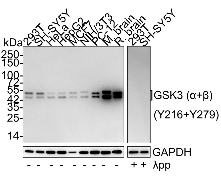
Western blot analysis of Phospho-GSK3 (alpha + beta) (Y216 + Y279) on different lysates with Rabbit anti-Phospho-GSK3 (alpha + beta) (Y216 + Y279) antibody at 1/5,000 dilution. Lane 1: 293T cell lysate, Lane 2: SH-SY5Y cell lysate, Lane 3: HeLa cell lysate, Lane 4: HepG2 cell lysate, Lane 5: MCF7 cell lysate, Lane 6: NIH/3T3 cell lysate, Lane 7: PC-12 cell lysate, Lane 8: Mouse brain tissue lysate, Lane 9: Rat brain tissue lysate, Lane 10: 293T treated with λpp for 1 hour cell lysate, Lane 11: SH-SY5Y treated with λpp for 1 hour cell lysate, Lysates/proteins at 15 µg/Lane. Exposure time: 5 minutes; 4-20% SDS-PAGE gel. Proteins were transferred to a PVDF membrane and blocked with 5% NFDM/TBST for 1 hour at room temperature. The primary antibody at 1/5,000 dilution was used in 5% NFDM/TBST at 4℃ overnight. Goat Anti-Rabbit IgG - HRP Secondary Antibody at 1:50,000 dilution was used for 1 hour at room temperature.ICC/IF
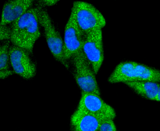
ICC staining Phospho-GSK3(alpha+beta)(Y216+Y279) in Hela cells (green). The nuclear counter stain is DAPI (blue). Cells were fixed in paraformaldehyde, permeabilised with 0.25% Triton X100/PBS.ICC/IF
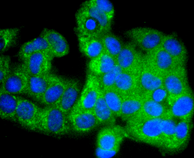
ICC staining Phospho-GSK3(alpha+beta)(Y216+Y279) in MCF-7 cells (green). The nuclear counter stain is DAPI (blue). Cells were fixed in paraformaldehyde, permeabilised with 0.25% Triton X100/PBS.IHC
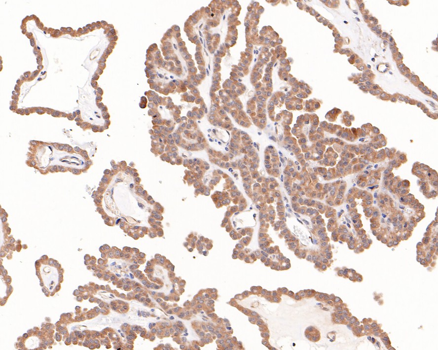
Immunohistochemical analysis of paraffin-embedded human thyroid carcinoma tissue with Rabbit anti-Phospho-GSK3 (alpha + beta) (Y216 + Y279) antibody at 1/800 dilution. The section was pre-treated using heat mediated antigen retrieval with sodium citrate buffer (pH 6.0) (high pressure) for 2 minutes. The tissues were blocked in 1% BSA for 20 minutes at room temperature, washed with ddH2O and PBS, and then probed with the primary antibody at 1/800 dilution for 1 hour at room temperature. The detection was performed using an HRP conjugated compact polymer system. DAB was used as the chromogen. Tissues were counterstained with hematoxylin and mounted with DPX.IHC
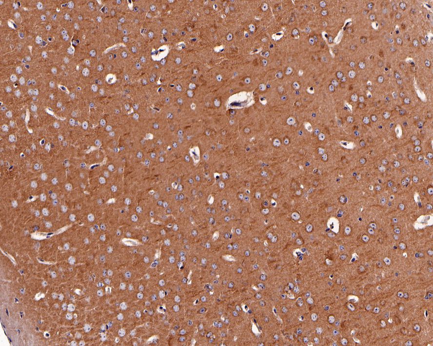
Immunohistochemical analysis of paraffin-embedded mouse brain tissue with Rabbit anti-Phospho-GSK3 (alpha + beta) (Y216 + Y279) antibody at 1/1,000 dilution. The section was pre-treated using heat mediated antigen retrieval with Tris-EDTA buffer (pH 9.0) for 20 minutes. The tissues were blocked in 1% BSA for 20 minutes at room temperature, washed with ddH2O and PBS, and then probed with the primary antibody at 1/1,000 dilution for 1 hour at room temperature. The detection was performed using an HRP conjugated compact polymer system. DAB was used as the chromogen. Tissues were counterstained with hematoxylin and mounted with DPX.IHC
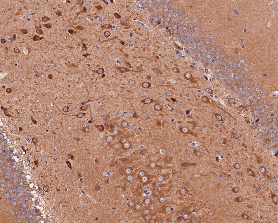
Immunohistochemical analysis of paraffin-embedded rat hippocampus tissue with Rabbit anti-Phospho-GSK3 (alpha + beta) (Y216 + Y279) antibody at 1/1,000 dilution. The section was pre-treated using heat mediated antigen retrieval with Tris-EDTA buffer (pH 9.0) for 20 minutes. The tissues were blocked in 1% BSA for 20 minutes at room temperature, washed with ddH2O and PBS, and then probed with the primary antibody at 1/1,000 dilution for 1 hour at room temperature. The detection was performed using an HRP conjugated compact polymer system. DAB was used as the chromogen. Tissues were counterstained with hematoxylin and mounted with DPX.FC
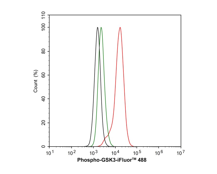
Flow cytometric analysis of HeLa cells labeling Phospho-GSK3 (alpha + beta) (Y216 + Y279). Cells were fixed and permeabilized. Then stained with the primary antibody (1μg/mL) (red) compared with Rabbit IgG Isotype Control (green). After incubation of the primary antibody at +4℃ for an hour, the cells were stained with a iFluor™ 488 conjugate-Goat anti-Rabbit IgG Secondary antibody at 1/1,000 dilution for 30 minutes at +4℃. Unlabelled sample was used as a control (cells without incubation with primary antibody; black).| Product Name | Phospho-GSK3 (alpha + beta) (Y216 + Y279) Recombinant Rabbit Monoclonal Antibody |
|---|---|
| Antibody Type | Primary Antibodies |
| Product description | Glycogen synthase kinase-3α (GSK-3α) and GSK-3β are highly similar isoforms of serine/ threonine kinases that regulate metabolic enzymes and transcription factors, which are responsible for coordinating processes such as glycogen synthesis and cell adhesion. GSK-3β activity is also required for nuclear activity of Rel dimers, which mediate an anti-apoptotic response to TNFα in mice. GSK-3 catalytic kinase activity is controlled through differential phosphorylation of serine/threonine residues, which have an inhibitory effect, and tyrosine residues, which have an activating effect. Growth factor stimulation of mammalian cells expressing GSK-3α and GSK-3β induces phosphorylation of Ser 21 and Ser 9, respectively, through a phosphatidylinositol 3-kinase (PI 3-K)-protein kinase B (PKB)-dependent pathway, thereby enhancing proliferative signals. Additionally, GSK-3 physically associates with cAMP-dependent protein kinase A (PKA), which phosphorylates Ser 21 of GSK-3α or Ser 9 of GSK-3β and inactivates both forms. GSK-3α/β is positively regulated by phosphorylation on Tyr 279 and Tyr 216, respectively. Activated GSK-3α/β participates in energy metabolism, neuronal cell development, and body pattern formation. Tyrosine dephosphorylation of GSK-3 is involved in its extracellular signal-dependent inactivation. |
| Immunogen | Synthetic phospho-peptide corresponding to residues surrounding Tyr216 and 279 of human GSK3(alpha+beta). |
| Modification | p-Y216+Y279 |
| Clonality | Monoclonal |
|---|---|
| Isotype | IgG |
| Host Species | Recombinant rabbit |
| Tested Applications | FCICC/IFIHCWB |
| WB:1:500-1:1000 IHC:1:200-1:800 ICC/IF:1:100-1:500 FC:1:1000 |
|
| Species Reactivity | HumanMouseRat |
| Concentration | 1mg/ml |
| Purification | Protein A |
| Alternative Names | Factor A antibody Glycogen synthase kinase 3 alpha antibody Glycogen synthase kinase 3 beta antibody GSK3 alpha antibody GSK3 beta antibody GSK3B antibody |
|---|---|
| Molecular Weight(MW) | 47/51kDa |
| Cellular Localization | Cytoplasm, Nucleus, Cell membrane. |
WB
Western blot analysis of Phospho-GSK3 (alpha + beta) (Y216 + Y279) on different lysates with Rabbit anti-Phospho-GSK3 (alpha + beta) (Y216 + Y279) antibody at 1/5,000 dilution. Lane 1: 293T cell lysate, Lane 2: SH-SY5Y cell lysate, Lane 3: HeLa cell lysate, Lane 4: HepG2 cell lysate, Lane 5: MCF7 cell lysate, Lane 6: NIH/3T3 cell lysate, Lane 7: PC-12 cell lysate, Lane 8: Mouse brain tissue lysate, Lane 9: Rat brain tissue lysate, Lane 10: 293T treated with λpp for 1 hour cell lysate, Lane 11: SH-SY5Y treated with λpp for 1 hour cell lysate, Lysates/proteins at 15 µg/Lane. Exposure time: 5 minutes; 4-20% SDS-PAGE gel. Proteins were transferred to a PVDF membrane and blocked with 5% NFDM/TBST for 1 hour at room temperature. The primary antibody at 1/5,000 dilution was used in 5% NFDM/TBST at 4℃ overnight. Goat Anti-Rabbit IgG - HRP Secondary Antibody at 1:50,000 dilution was used for 1 hour at room temperature.
ICC/IF
ICC staining Phospho-GSK3(alpha+beta)(Y216+Y279) in Hela cells (green). The nuclear counter stain is DAPI (blue). Cells were fixed in paraformaldehyde, permeabilised with 0.25% Triton X100/PBS.
ICC/IF
ICC staining Phospho-GSK3(alpha+beta)(Y216+Y279) in MCF-7 cells (green). The nuclear counter stain is DAPI (blue). Cells were fixed in paraformaldehyde, permeabilised with 0.25% Triton X100/PBS.
IHC
Immunohistochemical analysis of paraffin-embedded human thyroid carcinoma tissue with Rabbit anti-Phospho-GSK3 (alpha + beta) (Y216 + Y279) antibody at 1/800 dilution. The section was pre-treated using heat mediated antigen retrieval with sodium citrate buffer (pH 6.0) (high pressure) for 2 minutes. The tissues were blocked in 1% BSA for 20 minutes at room temperature, washed with ddH2O and PBS, and then probed with the primary antibody at 1/800 dilution for 1 hour at room temperature. The detection was performed using an HRP conjugated compact polymer system. DAB was used as the chromogen. Tissues were counterstained with hematoxylin and mounted with DPX.
IHC
Immunohistochemical analysis of paraffin-embedded mouse brain tissue with Rabbit anti-Phospho-GSK3 (alpha + beta) (Y216 + Y279) antibody at 1/1,000 dilution. The section was pre-treated using heat mediated antigen retrieval with Tris-EDTA buffer (pH 9.0) for 20 minutes. The tissues were blocked in 1% BSA for 20 minutes at room temperature, washed with ddH2O and PBS, and then probed with the primary antibody at 1/1,000 dilution for 1 hour at room temperature. The detection was performed using an HRP conjugated compact polymer system. DAB was used as the chromogen. Tissues were counterstained with hematoxylin and mounted with DPX.
IHC
Immunohistochemical analysis of paraffin-embedded rat hippocampus tissue with Rabbit anti-Phospho-GSK3 (alpha + beta) (Y216 + Y279) antibody at 1/1,000 dilution. The section was pre-treated using heat mediated antigen retrieval with Tris-EDTA buffer (pH 9.0) for 20 minutes. The tissues were blocked in 1% BSA for 20 minutes at room temperature, washed with ddH2O and PBS, and then probed with the primary antibody at 1/1,000 dilution for 1 hour at room temperature. The detection was performed using an HRP conjugated compact polymer system. DAB was used as the chromogen. Tissues were counterstained with hematoxylin and mounted with DPX.
FC
Flow cytometric analysis of HeLa cells labeling Phospho-GSK3 (alpha + beta) (Y216 + Y279). Cells were fixed and permeabilized. Then stained with the primary antibody (1μg/mL) (red) compared with Rabbit IgG Isotype Control (green). After incubation of the primary antibody at +4℃ for an hour, the cells were stained with a iFluor™ 488 conjugate-Goat anti-Rabbit IgG Secondary antibody at 1/1,000 dilution for 30 minutes at +4℃. Unlabelled sample was used as a control (cells without incubation with primary antibody; black).| Application Notes | WB:1:500-1:1000 IHC:1:200-1:800 ICC/IF:1:100-1:500 FC:1:1000 |
|---|
| Form | Liquid |
|---|---|
| Storage Instructions | Store at +4℃ after thawing. Aliquot store at -20℃ or -80℃. Avoid repeated freeze / thaw cycles. |
| Storage Buffer | 1*TBS (pH7.4), 1%BSA, 40%Glycerol. Preservative: 0.05% Sodium Azide. |


