WB
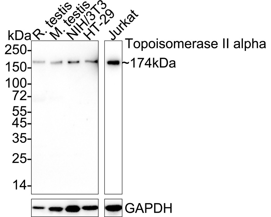
Western blot analysis of Topoisomerase II alpha on different lysates with Rabbit anti-Topoisomerase II alpha antibody at 1/1,000 dilution. Lane 1: Rat testis tissue lysate (40 µg/Lane), Lane 2: Mouse testis tissue lysate (40 µg/Lane), Lane 3: NIH/3T3 cell lysate (20 µg/Lane), Lane 4: HT-29 cell lysate (20 µg/Lane), Lane 5: Jurkat cell lysate (20 µg/Lane), Exposure time: Lane 1-4: 3 minutes; Lane 5: 18 seconds; 4-20% SDS-PAGE gel. Proteins were transferred to a PVDF membrane and blocked with 5% NFDM/TBST for 1 hour at room temperature. The primary antibody at 1/1,000 dilution was used in 5% NFDM/TBST at 4℃ overnight. Goat Anti-Rabbit IgG - HRP Secondary Antibody at 1/50,000 dilution was used for 1 hour at room temperature.IHC
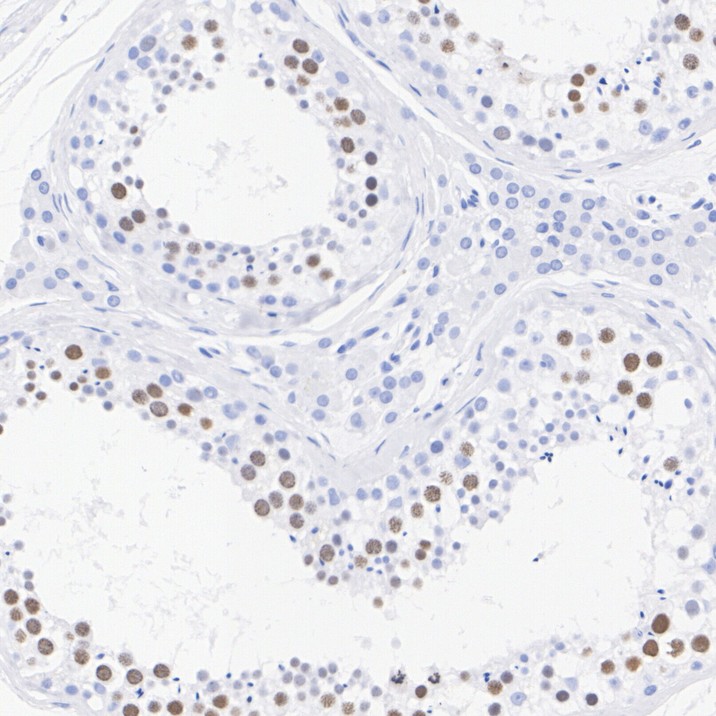
Immunohistochemical analysis of paraffin-embedded human testis tissue with Rabbit anti-Topoisomerase II alpha antibody at 1/1,000 dilution. The section was pre-treated using heat mediated antigen retrieval with sodium citrate buffer (pH 6.0) (high pressure) for 2 minutes. The tissues were blocked in 1% BSA for 20 minutes at room temperature, washed with ddH2O and PBS, and then probed with the primary antibody at 1/1,000 dilution for 1 hour at room temperature. The detection was performed using an HRP conjugated compact polymer system. DAB was used as the chromogen. Tissues were counterstained with hematoxylin and mounted with DPX.IHC
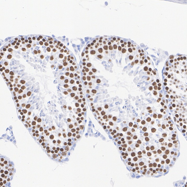
Immunohistochemical analysis of paraffin-embedded mouse testis tissue with Rabbit anti-Topoisomerase II alpha antibody at 1/1,000 dilution. The section was pre-treated using heat mediated antigen retrieval with sodium citrate buffer (pH 6.0) (high pressure) for 2 minutes. The tissues were blocked in 1% BSA for 20 minutes at room temperature, washed with ddH2O and PBS, and then probed with the primary antibody at 1/1,000 dilution for 1 hour at room temperature. The detection was performed using an HRP conjugated compact polymer system. DAB was used as the chromogen. Tissues were counterstained with hematoxylin and mounted with DPX.IHC
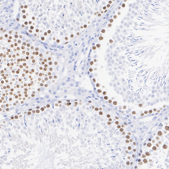
Immunohistochemical analysis of paraffin-embedded rat testis tissue with Rabbit anti-Topoisomerase II alpha antibody at 1/1,000 dilution. The section was pre-treated using heat mediated antigen retrieval with sodium citrate buffer (pH 6.0) (high pressure) for 2 minutes. The tissues were blocked in 1% BSA for 20 minutes at room temperature, washed with ddH2O and PBS, and then probed with the primary antibody at 1/1,000 dilution for 1 hour at room temperature. The detection was performed using an HRP conjugated compact polymer system. DAB was used as the chromogen. Tissues were counterstained with hematoxylin and mounted with DPX.| Product Name | Topoisomerase II alpha Recombinant Rabbit Monoclonal Antibody |
|---|---|
| Antibody Type | Primary Antibodies |
| Product description | DNA topoisomerase I and II (Topo I and Topo II) are nuclear enzymes that regulate the topological structure of DNA in eukaryotic cells by transiently breaking and rejoining DNA strands. Eukaryotic topoisomerases are capable of relaxing both positive and negative supercoils, whereas prokaryotic topoisomerases relax only negative supercoils. DNA topoisomerases play a role in DNA replication, recombination, and transcription and have been identified as targets of numerous anticancer drugs. Topo I, a ubiquitously expressed, soluble enzyme, acts by introducing a transient break in one strand of DNA, while Topo II acts by making a transient double-strand break. Topo II is encoded by two different genes to generate two distinct isoforms that are designated Topo IIα and Topo IIβ. Topo IIβ and Topo IIα, are largely homologous at their N-terminal three quarters, however, the C-terminal segments are considerably divergent, suggesting that these regions may mediate different cellular functions and account for the observed differential tissue expression patterns of the two isoforms. |
| Immunogen | Synthetic peptide within Human Topoisomerase Ⅱ alphaaa 1,482-1,531 / 1,531. |
| Clonality | Monoclonal |
|---|---|
| Isotype | IgG |
| Host Species | Recombinant rabbit |
| Tested Applications | IHCIPWB |
| WB:1:500-1:2000 IHC:1:200-1:1000 |
|
| Species Reactivity | HumanMouseRat |
| Concentration | 1mg/ml |
| Purification | Protein A |
| Alternative Names | alpha isozyme antibody ATP hydrolyzing DNA topoisomerase II alfa antibody DNA gyrase antibody DNA topoisomerase (ATP hydrolyzing) antibody DNA topoisomerase 2 alpha antibody DNA topoisomerase 2-alpha antibody DNA topoisomerase II 170 kD antibody DNA topoisomerase II alpha isozyme antibody DNA topoisomerase II antibody DNA Topoisomerase2 antibody TOP 2A antibody TOP2 antibody TOP2A antibody TOP2A_HUMAN antibody Topoisomerase DNA II alpha 170kDa antibody TP2A antibody |
|---|---|
| Molecular Weight(MW) | 174 kDa |
| Cellular Localization | Nucleoplasm, Cytoplasm. |
WB
Western blot analysis of Topoisomerase II alpha on different lysates with Rabbit anti-Topoisomerase II alpha antibody at 1/1,000 dilution. Lane 1: Rat testis tissue lysate (40 µg/Lane), Lane 2: Mouse testis tissue lysate (40 µg/Lane), Lane 3: NIH/3T3 cell lysate (20 µg/Lane), Lane 4: HT-29 cell lysate (20 µg/Lane), Lane 5: Jurkat cell lysate (20 µg/Lane), Exposure time: Lane 1-4: 3 minutes; Lane 5: 18 seconds; 4-20% SDS-PAGE gel. Proteins were transferred to a PVDF membrane and blocked with 5% NFDM/TBST for 1 hour at room temperature. The primary antibody at 1/1,000 dilution was used in 5% NFDM/TBST at 4℃ overnight. Goat Anti-Rabbit IgG - HRP Secondary Antibody at 1/50,000 dilution was used for 1 hour at room temperature.
IHC
Immunohistochemical analysis of paraffin-embedded human testis tissue with Rabbit anti-Topoisomerase II alpha antibody at 1/1,000 dilution. The section was pre-treated using heat mediated antigen retrieval with sodium citrate buffer (pH 6.0) (high pressure) for 2 minutes. The tissues were blocked in 1% BSA for 20 minutes at room temperature, washed with ddH2O and PBS, and then probed with the primary antibody at 1/1,000 dilution for 1 hour at room temperature. The detection was performed using an HRP conjugated compact polymer system. DAB was used as the chromogen. Tissues were counterstained with hematoxylin and mounted with DPX.
IHC
Immunohistochemical analysis of paraffin-embedded mouse testis tissue with Rabbit anti-Topoisomerase II alpha antibody at 1/1,000 dilution. The section was pre-treated using heat mediated antigen retrieval with sodium citrate buffer (pH 6.0) (high pressure) for 2 minutes. The tissues were blocked in 1% BSA for 20 minutes at room temperature, washed with ddH2O and PBS, and then probed with the primary antibody at 1/1,000 dilution for 1 hour at room temperature. The detection was performed using an HRP conjugated compact polymer system. DAB was used as the chromogen. Tissues were counterstained with hematoxylin and mounted with DPX.
IHC
Immunohistochemical analysis of paraffin-embedded rat testis tissue with Rabbit anti-Topoisomerase II alpha antibody at 1/1,000 dilution. The section was pre-treated using heat mediated antigen retrieval with sodium citrate buffer (pH 6.0) (high pressure) for 2 minutes. The tissues were blocked in 1% BSA for 20 minutes at room temperature, washed with ddH2O and PBS, and then probed with the primary antibody at 1/1,000 dilution for 1 hour at room temperature. The detection was performed using an HRP conjugated compact polymer system. DAB was used as the chromogen. Tissues were counterstained with hematoxylin and mounted with DPX.| Application Notes | WB:1:500-1:2000 IHC:1:200-1:1000 |
|---|
| Form | Liquid |
|---|---|
| Storage Instructions | Store at +4℃ after thawing. Aliquot store at -20℃ or -80℃. Avoid repeated freeze / thaw cycles. |
| Storage Buffer | 1*TBS (pH7.4), 1%BSA, 40%Glycerol. Preservative: 0.05% Sodium Azide. |


