Application
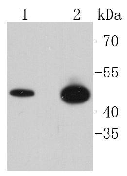
Fig1: Western blot analysis of Phospho-GSK3 beta(Ser 9) on different lysates using anti-Phospho-GSK3 beta(Ser 9) antibody at 1/1,000 dilution. Positive control: Lane 1: Hela Lane 2: MCF-7Application
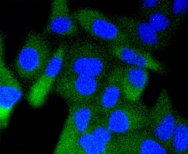
Fig2: ICC staining Phospho-GSK3 beta(Ser 9) in Hela cells (green). The nuclear counter stain is DAPI (blue). Cells were fixed in paraformaldehyde, permeabilised with 0.25% Triton X100/PBS.Application
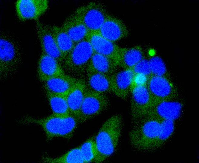
Fig3: ICC staining Phospho-GSK3 beta(Ser 9) in MCF-7 cells (green). The nuclear counter stain is DAPI (blue). Cells were fixed in paraformaldehyde, permeabilised with 0.25% Triton X100/PBS.Application
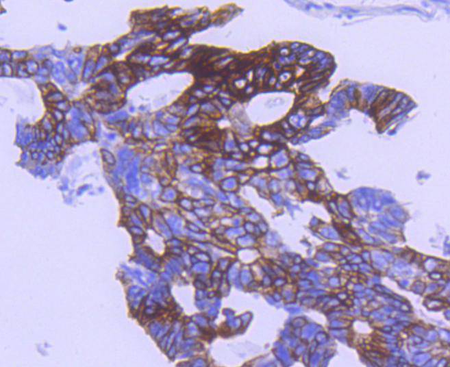
Fig4: Immunohistochemical analysis of paraffin-embedded human colon cancer tissue using anti-Phospho-GSK3 beta(Ser 9) antibody. Counter stained with hematoxylin.Application
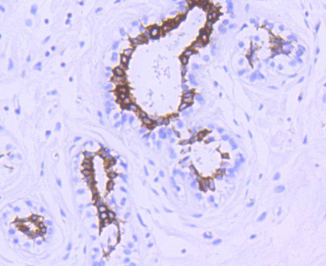
Fig5: Immunohistochemical analysis of paraffin-embedded human breast tissue using anti-Phospho-GSK3 beta(Ser 9) antibody. Counter stained with hematoxylin.Application
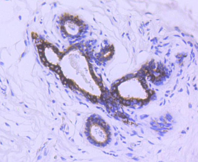
Fig6: Immunohistochemical analysis of paraffin-embedded human breast carcinoma tissue using anti-Phospho-GSK3 beta(Ser 9) antibody. Counter stained with hematoxylin.Application
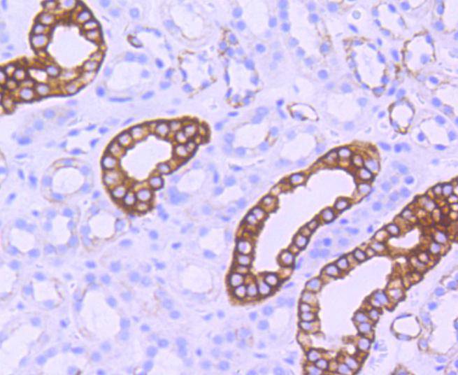
Fig7: Immunohistochemical analysis of paraffin-embedded human kidney tissue using anti-Phospho-GSK3 beta(Ser 9) antibody. Counter stained with hematoxylin.Application
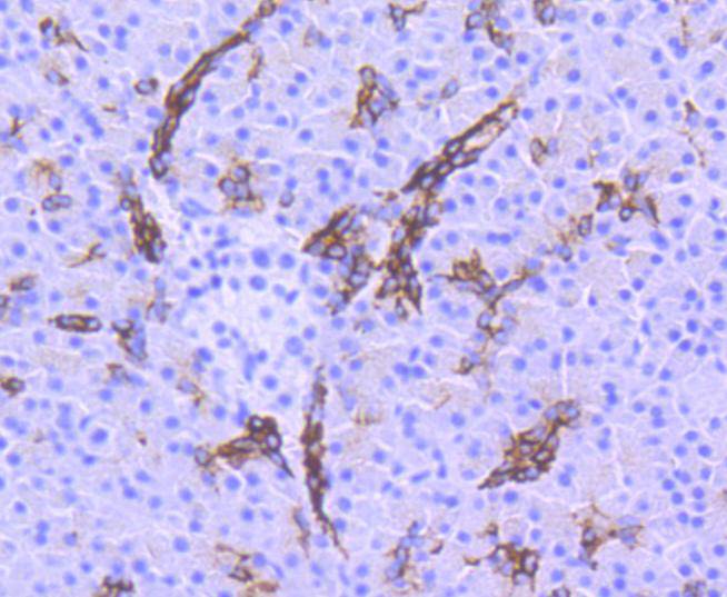
Fig8: Immunohistochemical analysis of paraffin-embedded human pancreas tissue using anti-Phospho-GSK3 beta(Ser 9) antibody. Counter stained with hematoxylin.| Product Name | Phospho-GSK3 beta(Ser 9) [SY02-71] |
|---|---|
| Antibody Type | Primary Antibodies |
| Antigen Alias | Glycogen Synthase Kinase 3 Beta antibody Glycogen synthase kinase-3 beta antibody GSK 3 beta antibody GSK-3 beta antibody GSK3B antibody GSK3B_HUMAN antibody GSK3beta isoform antibody Serine/threonine-protein kinase GSK3B antibody |
| Product description | Glycogen synthase kinase-3α (GSK-3α) and GSK-3β are highly similar isoforms of serine/ threonine kinases that regulate metabolic enzymes and transcription factors, which are responsible for coordinating processes such as glycogen synthesis and cell adhesion. GSK-3β activity is also required for nuclear activity of Rel dimers, which mediate an anti-apoptotic response to TNFα in mice. GSK-3 catalytic kinase activity is controlled through differential phosphorylation of serine/threonine residues, which have an inhibitory effect, and tyrosine residues, which have an activating effect. Growth factor stimulation of mammalian cells expressing GSK-3α and GSK-3β induces phosphorylation of Ser 21 and Ser 9, respectively, through a phosphatidylinositol 3-kinase (PI 3-K)-protein kinase B (PKB)-dependent pathway, thereby enhancing proliferative signals. Additionally, GSK-3 physically associates with cAMP-dependent protein kinase A (PKA), which phosphorylates Ser 21 of GSK-3α or Ser 9 of GSK-3β and inactivates both forms. GSK-3α/β is positively regulated by phosphorylation on Tyr 279 and Tyr 216, respectively. Activated GSK-3α/β participates in energy metabolism, neuronal cell development, and body pattern formation. Tyrosine dephosphorylation of GSK-3 is involved in its extracellular signal-dependent inactivation. |
| Immunogen | Synthetic phospho-peptide corresponding to residues surrounding Ser9 of human GSK3 beta. |
| Modification | p-Ser 9 |
| Clonality | Monoclonal |
|---|---|
| Isotype | IgG |
| Host Species | Recombinant rabbit |
| Tested Applications | WBICC/IFIHC |
| WB:1:1,000-1:2,000 ICC:1:50-1:200 IHC:1:50-1:200 |
|
| Species Reactivity | Human |
| Concentration | 1mg/ml |
| Alternative Names | Glycogen Synthase Kinase 3 Beta antibody Glycogen synthase kinase-3 beta antibody GSK 3 beta antibody GSK-3 beta antibody GSK3B antibody GSK3B_HUMAN antibody GSK3beta isoform antibody Serine/threonine-protein kinase GSK3B antibody |
|---|---|
| Molecular Weight(MW) | 47 kDa |
| Cellular Localization | Cytoplasm, Nucleus, Cell membrane. |
| SwissProt ID | P49841 |
|---|
Application
Fig1: Western blot analysis of Phospho-GSK3 beta(Ser 9) on different lysates using anti-Phospho-GSK3 beta(Ser 9) antibody at 1/1,000 dilution. Positive control: Lane 1: Hela Lane 2: MCF-7
Application
Fig2: ICC staining Phospho-GSK3 beta(Ser 9) in Hela cells (green). The nuclear counter stain is DAPI (blue). Cells were fixed in paraformaldehyde, permeabilised with 0.25% Triton X100/PBS.
Application
Fig3: ICC staining Phospho-GSK3 beta(Ser 9) in MCF-7 cells (green). The nuclear counter stain is DAPI (blue). Cells were fixed in paraformaldehyde, permeabilised with 0.25% Triton X100/PBS.
Application
Fig4: Immunohistochemical analysis of paraffin-embedded human colon cancer tissue using anti-Phospho-GSK3 beta(Ser 9) antibody. Counter stained with hematoxylin.
Application
Fig5: Immunohistochemical analysis of paraffin-embedded human breast tissue using anti-Phospho-GSK3 beta(Ser 9) antibody. Counter stained with hematoxylin.
Application
Fig6: Immunohistochemical analysis of paraffin-embedded human breast carcinoma tissue using anti-Phospho-GSK3 beta(Ser 9) antibody. Counter stained with hematoxylin.
Application
Fig7: Immunohistochemical analysis of paraffin-embedded human kidney tissue using anti-Phospho-GSK3 beta(Ser 9) antibody. Counter stained with hematoxylin.
Application
Fig8: Immunohistochemical analysis of paraffin-embedded human pancreas tissue using anti-Phospho-GSK3 beta(Ser 9) antibody. Counter stained with hematoxylin.| Positive Control | MCF-7, Hela, human colon cancer tissue, human kidney tissue, human breast tissue, human pancreas tissue, human breast carcinoma tissue. |
|---|---|
| Application Notes | WB:1:1,000-1:2,000 ICC:1:50-1:200 IHC:1:50-1:200 |
| Form | Liquid |
|---|---|
| Storage Instructions | Store at +4℃ after thawing. Aliquot store at -20℃ or -80℃. Avoid repeated freeze / thaw cycles. |
| Storage Buffer | 1*TBS (pH7.4), 1%BSA, 40%Glycerol. Preservative: 0.05% Sodium Azide. |


