Application
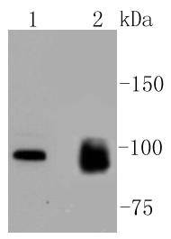
Fig1: Western blot analysis of MKLP1 on different lysates using anti-MKLP1 antibody at 1/1,000 dilution. Positive control: Lane 1: A549 Lane 2: HelaApplication
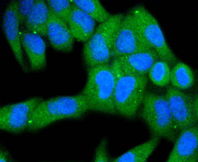
Fig2: ICC staining MKLP1 in Hela cells (green). The nuclear counter stain is DAPI (blue). Cells were fixed in paraformaldehyde, permeabilised with 0.25% Triton X100/PBS.Application
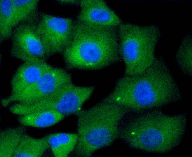
Fig3: ICC staining MKLP1 in A549 cells (green). The nuclear counter stain is DAPI (blue). Cells were fixed in paraformaldehyde, permeabilised with 0.25% Triton X100/PBS.| Product Name | MKLP1 [SY02-74] |
|---|---|
| Antibody Type | Primary Antibodies |
| Antigen Alias | CHO 1 antibody CHO1 antibody KIF 23 antibody KIF23 antibody KIF23_HUMAN antibody Kinesin family member 23 antibody Kinesin like 5 antibody Kinesin like protein 5 antibody Kinesin like protein KIF 23 antibody Kinesin like protein KIF23 antibody Kinesin-like protein 5 antibody Kinesin-like protein KIF23 antibody KNS L5 antibody KNSL 5 antibody KNSL5 antibody Mitotic kinesin like 1 antibody Mitotic kinesin like protein 1 antibody Mitotic kinesin-like protein 1 antibody MKLP 1 antibody |
| Product description | The monoclonal antibody CHO1 detects a spindle antigen required for mitotic progression. Screening a HeLa cell cDNA expression library with this antibody has been shown to yield a cDNA predicted to encode a protein significantly related within its amino terminal half to the motor ends of members of the kinesin superfamily. Since this similarity does not extend further, it has been suggested that the CHO1 antigen, now designated MKLP-1 (mitotic kinesin-like protein-1), represents a novel kinesin. Sequence analysis has also been shown to predict that MKLP-1 possesses features typical of nuclear proteins. Immunocytological studies have demonstrated that MKLP-1 moves from the nucleus early in mitosis and then to the midbody after cytokinesis. MKLP-1 has been shown to bundle antiparallel microtubules in vitro and to move them at rates comparable to spindle elongation in vivo. A hamster homolog of MKLP-1, designated CHO1 antigen, has also been isolated. Although apparently functionally equivalent with respect to microtubule bundling activity, there are significant differences between the human and hamster proteins at their C-termini, possibly due to alternative splicing or the presence of more than one MKLP-1 locus. |
| Immunogen | recombinant protein |
| Clonality | Monoclonal |
|---|---|
| Isotype | IgG |
| Host Species | Recombinant rabbit |
| Tested Applications | WBICC/IF |
| WB:1:1,000-1:5,000 ICC:1:50-1:200 |
|
| Species Reactivity | HumanMouseRat |
| Concentration | 1mg/ml |
| Alternative Names | CHO 1 antibody CHO1 antibody KIF 23 antibody KIF23 antibody KIF23_HUMAN antibody Kinesin family member 23 antibody Kinesin like 5 antibody Kinesin like protein 5 antibody Kinesin like protein KIF 23 antibody Kinesin like protein KIF23 antibody Kinesin-like protein 5 antibody Kinesin-like protein KIF23 antibody KNS L5 antibody KNSL 5 antibody KNSL5 antibody Mitotic kinesin like 1 antibody Mitotic kinesin like protein 1 antibody Mitotic kinesin-like protein 1 antibody MKLP 1 antibody |
|---|---|
| Molecular Weight(MW) | 98 kDa |
| Cellular Localization | Cytoplasm, Nucleus, Midbody. |
Application
Fig1: Western blot analysis of MKLP1 on different lysates using anti-MKLP1 antibody at 1/1,000 dilution. Positive control: Lane 1: A549 Lane 2: Hela
Application
Fig2: ICC staining MKLP1 in Hela cells (green). The nuclear counter stain is DAPI (blue). Cells were fixed in paraformaldehyde, permeabilised with 0.25% Triton X100/PBS.
Application
Fig3: ICC staining MKLP1 in A549 cells (green). The nuclear counter stain is DAPI (blue). Cells were fixed in paraformaldehyde, permeabilised with 0.25% Triton X100/PBS.| Positive Control | A549, Hela. |
|---|---|
| Application Notes | WB:1:1,000-1:5,000 ICC:1:50-1:200 |
| Form | Liquid |
|---|---|
| Storage Instructions | Store at +4℃ after thawing. Aliquot store at -20℃ or -80℃. Avoid repeated freeze / thaw cycles. |
| Storage Buffer | 1*TBS (pH7.4), 1%BSA, 40%Glycerol. Preservative: 0.05% Sodium Azide. |


