Application
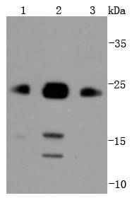
Fig1: Western blot analysis of Bim on different lysates using anti-Bim antibody at 1/1,000 dilution. Positive control: Lane 1: Raji Lane 2: Jurkat Lane 3: A431Application
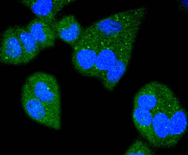
Fig2: ICC staining Bim in Hela cells (green). The nuclear counter stain is DAPI (blue). Cells were fixed in paraformaldehyde, permeabilised with 0.25% Triton X100/PBS.Application
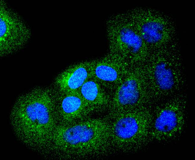
Fig3: ICC staining Bim in A431 cells (green). The nuclear counter stain is DAPI (blue). Cells were fixed in paraformaldehyde, permeabilised with 0.25% Triton X100/PBS.Application
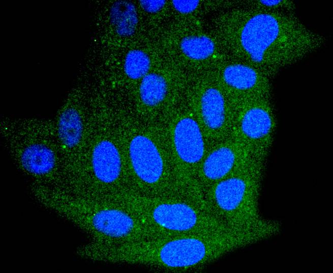
Fig4: ICC staining Bim in HepG2 cells (green). The nuclear counter stain is DAPI (blue). Cells were fixed in paraformaldehyde, permeabilised with 0.25% Triton X100/PBS.Application
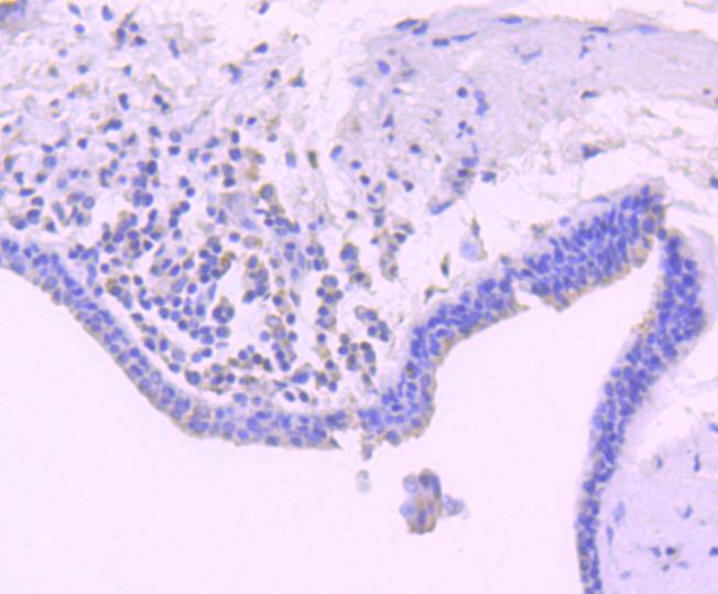
Fig5: Immunohistochemical analysis of paraffin-embedded human breast carcinoma tissue using anti-Bim antibody. Counter stained with hematoxylin.| Product Name | Bim [SU0318] |
|---|---|
| Antibody Type | Primary Antibodies |
| Antigen Alias | BCL2 like 11 antibody B2L11_HUMAN antibody BAM antibody Bcl 2 interacting protein Bim antibody Bcl 2 related ovarian death agonist antibody Bcl-2-like protein 11 antibody BCL2 interacting mediator of cell death antibody BCL2 like 11 (apoptosis facilitator) antibody BCL2 like protein 11 antibody Bcl2-interacting mediator of cell death antibody Bcl2-L-11 antibody Bcl2l11 antibody BIM alpha6 antibody BIM antibody BIM beta6 antibody BIM beta7 antibody BimEL antibody BimL antibody BOD antibody |
| Product description | Pro-apototic Bcl-2 family members promote cell death by neutralizing their anti-apoptotic relatives, which otherwise maintain cell viability by regulating caspase activity. Bim belongs to the BH3-only subgroup of Bcl-2 related proteins, and exists in three distinct isoforms, BimS (short), BimL (long) and BimEL (extra long). ERK1/2 phosphorylates BimEL, resulting in rapid degradation of the isoform via the proteasome pathway. At least three sites for ERK1/2 phosphorylation exist on BimEL, whereas ERK1/2 does not effect BimS and BimL, implying a unique role for BimEL in cell survival signaling. |
| Immunogen | recombinant protein |
| Clonality | Monoclonal |
|---|---|
| Isotype | IgG |
| Host Species | Recombinant rabbit |
| Tested Applications | WBICCIHCIP |
| WB:1:1,000-1:2,000 ICC:1:50-1:200 IHC:1:50-1:200 |
|
| Species Reactivity | Human |
| Concentration | 1mg/ml |
| Alternative Names | BCL2 like 11 antibody B2L11_HUMAN antibody BAM antibody Bcl 2 interacting protein Bim antibody Bcl 2 related ovarian death agonist antibody Bcl-2-like protein 11 antibody BCL2 interacting mediator of cell death antibody BCL2 like 11 (apoptosis facilitator) antibody BCL2 like protein 11 antibody Bcl2-interacting mediator of cell death antibody Bcl2-L-11 antibody Bcl2l11 antibody BIM alpha6 antibody BIM antibody BIM beta6 antibody BIM beta7 antibody BimEL antibody BimL antibody BOD antibody |
|---|---|
| Molecular Weight(MW) | 22/16/13 kDa |
| Cellular Localization | Endomembrane system, Mitochondrion. |
| SwissProt ID | O43521 |
|---|
Application
Fig1: Western blot analysis of Bim on different lysates using anti-Bim antibody at 1/1,000 dilution. Positive control: Lane 1: Raji Lane 2: Jurkat Lane 3: A431
Application
Fig2: ICC staining Bim in Hela cells (green). The nuclear counter stain is DAPI (blue). Cells were fixed in paraformaldehyde, permeabilised with 0.25% Triton X100/PBS.
Application
Fig3: ICC staining Bim in A431 cells (green). The nuclear counter stain is DAPI (blue). Cells were fixed in paraformaldehyde, permeabilised with 0.25% Triton X100/PBS.
Application
Fig4: ICC staining Bim in HepG2 cells (green). The nuclear counter stain is DAPI (blue). Cells were fixed in paraformaldehyde, permeabilised with 0.25% Triton X100/PBS.
Application
Fig5: Immunohistochemical analysis of paraffin-embedded human breast carcinoma tissue using anti-Bim antibody. Counter stained with hematoxylin.| Positive Control | A431, Jurkat, Hela, HepG2, Raji, human breast carcinoma tissue. |
|---|---|
| Application Notes | WB:1:1,000-1:2,000 ICC:1:50-1:200 IHC:1:50-1:200 |
| Form | Liquid |
|---|---|
| Storage Instructions | Store at +4℃ after thawing. Aliquot store at -20℃ or -80℃. Avoid repeated freeze / thaw cycles. |
| Storage Buffer | 1*TBS (pH7.4), 1%BSA, 40%Glycerol. Preservative: 0.05% Sodium Azide. |


