WB
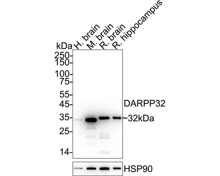
Western blot analysis of DARPP32 on different lysates with Rabbit anti-DARPP32 antibody at 1/2,000 dilution. Lane 1: Human brain tissue lysate (40 µg/Lane), Lane 2: Mouse brain tissue lysate (40 µg/Lane), Lane 3: Rat brain tissue lysate (40 µg/Lane), Lane 4: Rat hippocampus tissue lysate (40 µg/Lane), Exposure time: 12 seconds; 4-20% SDS-PAGE gel. Proteins were transferred to a PVDF membrane and blocked with 5% NFDM/TBST for 1 hour at room temperature. The primary antibody at 1/2,000 dilution was used in 5% NFDM/TBST at 4℃ overnight. Goat Anti-Rabbit IgG - HRP Secondary Antibody at 1/50,000 dilution was used for 1 hour at room temperature.IHC
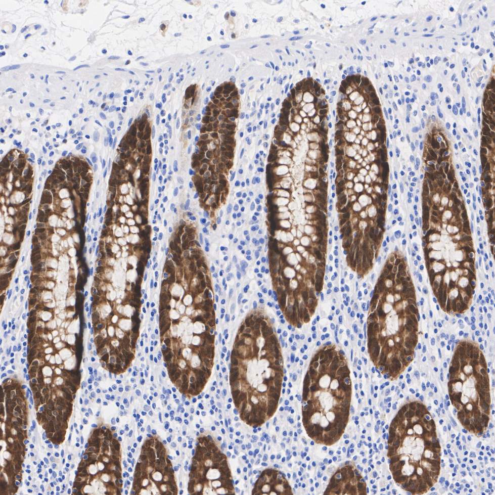
Immunohistochemical analysis of paraffin-embedded human colon tissue with Rabbit anti-DARPP32 antibody at 1/500 dilution. The section was pre-treated using heat mediated antigen retrieval with Tris-EDTA buffer (pH 9.0) for 20 minutes. The tissues were blocked in 1% BSA for 20 minutes at room temperature, washed with ddH2O and PBS, and then probed with the primary antibody at 1/500 dilution for 1 hour at room temperature. The detection was performed using an HRP conjugated compact polymer system. DAB was used as the chromogen. Tissues were counterstained with hematoxylin and mounted with DPX.IHC
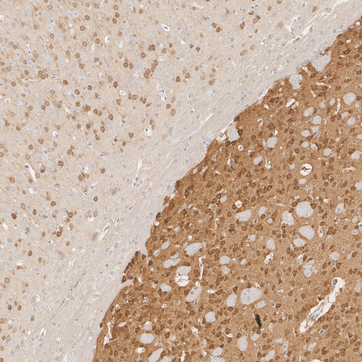
Immunohistochemical analysis of paraffin-embedded mouse forebrain tissue with Rabbit anti-DARPP32 antibody at 1/1,000 dilution. The section was pre-treated using heat mediated antigen retrieval with Tris-EDTA buffer (pH 9.0) for 20 minutes. The tissues were blocked in 1% BSA for 20 minutes at room temperature, washed with ddH2O and PBS, and then probed with the primary antibody at 1/1,000 dilution for 1 hour at room temperature. The detection was performed using an HRP conjugated compact polymer system. DAB was used as the chromogen. Tissues were counterstained with hematoxylin and mounted with DPX.IHC
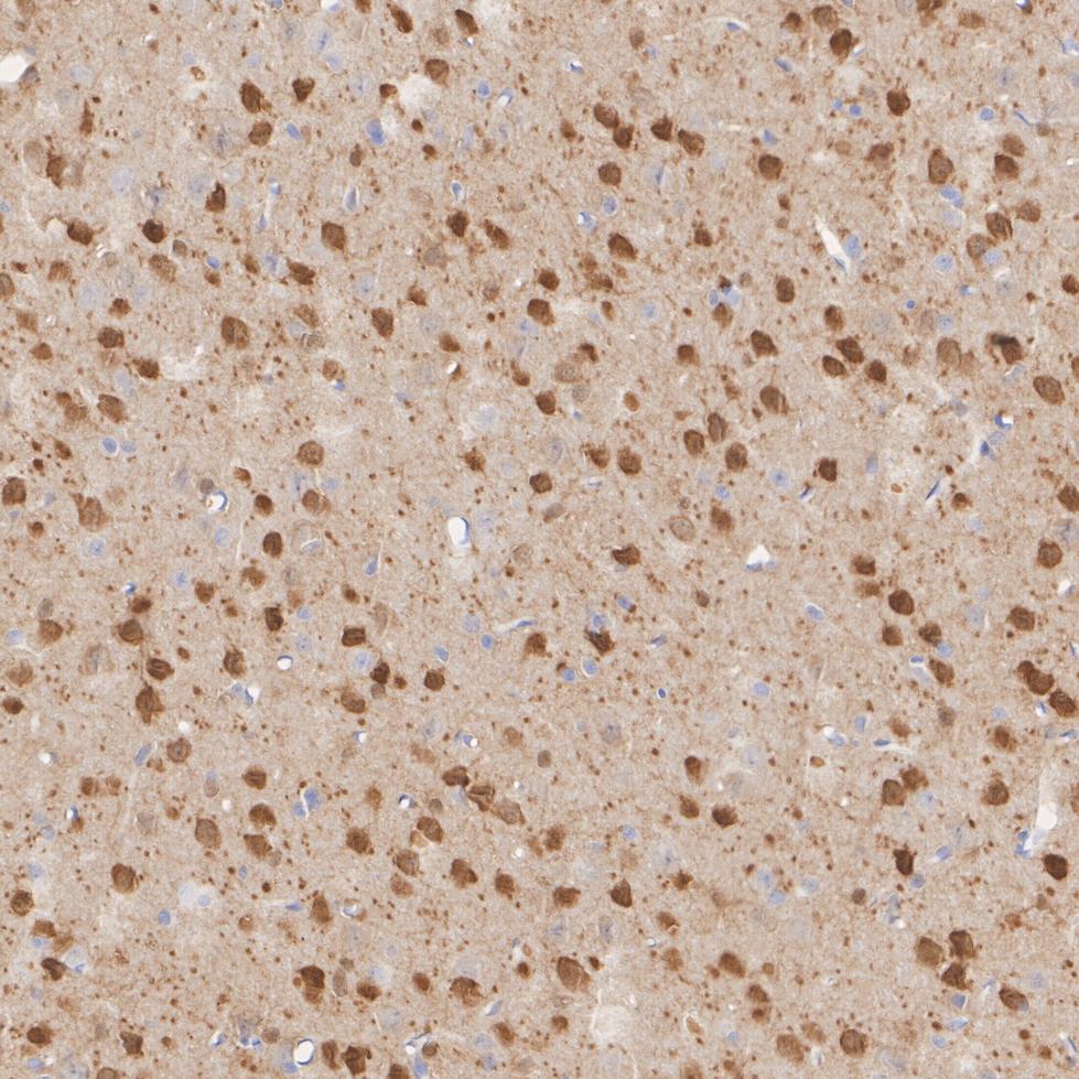
Immunohistochemical analysis of paraffin-embedded rat brian tissue with Rabbit anti-DARPP32 antibody at 1/1,000 dilution. The section was pre-treated using heat mediated antigen retrieval with Tris-EDTA buffer (pH 9.0) for 20 minutes. The tissues were blocked in 1% BSA for 20 minutes at room temperature, washed with ddH2O and PBS, and then probed with the primary antibody at 1/1,000 dilution for 1 hour at room temperature. The detection was performed using an HRP conjugated compact polymer system. DAB was used as the chromogen. Tissues were counterstained with hematoxylin and mounted with DPX.IF-F
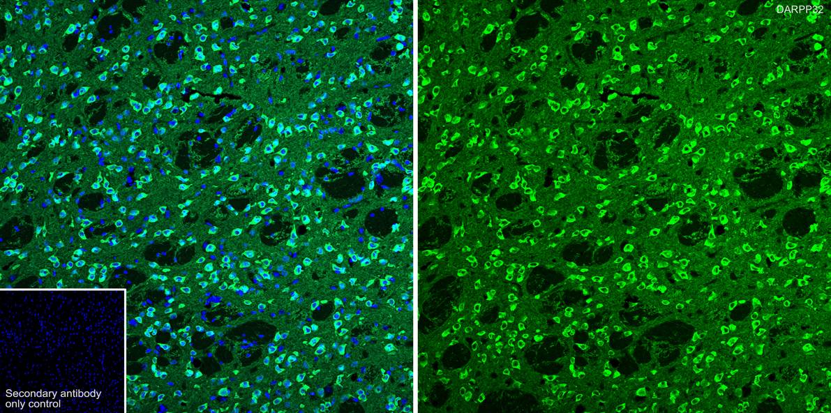
Species: Mouse, Site: Striatum, Sample: Frozen section, Antibody concentration: 1/500, Antigen retrieval: Recommend. The section was pre-treated using heat mediated antigen retrieval with sodium citrate buffer (pH 6.0) for about 2 minutes in microwave oven.IF-F
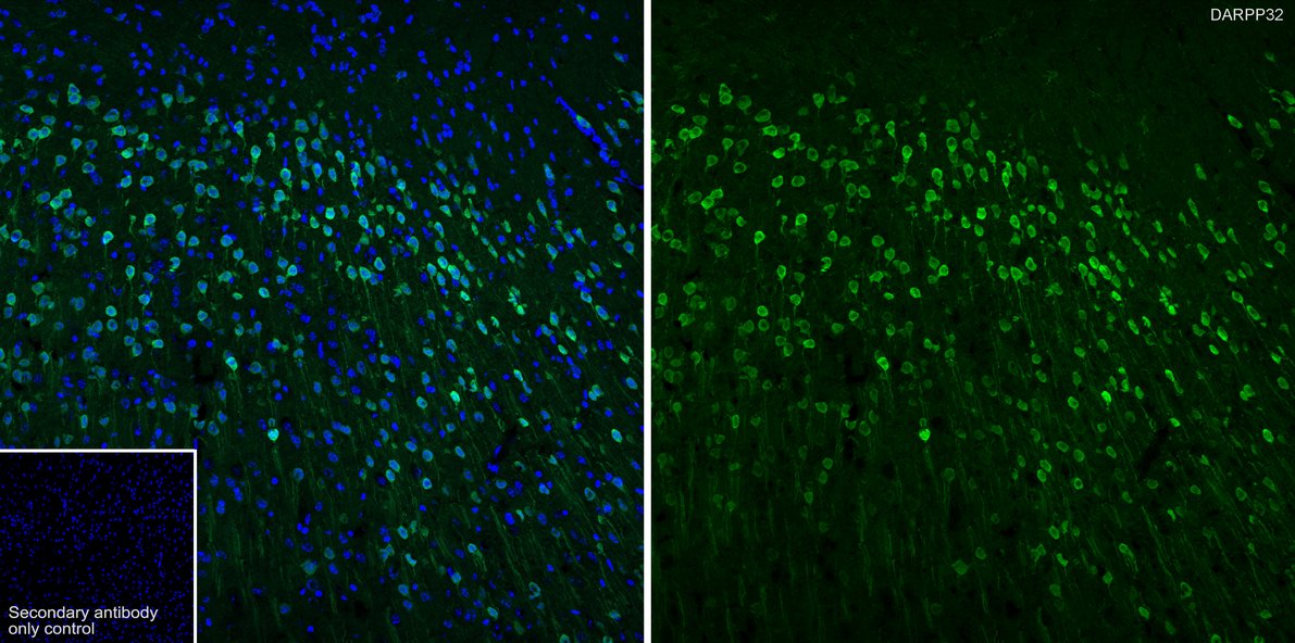
Species: Mouse, Site: Cerebral cortex, Sample: Frozen section, Antibody concentration: 1/500, Antigen retrieval: Recommend. The section was pre-treated using heat mediated antigen retrieval with sodium citrate buffer (pH 6.0) for about 2 minutes in microwave oven.IF-F
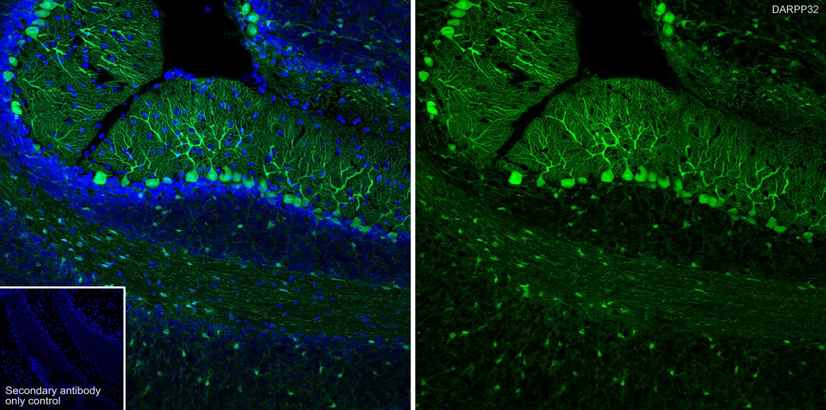
Species: Mouse, Site: Cerebellum, Sample: Frozen section, Antibody concentration: 1/500, Antigen retrieval: Recommend. The section was pre-treated using heat mediated antigen retrieval with sodium citrate buffer (pH 6.0) for about 2 minutes in microwave oven.| Product Name | DARPP32 Recombinant Rabbit Monoclonal Antibody |
|---|---|
| Antibody Type | Primary Antibodies |
| Product description | Dopaminergic signaling pathways, which are essential for multiple brain functions, are abnormal in several neurological disorders, such as schizophrenia, Parkinson's disease and drug abuse. DARPP-32 (for dopamine and adenosine 3',5'-monophosphate-regulated phosphoprotein) is abundant in neurons that receive dopaminergic input. Activation of PKA and the consequent phosphorylation of DARPP-32 on threonine occurs in response to dopamine acting upon D1-like receptors. Dopamine interaction with D2-like receptors results in the inhibition of PKA activation, the activation of protein phosphatase 2B and the consequent dephosphorylation of DARPP-32. Neurotransmitters other than dopamine may also be able to stimulate the phosphorylation or dephosphorylation of DARPP-32. Phosphorylated DARPP-32 is a potent inhibitor of PP-1. |
| Immunogen | Recombinant protein within Human DARPP32 aa 1-94 / 204. |
| Clonality | Monoclonal |
|---|---|
| Isotype | IgG |
| Host Species | Recombinant rabbit |
| Tested Applications | IF-FIHCWB |
| WB:1:1000-1:5000 IHC:1:500-1:1000 IF-F:1:500 |
|
| Species Reactivity | HumanMouseRat |
| Concentration | 1mg/ml |
| Purification | Protein A |
| Alternative Names | DARPP 32 antibody DARPP-32 antibody Dopamine and cAMP regulated neuronal phosphoprotein 32 antibody Dopamine and cAMP regulated neuronal phosphoprotein antibody Dopamine and cAMP regulated phosphoprotein antibody Dopamine and cAMP regulated phosphoprotein DARPP 32 antibody Dopamine and cAMP regulated phosphoprotein DARPP32 antibody Dopamine- and cAMP-regulated neuronal phosphoprotein antibody FLJ20940 antibody IPPD antibody Neuronal phosphoprotein DARPP 32 antibody PPP1R1B antibody PPR1B_HUMAN antibody Protein phosphatase 1 regulatory (inhibitor) subunit 1B antibody Protein phosphatase 1 regulatory inhibitor subunit 1B antibody Protein phosphatase 1 regulatory subunit 1B antibody |
|---|---|
| Molecular Weight(MW) | 23kDa(Observed band size: 32kDa) |
| Cellular Localization | Cytoplasm. |
WB
Western blot analysis of DARPP32 on different lysates with Rabbit anti-DARPP32 antibody at 1/2,000 dilution. Lane 1: Human brain tissue lysate (40 µg/Lane), Lane 2: Mouse brain tissue lysate (40 µg/Lane), Lane 3: Rat brain tissue lysate (40 µg/Lane), Lane 4: Rat hippocampus tissue lysate (40 µg/Lane), Exposure time: 12 seconds; 4-20% SDS-PAGE gel. Proteins were transferred to a PVDF membrane and blocked with 5% NFDM/TBST for 1 hour at room temperature. The primary antibody at 1/2,000 dilution was used in 5% NFDM/TBST at 4℃ overnight. Goat Anti-Rabbit IgG - HRP Secondary Antibody at 1/50,000 dilution was used for 1 hour at room temperature.
IHC
Immunohistochemical analysis of paraffin-embedded human colon tissue with Rabbit anti-DARPP32 antibody at 1/500 dilution. The section was pre-treated using heat mediated antigen retrieval with Tris-EDTA buffer (pH 9.0) for 20 minutes. The tissues were blocked in 1% BSA for 20 minutes at room temperature, washed with ddH2O and PBS, and then probed with the primary antibody at 1/500 dilution for 1 hour at room temperature. The detection was performed using an HRP conjugated compact polymer system. DAB was used as the chromogen. Tissues were counterstained with hematoxylin and mounted with DPX.
IHC
Immunohistochemical analysis of paraffin-embedded mouse forebrain tissue with Rabbit anti-DARPP32 antibody at 1/1,000 dilution. The section was pre-treated using heat mediated antigen retrieval with Tris-EDTA buffer (pH 9.0) for 20 minutes. The tissues were blocked in 1% BSA for 20 minutes at room temperature, washed with ddH2O and PBS, and then probed with the primary antibody at 1/1,000 dilution for 1 hour at room temperature. The detection was performed using an HRP conjugated compact polymer system. DAB was used as the chromogen. Tissues were counterstained with hematoxylin and mounted with DPX.
IHC
Immunohistochemical analysis of paraffin-embedded rat brian tissue with Rabbit anti-DARPP32 antibody at 1/1,000 dilution. The section was pre-treated using heat mediated antigen retrieval with Tris-EDTA buffer (pH 9.0) for 20 minutes. The tissues were blocked in 1% BSA for 20 minutes at room temperature, washed with ddH2O and PBS, and then probed with the primary antibody at 1/1,000 dilution for 1 hour at room temperature. The detection was performed using an HRP conjugated compact polymer system. DAB was used as the chromogen. Tissues were counterstained with hematoxylin and mounted with DPX.
IF-F
Species: Mouse, Site: Striatum, Sample: Frozen section, Antibody concentration: 1/500, Antigen retrieval: Recommend. The section was pre-treated using heat mediated antigen retrieval with sodium citrate buffer (pH 6.0) for about 2 minutes in microwave oven.
IF-F
Species: Mouse, Site: Cerebral cortex, Sample: Frozen section, Antibody concentration: 1/500, Antigen retrieval: Recommend. The section was pre-treated using heat mediated antigen retrieval with sodium citrate buffer (pH 6.0) for about 2 minutes in microwave oven.
IF-F
Species: Mouse, Site: Cerebellum, Sample: Frozen section, Antibody concentration: 1/500, Antigen retrieval: Recommend. The section was pre-treated using heat mediated antigen retrieval with sodium citrate buffer (pH 6.0) for about 2 minutes in microwave oven.| Application Notes | WB:1:1000-1:5000 IHC:1:500-1:1000 IF-F:1:500 |
|---|
| Form | Liquid |
|---|---|
| Storage Instructions | Store at +4℃ after thawing. Aliquot store at -20℃ or -80℃. Avoid repeated freeze / thaw cycles. |
| Storage Buffer | 1*TBS (pH7.4), 1%BSA, 40%Glycerol. Preservative: 0.05% Sodium Azide. |


