WB
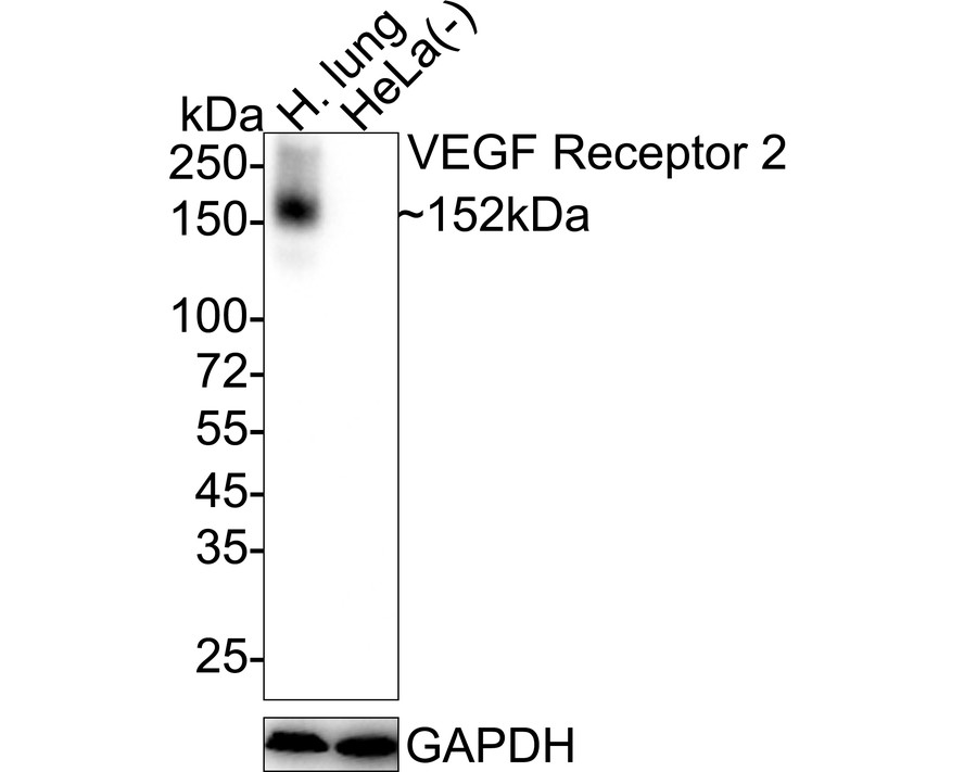
Western blot analysis of VEGF Receptor 2 on different lysates with Rabbit anti-VEGF Receptor 2 antibody at 1/1,000 dilution. Lane 1: Human lung tissue lysate, Lane 2: HeLa cell lysate (negative), Lysates/proteins at 20 µg/Lane. Exposure time: 1 minute 18 seconds; 4-20% SDS-PAGE gel. Proteins were transferred to a PVDF membrane and blocked with 5% NFDM/TBST for 1 hour at room temperature. The primary antibody at 1/1,000 dilution was used in 5% NFDM/TBST at 4℃ overnight. Goat Anti-Rabbit IgG - HRP Secondary Antibody at 1/50,000 dilution was used for 1 hour at room temperature.IHC
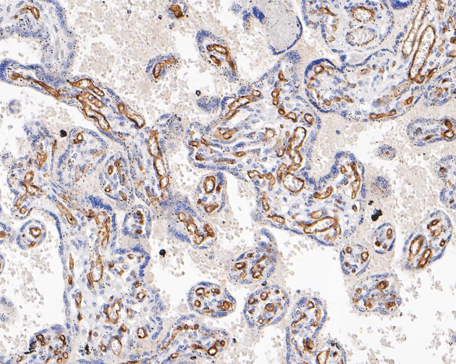
Immunohistochemical analysis of paraffin-embedded human placenta tissue with Rabbit anti-VEGF Receptor 2 antibody at 1/200 dilution. The section was pre-treated using heat mediated antigen retrieval with sodium citrate buffer (pH 6.0) (high pressure) for 2 minutes. The tissues were blocked in 1% BSA for 20 minutes at room temperature, washed with ddH2O and PBS, and then probed with the primary antibody at 1/200 dilution for 1 hour at room temperature. The detection was performed using an HRP conjugated compact polymer system. DAB was used as the chromogen. Tissues were counterstained with hematoxylin and mounted with DPX.IHC
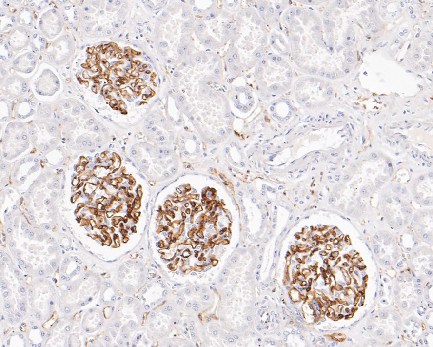
Immunohistochemical analysis of paraffin-embedded human kidney tissue with Rabbit anti-VEGF Receptor 2 antibody at 1/200 dilution. The section was pre-treated using heat mediated antigen retrieval with sodium citrate buffer (pH 6.0) (high pressure) for 2 minutes. The tissues were blocked in 1% BSA for 20 minutes at room temperature, washed with ddH2O and PBS, and then probed with the primary antibody at 1/200 dilution for 1 hour at room temperature. The detection was performed using an HRP conjugated compact polymer system. DAB was used as the chromogen. Tissues were counterstained with hematoxylin and mounted with DPX.ICC/IF
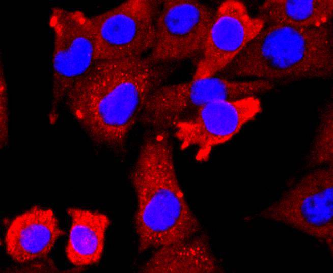
ICC staining VEGF Receptor 2 in PMVEC cells (red). The nuclear counter stain is DAPI (blue). Cells were fixed in paraformaldehyde, permeabilised with 0.25% Triton X100/PBS.| Product Name | VEGF Receptor 2 Recombinant Rabbit Monoclonal Antibody |
|---|---|
| Antibody Type | Primary Antibodies |
| Product description | Three cell membrane receptor tyrosine kinases, Flt (also designated VEGF-R1) (1-3), Flk-1 (also designated VEGF-R2) (4-6) and Flt-4, putatively involved in the growth of endothelial cells, are characterized by the presence of seven immunoglobulin-like sequences in their extracellular domain. These receptors exhibit high degrees of sequence relatedness to each other as well as lesser degrees of relatedness to the class III receptors including CSF-1/Fms, PDGR, SLFR/Kit and Flt-3/Flk-2. Two members of this receptor class, Flt-1 and Flk-1, have been shown to represent high affinity receptors for vascular endothelial growth factors (VEGFs) (3,5). On the basis of structural similarity to Flt and Flk-1, it has been speculated that Flt-4 might represent a third receptor for either VEGF or a VEGF-related ligand. |
| Immunogen | Recombinant protein within Human VEGF Receptor 2 aa 30-630 / 1,356. |
| Clonality | Monoclonal |
|---|---|
| Isotype | IgG |
| Host Species | Recombinant rabbit |
| Tested Applications | ICC/IFIHCWB |
| WB:1:500-1:1000 IHC:1:200 ICC/IF:1:50-1:200 |
|
| Species Reactivity | HumanRat |
| Concentration | 1mg/ml |
| Purification | Protein A |
| Alternative Names | CD309 antibody
CD309 antigen antibody
EC 2.7.10.1 antibody
Fetal liver kinase 1 antibody
FLK-1 antibody
FLK1 antibody
FLK1 mouse homolog of antibody Kdr antibody Kinase insert domain receptor (a type III receptor tyrosine kinase) antibody Kinase insert domain receptor antibody KRD1 antibody Ly73 antibody Protein tyrosine kinase receptor FLK1 antibody Protein-tyrosine kinase receptor flk-1 antibody soluble VEGFR2 antibody Tyrosine kinase growth factor receptor antibody Vascular endothelial growth factor receptor 2 antibody VEGFR 2 antibody VEGFR antibody VEGFR-2 antibody VEGFR2 antibody VGFR2_HUMAN antibody |
|---|---|
| Molecular Weight(MW) | 152 kDa |
| Cellular Localization | Cytoplasm, Nucleus, Cell junction, Endoplasmic reticulum, Cell membrane, Early endosome, Secreted. |
| SwissProt ID | P35968 |
|---|
WB
Western blot analysis of VEGF Receptor 2 on different lysates with Rabbit anti-VEGF Receptor 2 antibody at 1/1,000 dilution. Lane 1: Human lung tissue lysate, Lane 2: HeLa cell lysate (negative), Lysates/proteins at 20 µg/Lane. Exposure time: 1 minute 18 seconds; 4-20% SDS-PAGE gel. Proteins were transferred to a PVDF membrane and blocked with 5% NFDM/TBST for 1 hour at room temperature. The primary antibody at 1/1,000 dilution was used in 5% NFDM/TBST at 4℃ overnight. Goat Anti-Rabbit IgG - HRP Secondary Antibody at 1/50,000 dilution was used for 1 hour at room temperature.
IHC
Immunohistochemical analysis of paraffin-embedded human placenta tissue with Rabbit anti-VEGF Receptor 2 antibody at 1/200 dilution. The section was pre-treated using heat mediated antigen retrieval with sodium citrate buffer (pH 6.0) (high pressure) for 2 minutes. The tissues were blocked in 1% BSA for 20 minutes at room temperature, washed with ddH2O and PBS, and then probed with the primary antibody at 1/200 dilution for 1 hour at room temperature. The detection was performed using an HRP conjugated compact polymer system. DAB was used as the chromogen. Tissues were counterstained with hematoxylin and mounted with DPX.
IHC
Immunohistochemical analysis of paraffin-embedded human kidney tissue with Rabbit anti-VEGF Receptor 2 antibody at 1/200 dilution. The section was pre-treated using heat mediated antigen retrieval with sodium citrate buffer (pH 6.0) (high pressure) for 2 minutes. The tissues were blocked in 1% BSA for 20 minutes at room temperature, washed with ddH2O and PBS, and then probed with the primary antibody at 1/200 dilution for 1 hour at room temperature. The detection was performed using an HRP conjugated compact polymer system. DAB was used as the chromogen. Tissues were counterstained with hematoxylin and mounted with DPX.
ICC/IF
ICC staining VEGF Receptor 2 in PMVEC cells (red). The nuclear counter stain is DAPI (blue). Cells were fixed in paraformaldehyde, permeabilised with 0.25% Triton X100/PBS.| Application Notes | WB:1:500-1:1000 IHC:1:200 ICC/IF:1:50-1:200 |
|---|
| Form | Liquid |
|---|---|
| Storage Instructions | Store at +4℃ after thawing. Aliquot store at -20℃ or -80℃. Avoid repeated freeze / thaw cycles. |
| Storage Buffer | 1*TBS (pH7.4), 1%BSA, 40%Glycerol. Preservative: 0.05% Sodium Azide. |


