Application
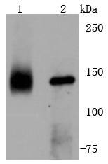
Fig1: Western blot analysis of TrkA on different lysates using anti-TrkA antibody at 1/1,000 dilution. Positive control: Lane 1: Mouse brain Lane 2: SH-SY-5YApplication
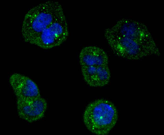
Fig2: ICC staining TrkA in Hela cells (green). The nuclear counter stain is DAPI (blue). Cells were fixed in paraformaldehyde, permeabilised with 0.25% Triton X100/PBS.Application
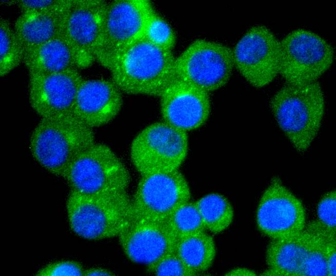
Fig3: ICC staining TrkA in N2A cells (green). The nuclear counter stain is DAPI (blue). Cells were fixed in paraformaldehyde, permeabilised with 0.25% Triton X100/PBS.Application
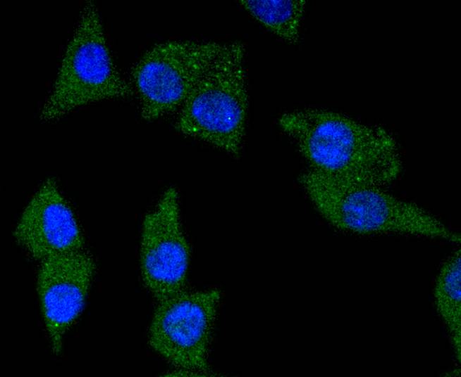
Fig4: ICC staining TrkA in SH-SY-5Y cells (green). The nuclear counter stain is DAPI (blue). Cells were fixed in paraformaldehyde, permeabilised with 0.25% Triton X100/PBS.Application
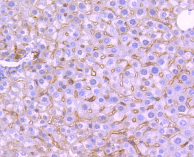
Fig5: Immunohistochemical analysis of paraffin-embedded mouse liver tissue using anti-TrkA antibody. Counter stained with hematoxylin.Application
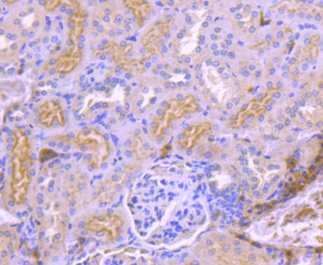
Fig6: Immunohistochemical analysis of paraffin-embedded mouse kidney tissue using anti-TrkA antibody. Counter stained with hematoxylin.Application
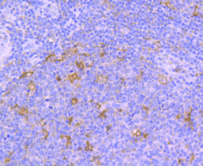
Fig7: Immunohistochemical analysis of paraffin-embedded human tonsil tissue using anti-TrkA antibody. Counter stained with hematoxylin.Application
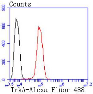
Fig8: Flow cytometric analysis of SH-SY-5Y cells with TrkA antibody at 1/50 dilution (red) compared with an unlabelled control (cells without incubation with primary antibody; black). Alexa Fluor 488-conjugated goat anti rabbit IgG was used as the secondary antibody.| Product Name | TrkA [JJ084-04] |
|---|---|
| Antibody Type | Primary Antibodies |
| Antigen Alias | gp140trk antibody High affinity nerve growth factor receptor antibody High affinity nerve growth factor receptor precursor antibody MTC antibody Neurotrophic tyrosine kinase receptor type 1 antibody NTRK1 antibody NTRK1_HUMAN antibody Oncogene TRK antibody p14-TrkA antibody p140 TrkA antibody p140-TrkA antibody Slow nerve growth antibody Trk A antibody TRK antibody Trk-A antibody TRK1 antibody TRK1-transforming tyrosine kinase protein antibody Tropomyosin-related kinase A antibody Tyrosine kinase receptor A antibody Tyrosine kinase receptor antibody |
| Product description | The family of Trk receptor tyrosine kinases consists of TrkA, TrkB, and TrkC. While the sequence of these family members is highly conserved, they are activated by different neurotrophins: TrkA by NGF, TrkB by BDNF or NT4, and TrkC by NT3. Neurotrophin signaling through these receptors regulates a number of physiological processes, such as cell survival, proliferation, neural development, and axon and dendrite growth and patterning. In the adult nervous system, the Trk receptors regulate synaptic strength and plasticity. TrkA regulates proliferation and is important for development and maturation of the nervous system. Point mutations, deletions, and chromosomal rearrangements (chimeras) cause ligand-independent receptor dimerization and activation of TrkA. TrkA is activated in many malignancies including breast, ovarian, prostate, and thyroid carcinomas. Research studies suggest that expression of TrkA in neuroblastomas may be a good prognostic marker as TrkA signals growth arrest and differentiation of cells originating from the neural crest. |
| Immunogen | recombinant protein |
| Clonality | Monoclonal |
|---|---|
| Isotype | IgG |
| Host Species | Recombinant rabbit |
| Tested Applications | WBICC/IFIHCIPFC |
| WB:1:1,000-1:2,000 ICC:1:50-1:200 IHC:1:50-1:200 FC:1:50-1:100 |
|
| Species Reactivity | HumanMouseRat |
| Concentration | 1mg/ml |
| Alternative Names | gp140trk antibody High affinity nerve growth factor receptor antibody High affinity nerve growth factor receptor precursor antibody MTC antibody Neurotrophic tyrosine kinase receptor type 1 antibody NTRK1 antibody NTRK1_HUMAN antibody Oncogene TRK antibody p14-TrkA antibody p140 TrkA antibody p140-TrkA antibody Slow nerve growth antibody Trk A antibody TRK antibody Trk-A antibody TRK1 antibody TRK1-transforming tyrosine kinase protein antibody Tropomyosin-related kinase A antibody Tyrosine kinase receptor A antibody Tyrosine kinase receptor antibody |
|---|---|
| Molecular Weight(MW) | 145 kDa |
| Cellular Localization | Cell membrane, Early endosome membrane, Late endosome membrane. |
Application
Fig1: Western blot analysis of TrkA on different lysates using anti-TrkA antibody at 1/1,000 dilution. Positive control: Lane 1: Mouse brain Lane 2: SH-SY-5Y
Application
Fig2: ICC staining TrkA in Hela cells (green). The nuclear counter stain is DAPI (blue). Cells were fixed in paraformaldehyde, permeabilised with 0.25% Triton X100/PBS.
Application
Fig3: ICC staining TrkA in N2A cells (green). The nuclear counter stain is DAPI (blue). Cells were fixed in paraformaldehyde, permeabilised with 0.25% Triton X100/PBS.
Application
Fig4: ICC staining TrkA in SH-SY-5Y cells (green). The nuclear counter stain is DAPI (blue). Cells were fixed in paraformaldehyde, permeabilised with 0.25% Triton X100/PBS.
Application
Fig5: Immunohistochemical analysis of paraffin-embedded mouse liver tissue using anti-TrkA antibody. Counter stained with hematoxylin.
Application
Fig6: Immunohistochemical analysis of paraffin-embedded mouse kidney tissue using anti-TrkA antibody. Counter stained with hematoxylin.
Application
Fig7: Immunohistochemical analysis of paraffin-embedded human tonsil tissue using anti-TrkA antibody. Counter stained with hematoxylin.
Application
Fig8: Flow cytometric analysis of SH-SY-5Y cells with TrkA antibody at 1/50 dilution (red) compared with an unlabelled control (cells without incubation with primary antibody; black). Alexa Fluor 488-conjugated goat anti rabbit IgG was used as the secondary antibody.| Positive Control | N2A, SH-SY-5Y, Hela, human tonsil tissue, mouse liver tissue, mouse brain tissue, mouse kidney tissue. |
|---|---|
| Application Notes | WB:1:1,000-1:2,000 ICC:1:50-1:200 IHC:1:50-1:200 FC:1:50-1:100 |
| Form | Liquid |
|---|---|
| Storage Instructions | Store at +4℃ after thawing. Aliquot store at -20℃ or -80℃. Avoid repeated freeze / thaw cycles. |
| Storage Buffer | 1*TBS (pH7.4), 1%BSA, 40%Glycerol. Preservative: 0.05% Sodium Azide. |


