WB
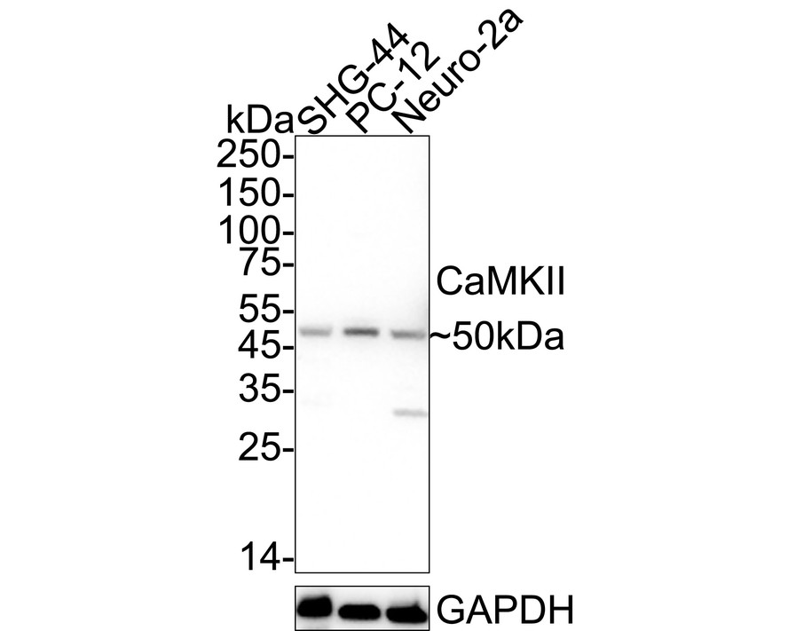
Western blot analysis of CaMKII on different lysates with Rabbit anti-CaMKII antibody at 1/1,000 dilution. Lane 1: SHG-44 cell lysate, Lane 2: PC-12 cell lysate, Lane 3: Neuro-2a cell lysate, Lysates/proteins at 30 µg/Lane. Exposure time: 14 seconds; 4-20% SDS-PAGE gel. Proteins were transferred to a PVDF membrane and blocked with 5% NFDM/TBST for 1 hour at room temperature. The primary antibody at 1/1,000 dilution was used in 5% NFDM/TBST at 4℃ overnight. Goat Anti-Rabbit IgG - HRP Secondary Antibody at 1/50,000 dilution was used for 1 hour at room temperature.IHC
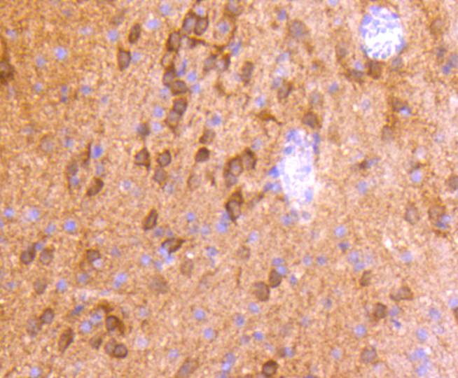
Immunohistochemical analysis of paraffin-embedded rat brain tissue using anti-CaMKⅡ antibody. Counter stained with hematoxylin.IHC
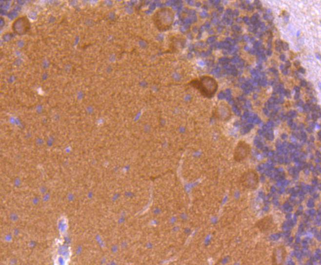
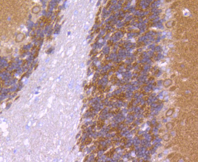
Immunohistochemical analysis of paraffin-embedded rat cerebellum tissue using anti-CaMKⅡ antibody. Counter stained with hematoxylin.IHC
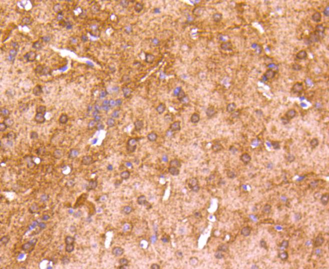
Immunohistochemical analysis of paraffin-embedded mouse brain tissue using anti-CaMKⅡ antibody. Counter stained with hematoxylin.IHC
Immunohistochemical analysis of paraffin-embedded mouse cerebellum tissue using anti-CaMKⅡ antibody. Counter stained with hematoxylin.ICC/IF
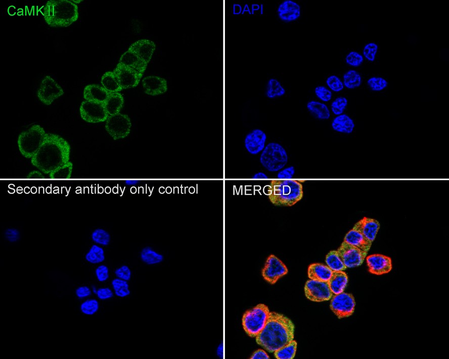
Immunocytochemistry analysis of PC-12 cells labeling CaMKII with Rabbit anti-CaMKII antibody at 1/100 dilution. Cells were fixed in 4% paraformaldehyde for 15 minutes at room temperature, permeabilized with 0.1% Triton X-100 in PBS for 15 minutes at room temperature, then blocked with 1% BSA in 10% negative goat serum for 1 hour at room temperature. Cells were then incubated with Rabbit anti-CaMKII antibody at 1/100 dilution in 1% BSA in PBST overnight at 4 ℃. Goat Anti-Rabbit IgG H&L (488) was used as the secondary antibody at 1/1,000 dilution. PBS instead of the primary antibody was used as the secondary antibody only control. Nuclear DNA was labelled in blue with DAPI. Beta tubulin (red) was stained at 1/100 dilution overnight at +4℃. Goat Anti-Mouse IgG H&L (594) was used as the secondary antibody at 1/1,000 dilution.IF-F
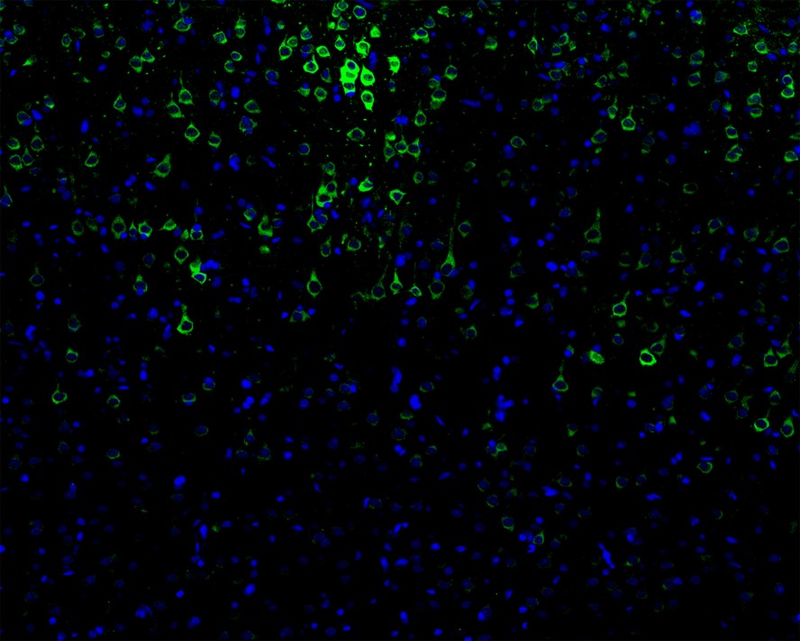
Immunofluorescence analysis of frozen mouse cerebral cortex tissue labeling CaMKⅡ with Rabbit anti-CaMKⅡ antibody. The tissues were blocked in 3% BSA for 30 minutes at room temperature, washed with PBS, and then probed with the primary antibody ((green) at 1/100 dilution overnight at 4℃, washed with PBS. Goat Anti-Rabbit IgG H&L (Alexa Fluor® 488) was used as the secondary antibody at 1/200 dilution. Nuclei were counterstained with DAPI (blue).FC
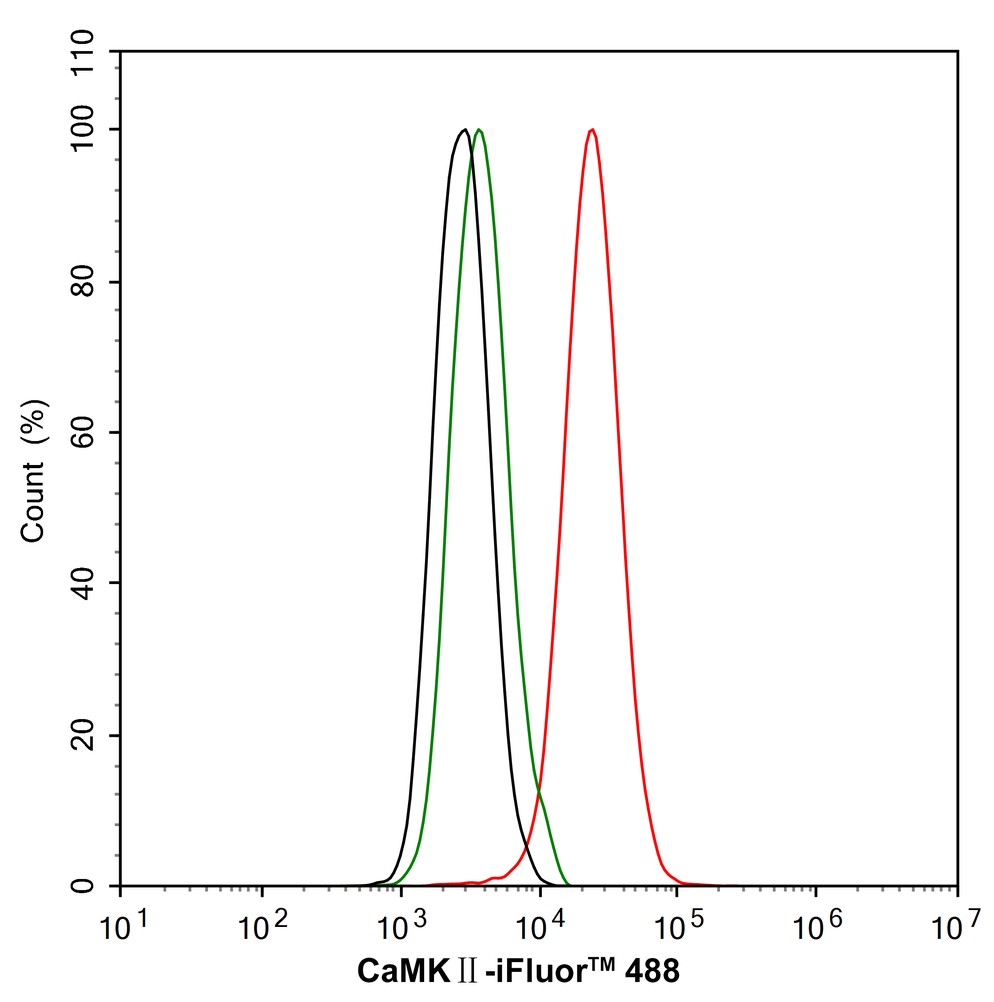
Flow cytometric analysis of PC-12 cells labeling CaMKII. Cells were fixed and permeabilized. Then stained with the primary antibody (1/1,000) (red) compared with Rabbit IgG Isotype Control (green). After incubation of the primary antibody at +4℃ for an hour, the cells were stained with a iFluor™ 488 conjugate-Goat anti-Rabbit IgG Secondary antibody at 1/1,000 dilution for 30 minutes at +4℃. Unlabelled sample was used as a control (cells without incubation with primary antibody; black).| Product Name | CaMKII Recombinant Rabbit Monoclonal Antibody |
|---|---|
| Antibody Type | Primary Antibodies |
| Product description | The Ca2+/calmodulin-dependent protein kinases (CaM kinases) comprise a structurally related subfamily of serine/threonine kinases which include CaMKI, CaMKII and CaMKIV. CaMKII is a ubiquitously expressed serine/threonine protein kinase that is activated by Ca2+and calmodulin (CaM) and has been implicated in regulation of the cell cycle and transcription. There are four CaMKII isozymes designated α, β, γ and δ, which may or may not be co-expressed in the same tissue type. CaMKIV is stimulated by Ca2+ and CaM but also requires phosphorylation by a CaMK for full activation. Stimulation of the T cell receptor CD3 signaling complex with an anti-CD3 monoclonal antibody leads to a 10-40 fold increase in CaMKIV activity. An additional kinase, CaMKK, functions to activate CaMKI through the specific phosphorylation of the regulatory Threonine residue at position 177. |
| Immunogen | Synthetic peptide within Human CaMKⅡ aa 201-250 / 478. |
| Clonality | Monoclonal |
|---|---|
| Isotype | IgG |
| Host Species | Recombinant rabbit |
| Tested Applications | FCICC/IFIF-FIHCWB |
| WB:1:500-1:2000 IHC:1:50-1:200 ICC/IF:1:100 IF-F:1:100 FC:1:1000 |
|
| Species Reactivity | HumanMouseRat |
| Concentration | 1mg/ml |
| Purification | Protein A |
| Alternative Names | Calcium/calmodulin dependent protein kinase II alpha antibody Calcium/calmodulin dependent protein kinase II beta antibody Calcium/calmodulin dependent protein kinase II delta antibody Calcium/calmodulin dependent protein kinase II gamma antibody Calcium/calmodulin-dependent protein kinase type II subunit alpha antibody CaM kinase II alpha antibody CaM kinase II antibody CaM kinase II beta antibody CaM kinase II delta antibody CaM kinase II gamma antibody CaM kinase II subunit alpha antibody CaMK-II subunit alpha antibody CAMK2 antibody Camk2a antibody CAMK2B antibody CAMK2D antibody CAMK2G antibody CAMKA antibody KCC2A_HUMAN antibody |
|---|---|
| Molecular Weight(MW) | 54kDa(Observed band size: 50kDa) |
| Cellular Localization | Cytoplasm, Sarcoplasmic reticulum membrane, Cell membrane, Cell junction. |
WB
Western blot analysis of CaMKII on different lysates with Rabbit anti-CaMKII antibody at 1/1,000 dilution. Lane 1: SHG-44 cell lysate, Lane 2: PC-12 cell lysate, Lane 3: Neuro-2a cell lysate, Lysates/proteins at 30 µg/Lane. Exposure time: 14 seconds; 4-20% SDS-PAGE gel. Proteins were transferred to a PVDF membrane and blocked with 5% NFDM/TBST for 1 hour at room temperature. The primary antibody at 1/1,000 dilution was used in 5% NFDM/TBST at 4℃ overnight. Goat Anti-Rabbit IgG - HRP Secondary Antibody at 1/50,000 dilution was used for 1 hour at room temperature.
IHC
Immunohistochemical analysis of paraffin-embedded rat brain tissue using anti-CaMKⅡ antibody. Counter stained with hematoxylin.
IHC
Immunohistochemical analysis of paraffin-embedded rat cerebellum tissue using anti-CaMKⅡ antibody. Counter stained with hematoxylin.
IHC
Immunohistochemical analysis of paraffin-embedded mouse brain tissue using anti-CaMKⅡ antibody. Counter stained with hematoxylin.
IHC
Immunohistochemical analysis of paraffin-embedded mouse cerebellum tissue using anti-CaMKⅡ antibody. Counter stained with hematoxylin.
ICC/IF
Immunocytochemistry analysis of PC-12 cells labeling CaMKII with Rabbit anti-CaMKII antibody at 1/100 dilution. Cells were fixed in 4% paraformaldehyde for 15 minutes at room temperature, permeabilized with 0.1% Triton X-100 in PBS for 15 minutes at room temperature, then blocked with 1% BSA in 10% negative goat serum for 1 hour at room temperature. Cells were then incubated with Rabbit anti-CaMKII antibody at 1/100 dilution in 1% BSA in PBST overnight at 4 ℃. Goat Anti-Rabbit IgG H&L (488) was used as the secondary antibody at 1/1,000 dilution. PBS instead of the primary antibody was used as the secondary antibody only control. Nuclear DNA was labelled in blue with DAPI. Beta tubulin (red) was stained at 1/100 dilution overnight at +4℃. Goat Anti-Mouse IgG H&L (594) was used as the secondary antibody at 1/1,000 dilution.
IF-F
Immunofluorescence analysis of frozen mouse cerebral cortex tissue labeling CaMKⅡ with Rabbit anti-CaMKⅡ antibody. The tissues were blocked in 3% BSA for 30 minutes at room temperature, washed with PBS, and then probed with the primary antibody ((green) at 1/100 dilution overnight at 4℃, washed with PBS. Goat Anti-Rabbit IgG H&L (Alexa Fluor® 488) was used as the secondary antibody at 1/200 dilution. Nuclei were counterstained with DAPI (blue).
FC
Flow cytometric analysis of PC-12 cells labeling CaMKII. Cells were fixed and permeabilized. Then stained with the primary antibody (1/1,000) (red) compared with Rabbit IgG Isotype Control (green). After incubation of the primary antibody at +4℃ for an hour, the cells were stained with a iFluor™ 488 conjugate-Goat anti-Rabbit IgG Secondary antibody at 1/1,000 dilution for 30 minutes at +4℃. Unlabelled sample was used as a control (cells without incubation with primary antibody; black).| Application Notes | WB:1:500-1:2000 IHC:1:50-1:200 ICC/IF:1:100 IF-F:1:100 FC:1:1000 |
|---|
| Form | Liquid |
|---|---|
| Storage Instructions | Store at +4℃ after thawing. Aliquot store at -20℃ or -80℃. Avoid repeated freeze / thaw cycles. |
| Storage Buffer | 1*TBS (pH7.4), 1%BSA, 40%Glycerol. Preservative: 0.05% Sodium Azide. |


