WB
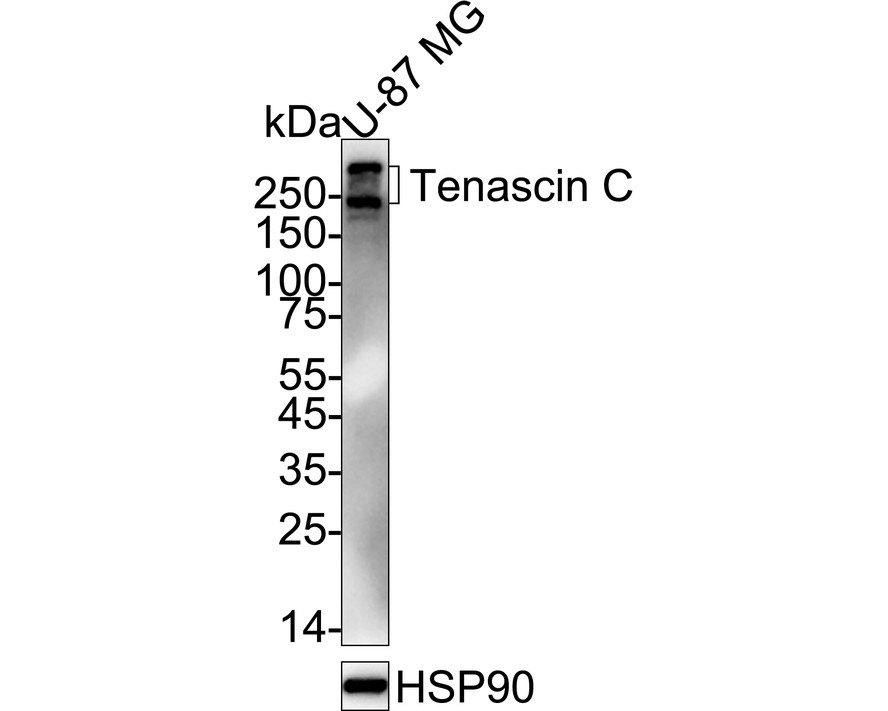
Western blot analysis of Tenascin C on U-87 MG cell lysates with Rabbit anti-Tenascin C antibody at 1/2,000 dilution. Lysates/proteins at 20 µg/Lane. Exposure time: 3 minutes; 4-20% SDS-PAGE gel. Proteins were transferred to a PVDF membrane and blocked with 5% NFDM/TBST for 1 hour at room temperature. The primary antibody at 1/2,000 dilution was used in 5% NFDM/TBST at 4℃ overnight. Goat Anti-Rabbit IgG - HRP Secondary Antibody at 1/50,000 dilution was used for 1 hour at room temperature.IHC
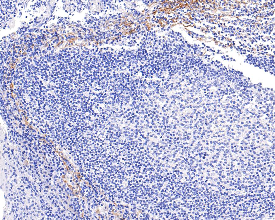
Immunohistochemical analysis of paraffin-embedded human tonsil tissue with Rabbit anti-Tenascin C antibody at 1/400 dilution. The section was pre-treated using heat mediated antigen retrieval with Tris-EDTA buffer (pH 9.0) for 20 minutes. The tissues were blocked in 1% BSA for 20 minutes at room temperature, washed with ddH2O and PBS, and then probed with the primary antibody at 1/400 dilution for 1 hour at room temperature. The detection was performed using an HRP conjugated compact polymer system. DAB was used as the chromogen. Tissues were counterstained with hematoxylin and mounted with DPX.IHC
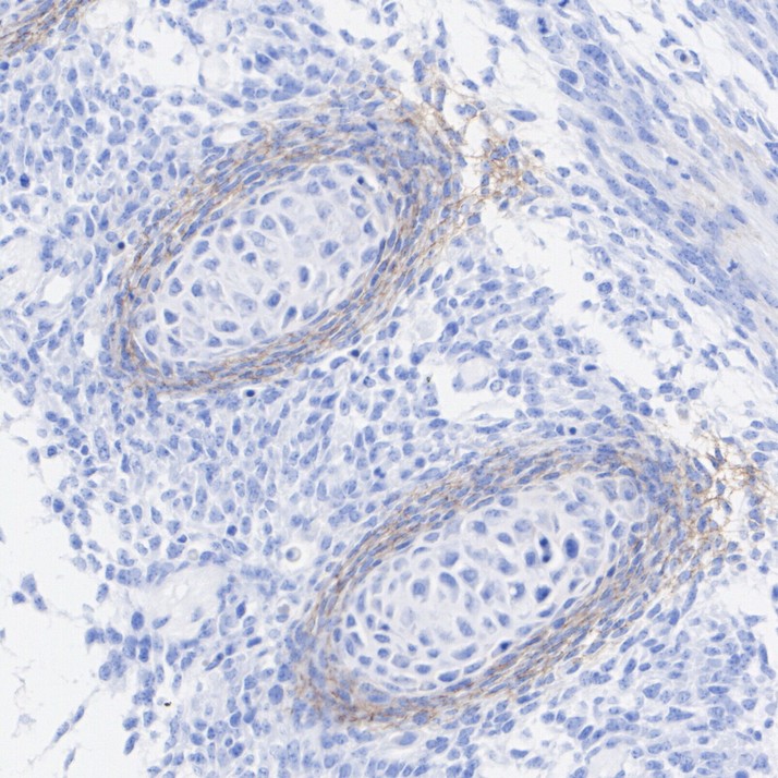
Immunohistochemical analysis of paraffin-embedded mouse embryonic cartilage tissue with Rabbit anti-Tenascin C antibody at 1/500 dilution. The section was pre-treated using heat mediated antigen retrieval with Tris-EDTA buffer (pH 9.0) for 20 minutes. The tissues were blocked in 1% BSA for 20 minutes at room temperature, washed with ddH2O and PBS, and then probed with the primary antibody at 1/500 dilution for 1 hour at room temperature. The detection was performed using an HRP conjugated compact polymer system. DAB was used as the chromogen. Tissues were counterstained with hematoxylin and mounted with DPX.IHC
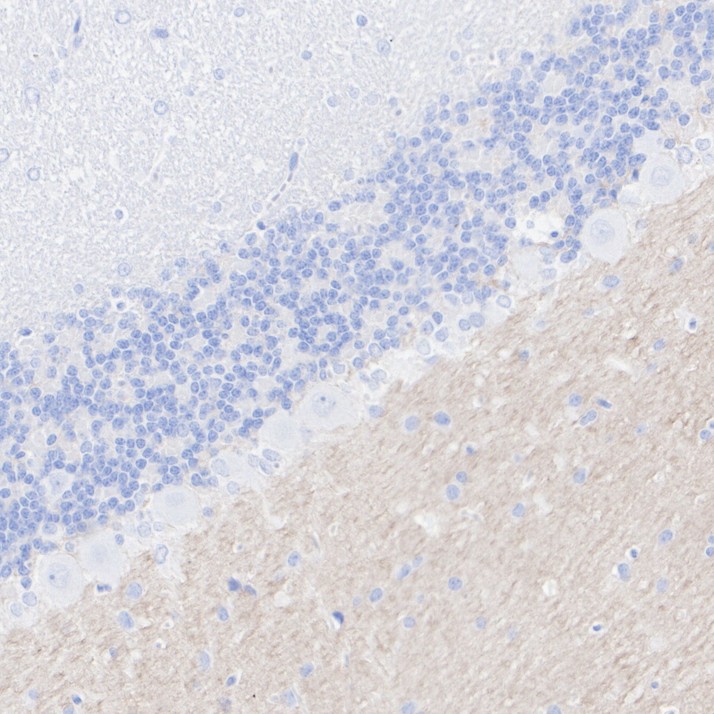
Immunohistochemical analysis of paraffin-embedded rat cerebellum tissue with Rabbit anti-Tenascin C antibody at 1/500 dilution. The section was pre-treated using heat mediated antigen retrieval with Tris-EDTA buffer (pH 9.0) for 20 minutes. The tissues were blocked in 1% BSA for 20 minutes at room temperature, washed with ddH2O and PBS, and then probed with the primary antibody at 1/500 dilution for 1 hour at room temperature. The detection was performed using an HRP conjugated compact polymer system. DAB was used as the chromogen. Tissues were counterstained with hematoxylin and mounted with DPX.IF-F
Species: Mouse, Site: Cerebellum, Sample: Frozen section, Antigen retrieval: Recommend. The section was pre-treated using heat mediated antigen retrieval with sodium citrate buffer (pH 6.0) for about 2 minutes in microwave oven.IF-P
Species: Mouse, Site: Cerebellum, Sample: Paraffin-embedded section.| Product Name | Tenascin C Recombinant Rabbit Monoclonal Antibody |
|---|---|
| Antibody Type | Primary Antibodies |
| Product description | The tenascin family of extracellular matrix proteins includes Tenascin-C (also designated cytotactin or Tenascin), Tenascin-R (also designated restrictin, TN-R or janusin) and Tenascin-X. Tenascin proteins function as substrate-adhesion molecules (SAMs) and are involved in regulating numerous developmental processes, such as morphogenetic cell migration and organogenesis. The tenascin family proteins arise from various splicing events in the region of coding for FNIII repeats. Tenascin-C and Tenascin-X are expressed in several tissues during embryogenesis and in adult tissues undergoing active remodeling, such as healing wounds and tumors. Tenascin-R is expressed on the surface of neurons and glial cells. |
| Immunogen | Synthetic peptide within Human Tenascin C aa 2,152-2,201 / 2,201. |
| Clonality | Monoclonal |
|---|---|
| Isotype | IgG |
| Host Species | Recombinant rabbit |
| Tested Applications | IF-FIF-PIHCWB |
| WB:1:2000 IHC:1:50-1:100 IF-F:1:500 IF-P:1:500 |
|
| Species Reactivity | HumanMouseRat |
| Concentration | 1mg/ml |
| Purification | Protein A |
| Alternative Names | 150 225 antibody Cytotactin antibody Glioma associated extracellular matrix antigen antibody Glioma-associated-extracellular matrix antigen antibody GMEM antibody GP 150 225 antibody GP 150-225 antibody GP antibody Hexabrachion antibody HXB antibody JI antibody MGC167029 antibody Miotendinous antigen antibody Myotendinous antigen antibody Neuronectin antibody TENA_HUMAN antibody Tenascin antibody Tenascin-C antibody Tenascin-C isoform 14/AD1/16 antibody TenascinC antibody TN antibody TN C antibody TN-C antibody TNC antibody |
|---|---|
| Molecular Weight(MW) | 241kDa(Observed band size: 241-350kDa) |
| Cellular Localization | Secreted.Extracellular matrix. |
| SwissProt ID | P24821 |
|---|
WB
Western blot analysis of Tenascin C on U-87 MG cell lysates with Rabbit anti-Tenascin C antibody at 1/2,000 dilution. Lysates/proteins at 20 µg/Lane. Exposure time: 3 minutes; 4-20% SDS-PAGE gel. Proteins were transferred to a PVDF membrane and blocked with 5% NFDM/TBST for 1 hour at room temperature. The primary antibody at 1/2,000 dilution was used in 5% NFDM/TBST at 4℃ overnight. Goat Anti-Rabbit IgG - HRP Secondary Antibody at 1/50,000 dilution was used for 1 hour at room temperature.
IHC
Immunohistochemical analysis of paraffin-embedded human tonsil tissue with Rabbit anti-Tenascin C antibody at 1/400 dilution. The section was pre-treated using heat mediated antigen retrieval with Tris-EDTA buffer (pH 9.0) for 20 minutes. The tissues were blocked in 1% BSA for 20 minutes at room temperature, washed with ddH2O and PBS, and then probed with the primary antibody at 1/400 dilution for 1 hour at room temperature. The detection was performed using an HRP conjugated compact polymer system. DAB was used as the chromogen. Tissues were counterstained with hematoxylin and mounted with DPX.
IHC
Immunohistochemical analysis of paraffin-embedded mouse embryonic cartilage tissue with Rabbit anti-Tenascin C antibody at 1/500 dilution. The section was pre-treated using heat mediated antigen retrieval with Tris-EDTA buffer (pH 9.0) for 20 minutes. The tissues were blocked in 1% BSA for 20 minutes at room temperature, washed with ddH2O and PBS, and then probed with the primary antibody at 1/500 dilution for 1 hour at room temperature. The detection was performed using an HRP conjugated compact polymer system. DAB was used as the chromogen. Tissues were counterstained with hematoxylin and mounted with DPX.
IHC
Immunohistochemical analysis of paraffin-embedded rat cerebellum tissue with Rabbit anti-Tenascin C antibody at 1/500 dilution. The section was pre-treated using heat mediated antigen retrieval with Tris-EDTA buffer (pH 9.0) for 20 minutes. The tissues were blocked in 1% BSA for 20 minutes at room temperature, washed with ddH2O and PBS, and then probed with the primary antibody at 1/500 dilution for 1 hour at room temperature. The detection was performed using an HRP conjugated compact polymer system. DAB was used as the chromogen. Tissues were counterstained with hematoxylin and mounted with DPX.
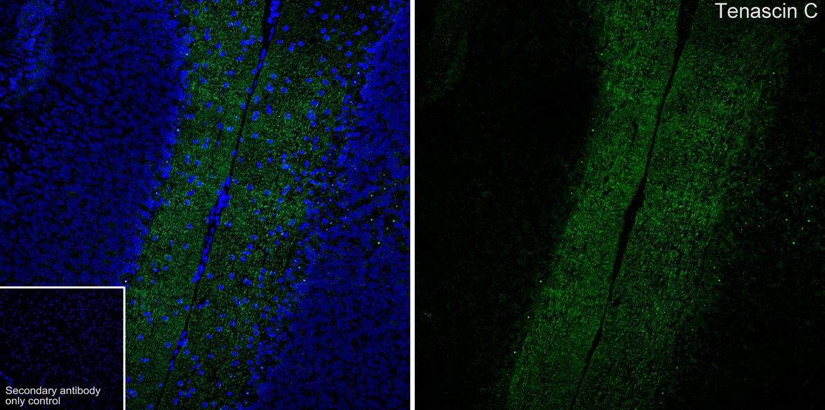
IF-F
Species: Mouse, Site: Cerebellum, Sample: Frozen section, Antigen retrieval: Recommend. The section was pre-treated using heat mediated antigen retrieval with sodium citrate buffer (pH 6.0) for about 2 minutes in microwave oven.
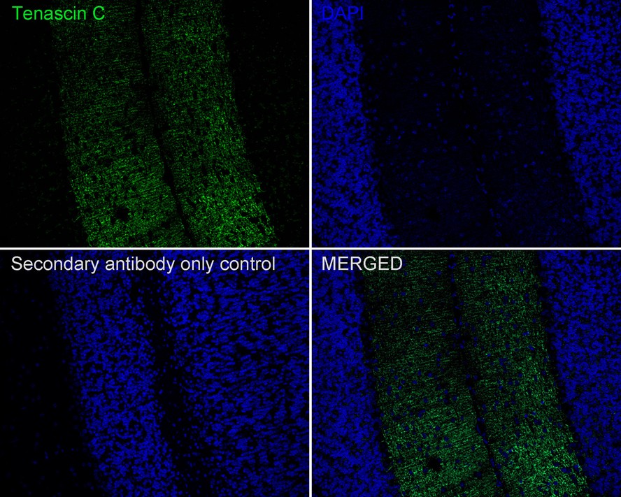
IF-P
Species: Mouse, Site: Cerebellum, Sample: Paraffin-embedded section.| Application Notes | WB:1:2000 IHC:1:50-1:100 IF-F:1:500 IF-P:1:500 |
|---|
| Form | Liquid |
|---|---|
| Storage Instructions | Store at +4℃ after thawing. Aliquot store at -20℃ or -80℃. Avoid repeated freeze / thaw cycles. |
| Storage Buffer | 1*TBS (pH7.4), 1%BSA, 40%Glycerol. Preservative: 0.05% Sodium Azide. |


